
Article Figures & Data
Figures
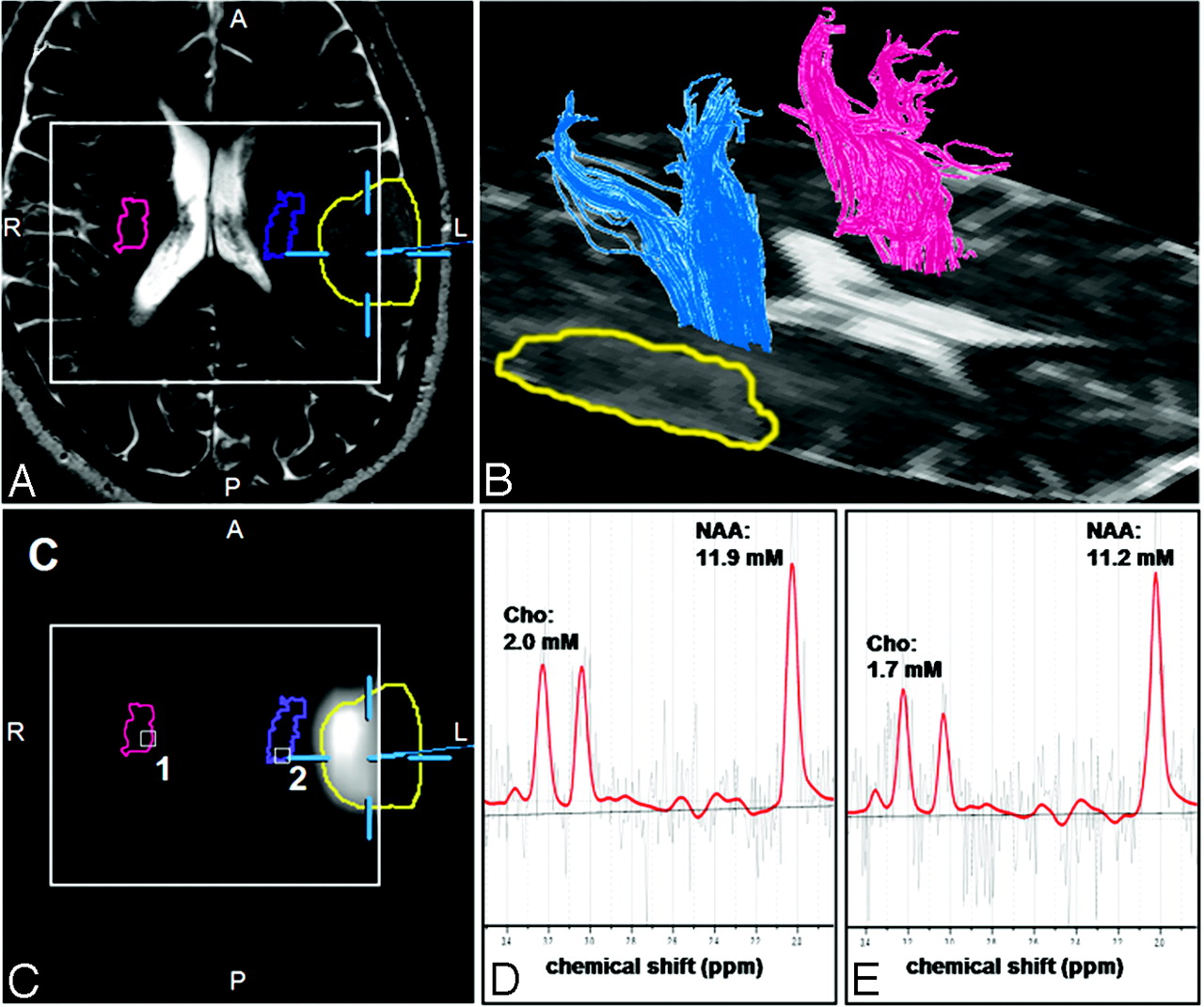
- Fig 1.
Results of the fiber-tracking procedure of the DTI data and the spectral analysis of the MRSI data for a patient having no sensorimotor deficits (patient 17 in Table 1). Screenshot from the planning workstation of a navigation system of an axial T2-weighted MR imaging (A) coregistered with a segmented metabolic Cho/NAA map (C). Overlaid on these images are the cross sections of the ipsilateral (blue) and contralateral (magenta) pyramidal tracts, the tumor segmented manually by a neurosurgeron (yellow), and the VOI (PRESS-box) of the MRSI experiment (white rectangle).
B, a 3D reconstruction of the ipsilateral (blue) and contralateral (magenta) pyramidal tracts depicted on the axial sections of the DTI dataset measured with a b-value = 0 s/mm2. LCModel fits (red line) of the MRSI data of voxel position 1 (D) and 2 (E) as depicted in C as white squares. Overlaid on these images are the molar concentrations for Cho, Cr, and NAA calculated by LCModel.
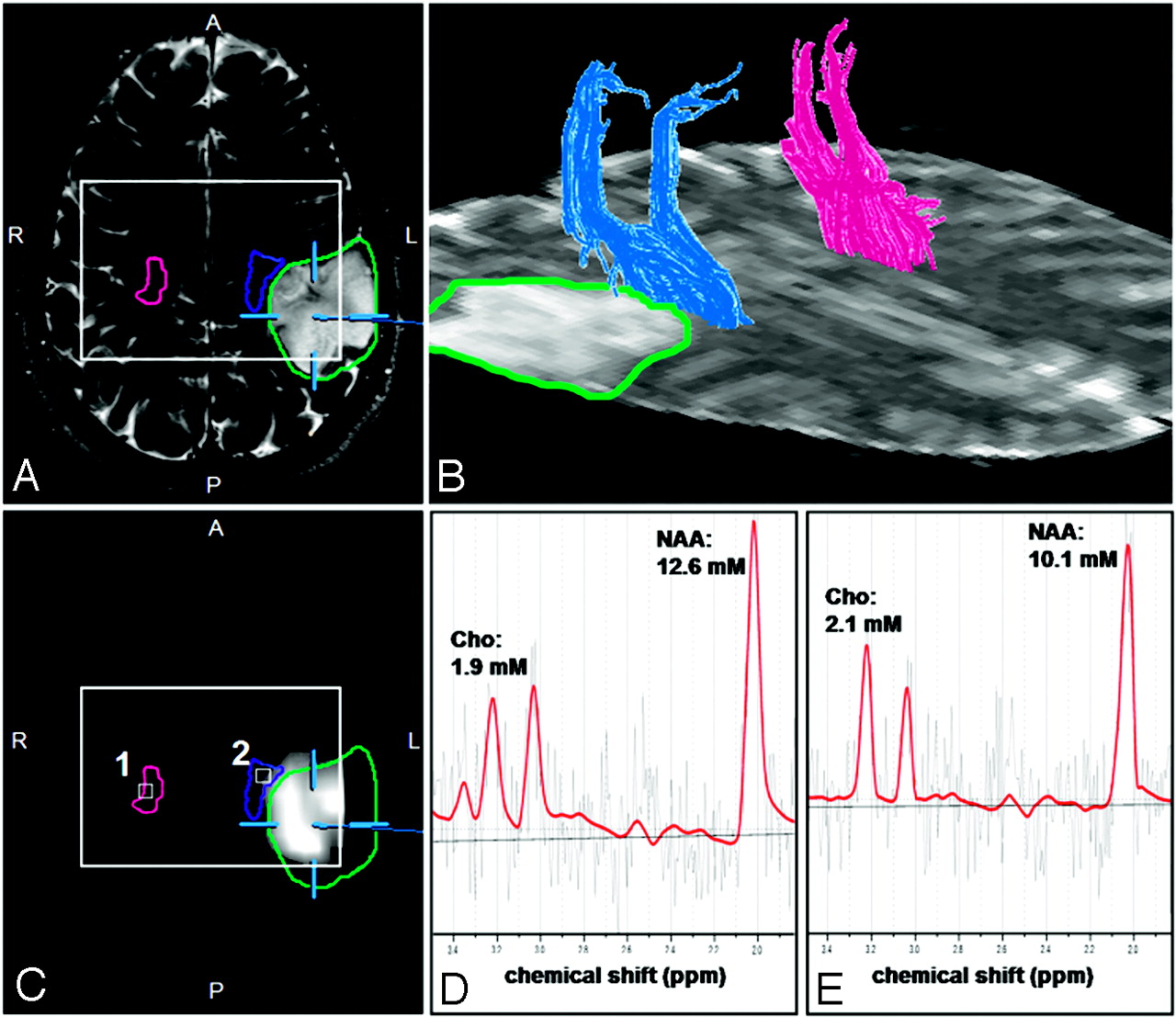
- Fig 2.
Results of the fiber-tracking procedure of the DTI data and the spectral analysis of the MRSI data for a patient with a hypesthesia in right arm (patient 14 in Table 1). Screenshot from the planning workstation of a navigation system of an axial T2-weighted MR imaging (A) coregistred with a segmented metabolic Cho/NAA map (C). Overlaid on these images are the cross sections of the ipsilateral (blue) and contralateral (magenta) pyramidal tracts, the tumor segmented manually by a neurosurgeon (green), and the PRESS-box of the MRSI experiment (white rectangle).
B, a 3D reconstruction of the ipsilateral (blue) and contralateral (magenta) pyramidal tracts depicted on the axial sections of the DTI dataset measured with a b-value = 0 s/mm2. LCModel fits (red line) of the MRSI data of voxel positions 1 (D) and 2 (E) as depicted in C as white squares. Overlaid on these images are the molar concentrations for Cho, Cr, and NAA calculated by LCModel.
- Fig 3.
Shown are boxplots of FpV, FA, and MD values of the whole ipsilateral and contralateral pyramidal tracts (A, B, and C, respectively). The horizontal lines are the medians, and the ends of the boxes are the lower and upper quartiles (25th and 75th percentiles). MD values are expressed in units × 10−3 mm2/s. The error bars depict the SD. Overlaid are the P values calculated from a 2-sided paired t test (ns = not significant).
- Fig 4.
Shown are boxplots of FpV, FA, and MD values for the section of the ipsilateral and contralateral pyramidal tracts (A, B, and C, respectively) corresponding with the MRSI section, respectively. Boxplots of molar concentrations of NAA, Cho, and the Cho/NAA ratio averaged over voxel positions located at the ipsilateral and contralateral pyramidal tract cross sections (D, E, and F, respectively). MD values are expressed in units × 10−3 mm2/s. NAA and Cho values are are expressed in millimoles per liter. Overlaid are the P values calculated from a 2-sided paired t test.
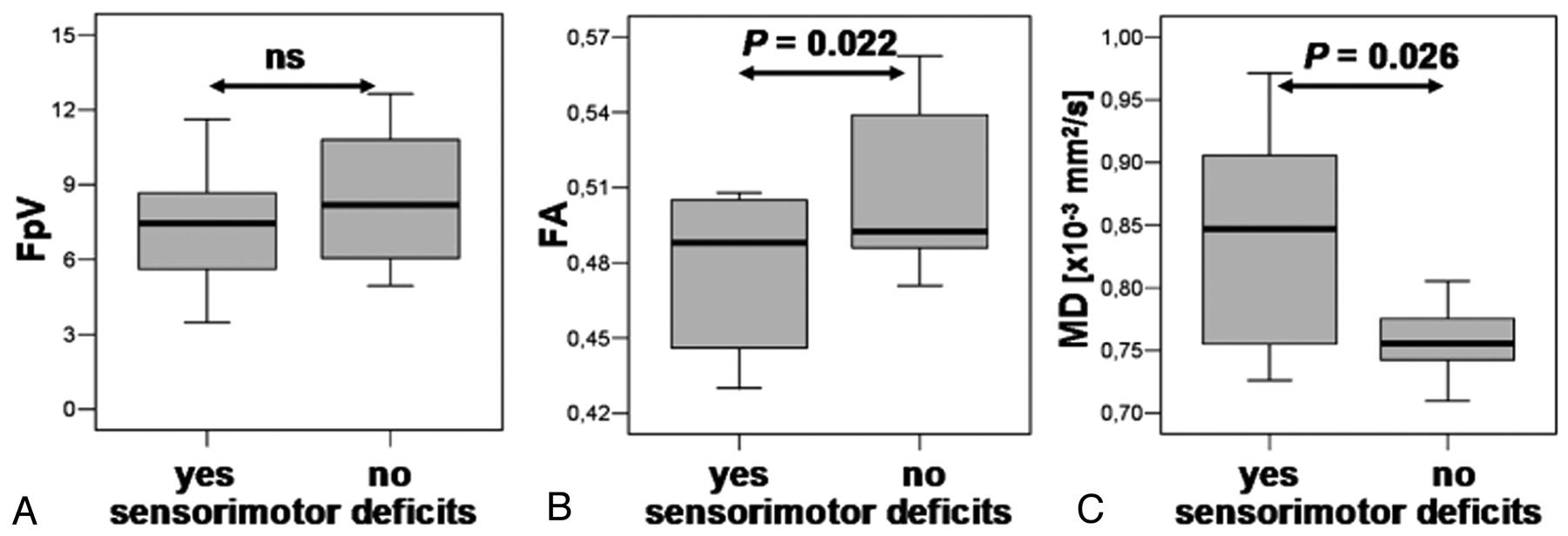
- Fig 5.
Boxplots of FpV, FA, and MD values of the section of the ipsilateral pyramidal tracts corresponding with the MRSI section for subgroups of patients without sensorimotor deficits, “no,” and patients with sensorimotor deficits, “yes” (A, B, and C, respectively). MD values are expressed in units × 10−3 mm2/s. Overlaid are the P values calculated from a 2-sided unpaired t test (ns = not significant).


Tables
- Table 1:
Type, grade, and location of the investigated brain tumors and preoperative sensorimotor deficits of the 20 patients
Patient Age (Years) Sex Clinical Diagnosis and Lesion Location Lesion Location Relative to PT Preoperative Sensorimotor Deficits 1 29 M Oligoastrocytoma III, right frontal Medial Paresthesia, left arm and leg 2 24 M Astrocytoma II, right frontal Lateral None 3 35 F Oligodendroglioma III, left frontal Anterior Paresthesia, right leg 4 29 F Astrocytoma II, right central Anterior None 5 39 F Oligoastrocytoma III, left frontal Anterior Hemiparesis, right 6 29 M Astrocytoma III, right frontal Anterior None 7 64 M GBM IV, right temporoparietal Posterior Hemiparesis, hemihypesthesia, left 8 29 M Oligoastrocytoma III, left temporal Lateral None 9 37 M Astrocytoma II, right frontotemporal Anterior None 10 30 M Astrocytoma III, right temporal Anterior None 11 53 F Oligoastrocytoma II, left central Anterior None 12 30 M Astrocytoma II, right postcentral Lateral Hypesthesia, left arm and leg 13 42 F Astrocytoma III, left central Medial None 14 29 M Astrocytoma II, left postcentral Lateral Hypesthesia, right arm 15 37 F Oligoastrocytoma III, right frontal Lateral None 16 39 F GBM IV, left frontal Medial None 17 63 M Astrocytoma III, left postcentral Lateral None 18 72 F GBM IV, left frontal Anterior Hemiparesis, hemihypesthesia, right 19 52 M Oligoastrocytoma III, right frontal Anterior None 20 50 F GBM IV, right temporal Lateral Hemiparesis, left Note:—M indicates male; F, female; GBM, glioblastoma multiforme; PT, pyramidal tract.
- Table 2:
P values for t tests between ipsilateral and contralateral parameters and for subgroups of patients
Sensorimotor Deficits Pyramidal Tract Total Pyramidal Tract Section MRSI FpV FA MD FpV FA MD NAA Cho Cho/NAA Yes (n = 8) .002 <.001 .04 .024 <.001 .003 .011 .002 .001 No (n = 12) <.001 <.001 NS <.001 .009 NS .033 NS .047 Note:—FpV indicates fibers per voxel; FA, fractional anisotropy; MD, mean diffusivity; NS, nonsignificant. Statistical significance indicated by P < .05 using 2-sided paired t tests.


