
Article Figures & Data
Figures
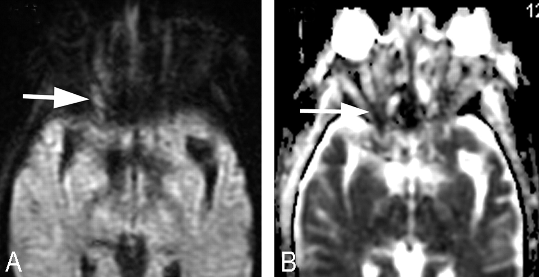
- Fig 1.
MR imaging performed 6 days after vision loss in the right eye. A, DWI shows subtle high signal intensity in the posterior right optic nerve (arrow). B, ADC map shows hypointense posterior right optic nerve (arrow).
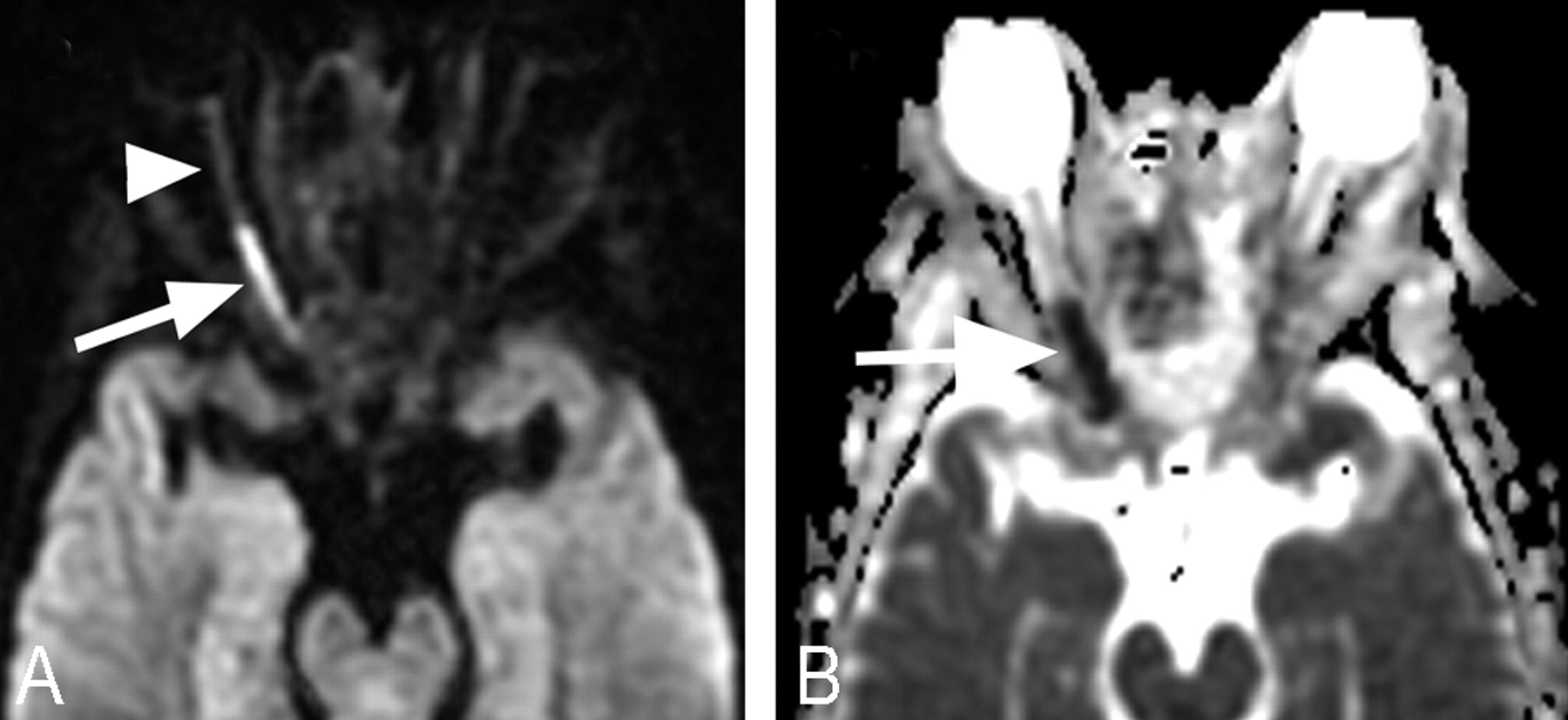
- Fig 2.
MR imaging performed 15 days after vision loss in the right eye. A, DWI demonstrates marked diffusion restriction in the posterior right optic nerve (arrow). Note that there is no obvious signal-intensity change of the retrobulbar portion of right optic nerve (arrowhead). B, ADC map shows a hypointense posterior right optic nerve (arrow).


In this issue


Jump to section
Related Articles
Cited By...
- MRI signs helpful in the differentiation of patients with anterior ischaemic optic neuropathy and optic neuritis
- Rhino-orbital-cerebral mucormycosis
- Hyperintense Optic Nerve Heads on Diffusion-Weighted Imaging: A Potential Imaging Sign of Papilledema
- Diffusion Changes in the Vitreous Humor of the Eye during Aging
- The "Black Turbinate" Sign: An Early MR Imaging Finding of Nasal Mucormycosis