Advertisement

Main menu

  • Home
  • Content
    • Current Issue
    • Accepted Manuscripts
    • Article Preview
    • Past Issue Archive
    • Video Articles
    • AJNR Case Collection
    • Case of the Week Archive
    • Case of the Month Archive
    • Classic Case Archive
  • Special Collections
    • AJNR Awards
    • Low-Field MRI
    • Alzheimer Disease
    • ASNR Foundation Special Collection
    • Photon-Counting CT
    • View All
  • Multimedia
    • AJNR Podcasts
    • AJNR SCANtastic
    • Trainee Corner
    • MRI Safety Corner
    • Imaging Protocols
  • For Authors
    • Submit a Manuscript
    • Submit a Video Article
    • Submit an eLetter to the Editor/Response
    • Manuscript Submission Guidelines
    • Statistical Tips
    • Fast Publishing of Accepted Manuscripts
    • Graphical Abstract Preparation
    • Imaging Protocol Submission
    • Author Policies
  • About Us
    • About AJNR
    • Editorial Board
    • Editorial Board Alumni
  • More
    • Become a Reviewer/Academy of Reviewers
    • Subscribers
    • Permissions
    • Alerts
    • Feedback
    • Advertisers
    • ASNR Home

User menu

  • Alerts
  • Log in

Search

  • Advanced search
American Journal of Neuroradiology

American Journal of Neuroradiology

ASHNR American Society of Functional Neuroradiology ASHNR American Society of Pediatric Neuroradiology ASSR
  • Alerts
  • Log in

Advanced Search

  • Home
  • Content
    • Current Issue
    • Accepted Manuscripts
    • Article Preview
    • Past Issue Archive
    • Video Articles
    • AJNR Case Collection
    • Case of the Week Archive
    • Case of the Month Archive
    • Classic Case Archive
  • Special Collections
    • AJNR Awards
    • Low-Field MRI
    • Alzheimer Disease
    • ASNR Foundation Special Collection
    • Photon-Counting CT
    • View All
  • Multimedia
    • AJNR Podcasts
    • AJNR SCANtastic
    • Trainee Corner
    • MRI Safety Corner
    • Imaging Protocols
  • For Authors
    • Submit a Manuscript
    • Submit a Video Article
    • Submit an eLetter to the Editor/Response
    • Manuscript Submission Guidelines
    • Statistical Tips
    • Fast Publishing of Accepted Manuscripts
    • Graphical Abstract Preparation
    • Imaging Protocol Submission
    • Author Policies
  • About Us
    • About AJNR
    • Editorial Board
    • Editorial Board Alumni
  • More
    • Become a Reviewer/Academy of Reviewers
    • Subscribers
    • Permissions
    • Alerts
    • Feedback
    • Advertisers
    • ASNR Home
  • Follow AJNR on Twitter
  • Visit AJNR on Facebook
  • Follow AJNR on Instagram
  • Join AJNR on LinkedIn
  • RSS Feeds

AJNR Awards, New Junior Editors, and more. Read the latest AJNR updates

Research ArticleSpine Imaging and Spine Image-Guided Interventions

Diffusion-Weighted Imaging of the Spine with a Non–Carr-Purcell-Meiboom-Gill Single-Shot Fast Spin-Echo Sequence: Initial Experience

A.Y. Oner, T. Tali, F. Celikyay, A. Celik and P. Le Roux
American Journal of Neuroradiology March 2007, 28 (3) 575-580;
A.Y. Oner
  • Find this author on Google Scholar
  • Find this author on PubMed
  • Search for this author on this site
T. Tali
  • Find this author on Google Scholar
  • Find this author on PubMed
  • Search for this author on this site
F. Celikyay
  • Find this author on Google Scholar
  • Find this author on PubMed
  • Search for this author on this site
A. Celik
  • Find this author on Google Scholar
  • Find this author on PubMed
  • Search for this author on this site
P. Le Roux
  • Find this author on Google Scholar
  • Find this author on PubMed
  • Search for this author on this site
  • Article
  • Figures & Data
  • Info & Metrics
  • Responses
  • References
  • PDF
Loading

Article Figures & Data

Figures

  • Tables
  • Fig 1.
    • Download figure
    • Open in new tab
    • Download powerpoint
    Fig 1.

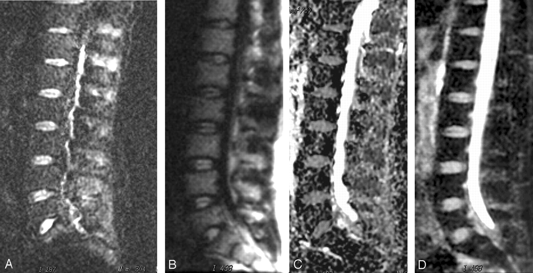
    Sagittal diffusion-weighted images in a 25-year-old man with normal spine MR imaging findings obtained at the same level with 2 different techniques: EPI DWI at b = 600 mm2/s (A) and non-CPMG SS-FSE DWI at b = 600 mm2/s (B) and corresponding apparent diffusion coefficient (ADC) maps (C and D, respectively). Increased signal intensity, with better background suppression is noted with the non-CPMG SS-FSE technique. ADC values for the L3 vertebra are (mean ± SD) 0.51 ± 0.17 and 0.37 ± 0.14 × 10⁻³ mm2/s for EPI and non-CPMG SS-FSE DWI, respectively.

  • Fig 2.
    • Download figure
    • Open in new tab
    • Download powerpoint
    Fig 2.

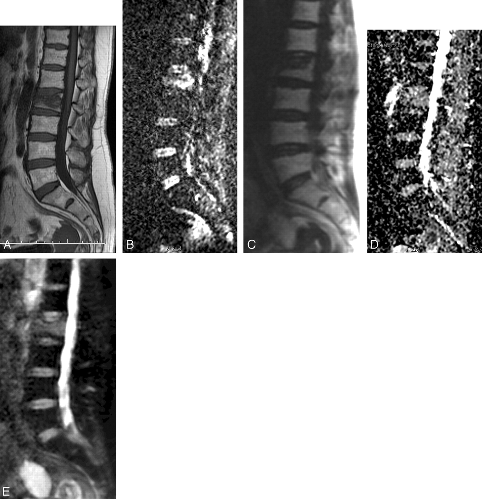
    A 46-year-old woman with L2 vertebral compression fracture. Sagittal T1-weighted image (A) and diffusion-weighted images obtained at the same level with 2 different techniques: EPI DWI at b = 600 mm2/s (B), non-CPMG SS-FSE DWI at b = 600 mm2/s (C), and corresponding apparent diffusion coefficient (ADC) maps (D and E, respectively). The compression is more readily appreciated on the non-CPMG SS-FSE images. ADC measured from L3 vertebral body is (mean ± SD × 10−3 mm2/s) 0.54 ± 0.14 and 0.38 ± 0.16 for EPI and non-CPMG SS-FSE DWI, respectively. ADC values measured from L2 vertebra are (mean ± SD) 1.63 ± 0.27 and 1.55 ± 0.24 × 10⁻³ mm2/s for EPI and non-CPMG SS-FSE DWI, respectively. This reflects the increased diffusion caused by the accompanying bone marrow edema.

  • Fig 3.
    • Download figure
    • Open in new tab
    • Download powerpoint
    Fig 3.

    A 53-year-old man with Brucella species spondylodiskitis involving the L3–L4 vertebrae. Sagittal postcontrast T1-weighted image (A) and diffusion-weighted images obtained at the same level with 2 different techniques: EPI DWI at b = 600 mm2/s (B), non-CPMG SS-FSE DWI at b = 600 mm2/s (C), and corresponding apparent diffusion coefficient (ADC) maps (D and E, respectively). Postcontrast T1-weighted image demonstrates contrast enhancement in the disk and adjacent vertebral body. Spinal involvement is more readily appreciated on non-CPMG SS-FSE images. ADC measured from L3 vertebral body is (mean ± SD) 1.57 ± 0.14 and 1.31 ± 0.17 × 10⁻³ mm2/s for EPI and non-CPMG SS-FSE DWI, respectively.

  • Fig 4.
    • Download figure
    • Open in new tab
    • Download powerpoint
    Fig 4.

    A 56-year-old man with small-cell lung cancer involving T12 and L4 vertebrae. Sagittal postcontrast T1-weighted image (A) and diffusion-weighted images obtained at the same level with 2 different techniques: EPI DWI at b = 600 mm2/s (B), non-CPMG SS-FSE DWI at b = 600 mm2/s (C), and corresponding apparent diffusion coefficient (ADC) maps (D and E, respectively). Postcontrast T1-weighted image demonstrates mild contrast enhancement at T12 and L4 vertebral body. High signal intensities over T12 and L4 vertebrae are observed on both diffusion images. ADC measured from T12 vertebral body is (mean ± SD) 0.74 ± 0.14 and 0.70 ± 0.17 × 10⁻³ mm2/s for EPI and non-CPMG SS-FSE DWI, respectively.

Tables

  • Figures
    • View popup
    Table 1:

    Mean SNR, ADC values and corresponding image quality scores of the normal lumbar vertebra measured by two different DWI sequences

    SequenceP
    EPI-DWINon-CPMG SS-FSE DWI
    SNR5.83 ± 2.211.68 ± 2.87<.01
    ADC (× 10−3 s/mm2)0.53 ± 0.150.35 ± 0.15<.01
    Image quality scores1.98 ± 0.183.03 ± 0.19<.05
    • Note:—SNR indicates signal-to-noise ratio; ADC, apparent diffusion coefficient; DWI, diffusion-weighted imaging; EPI, echo-planar imaging; non-CPMG, non–Carr-Purcell-Meiboom-Gill; SS-FSE, single-shot fast spin-echo.

      Data are presented as mean ± SD. ADC values were calculated for a “b” value of 600 mm2/s.

    • View popup
    Table 2:

    Mean CNR and ADC values measured by two different DWI sequences in spinal lesions

    Sequence
    EPI-DWINon-CPMG ssFSE DWI
    CNRADC(10−3 mm2/s)CNRADC(10−3 mm2/s)
    Metastases (n = 6)15.46 ± 5.760.72 ± 0.3115.96 ± 5.810.69 ± 0.30
    Spondylodiskitis (n = 6)4.02 ± 0.901.51 ± 0.255.26 ± 0.931.21 ± 0.24
    Compression fracture (n = 2)10.43 ± 2.951.61 ± 0.4610.78 ± 3.151.54 ± 0.36
    • Note:—CNR indicates contrast-to-noise ratio; ADC, apparent diffusion coefficient; DWI, diffusion-weighted imaging; EPI, echo-planar imaging; non-CPMG, non–Carr-Purcell-Meiboom-Gill; SS-FSE, single-shot fast spin-echo.

      Data are presented as mean ± SD. ADC values were calculated for a “b” value of 600 mm2/s.

PreviousNext
Back to top

In this issue

American Journal of Neuroradiology: 28 (3)
American Journal of Neuroradiology
Vol. 28, Issue 3
March 2007
  • Table of Contents
  • Index by author
Advertisement
Print
Download PDF
Email Article

Thank you for your interest in spreading the word on American Journal of Neuroradiology.

NOTE: We only request your email address so that the person you are recommending the page to knows that you wanted them to see it, and that it is not junk mail. We do not capture any email address.

Enter multiple addresses on separate lines or separate them with commas.
Diffusion-Weighted Imaging of the Spine with a Non–Carr-Purcell-Meiboom-Gill Single-Shot Fast Spin-Echo Sequence: Initial Experience
(Your Name) has sent you a message from American Journal of Neuroradiology
(Your Name) thought you would like to see the American Journal of Neuroradiology web site.
CAPTCHA
This question is for testing whether or not you are a human visitor and to prevent automated spam submissions.
Cite this article
A.Y. Oner, T. Tali, F. Celikyay, A. Celik, P. Le Roux
Diffusion-Weighted Imaging of the Spine with a Non–Carr-Purcell-Meiboom-Gill Single-Shot Fast Spin-Echo Sequence: Initial Experience
American Journal of Neuroradiology Mar 2007, 28 (3) 575-580;

Citation Manager Formats

  • BibTeX
  • Bookends
  • EasyBib
  • EndNote (tagged)
  • EndNote 8 (xml)
  • Medlars
  • Mendeley
  • Papers
  • RefWorks Tagged
  • Ref Manager
  • RIS
  • Zotero
0 Responses
Respond to this article
Share
Bookmark this article
Diffusion-Weighted Imaging of the Spine with a Non–Carr-Purcell-Meiboom-Gill Single-Shot Fast Spin-Echo Sequence: Initial Experience
A.Y. Oner, T. Tali, F. Celikyay, A. Celik, P. Le Roux
American Journal of Neuroradiology Mar 2007, 28 (3) 575-580;
del.icio.us logo Twitter logo Facebook logo Mendeley logo
  • Tweet Widget
  • Facebook Like
  • Google Plus One

Jump to section

  • Article
    • Abstract
    • Materials and Methods
    • Results
    • Discussion
    • Footnotes
    • References
  • Figures & Data
  • Info & Metrics
  • Responses
  • References
  • PDF

Related Articles

  • No related articles found.
  • PubMed
  • Google Scholar

Cited By...

  • No citing articles found.
  • Crossref
  • Google Scholar

This article has not yet been cited by articles in journals that are participating in Crossref Cited-by Linking.

More in this TOC Section

  • Characteristics of SIH Type I Culprit Lesions
  • Management Outcomes For VO Spine Biopsy
  • Advanced Imaging of Type 2 Spinal CSF Leaks
Show more Spine Imaging and Spine Image-Guided Interventions

Similar Articles

Advertisement

Indexed Content

  • Current Issue
  • Accepted Manuscripts
  • Article Preview
  • Past Issues
  • Editorials
  • Editor's Choice
  • Fellows' Journal Club
  • Letters to the Editor
  • Video Articles

Cases

  • Case Collection
  • Archive - Case of the Week
  • Archive - Case of the Month
  • Archive - Classic Case

More from AJNR

  • Trainee Corner
  • Imaging Protocols
  • MRI Safety Corner
  • Book Reviews

Multimedia

  • AJNR Podcasts
  • AJNR Scantastics

Resources

  • Turnaround Time
  • Submit a Manuscript
  • Submit a Video Article
  • Submit an eLetter to the Editor/Response
  • Manuscript Submission Guidelines
  • Statistical Tips
  • Fast Publishing of Accepted Manuscripts
  • Graphical Abstract Preparation
  • Imaging Protocol Submission
  • Evidence-Based Medicine Level Guide
  • Publishing Checklists
  • Author Policies
  • Become a Reviewer/Academy of Reviewers
  • News and Updates

About Us

  • About AJNR
  • Editorial Board
  • Editorial Board Alumni
  • Alerts
  • Permissions
  • Not an AJNR Subscriber? Join Now
  • Advertise with Us
  • Librarian Resources
  • Feedback
  • Terms and Conditions
  • AJNR Editorial Board Alumni

American Society of Neuroradiology

  • Not an ASNR Member? Join Now

© 2025 by the American Society of Neuroradiology All rights, including for text and data mining, AI training, and similar technologies, are reserved.
Print ISSN: 0195-6108 Online ISSN: 1936-959X

Powered by HighWire