
Abstract
SUMMARY: Facial skeletal changes associated with hyperparathyroidism assume 3 radiographic patterns: osteitis fibrosa cystica, fibrous dysplasia, and leontiasis ossea. The 3rd pattern is unique to renal osteodystrophy. We report a case of uremic leontiasis ossea with CT images illustrating significant hypertrophy of the jaws with serpiginous tunneling within the bone and poor visualization of the cortical bone.
Although the skeletal changes of hyperparathyroidism are well documented, they are rarely seen today because the disease is usually diagnosed by laboratory tests before the macroscopic skeletal abnormalities develop. Among the least commonly encountered skeletal changes are those that affect the facial bone. The purpose of this report was to illustrate with CT scans an unusual presentation of hyperparathyroidism affecting the facial bones, especially the hard palate.
Case Report
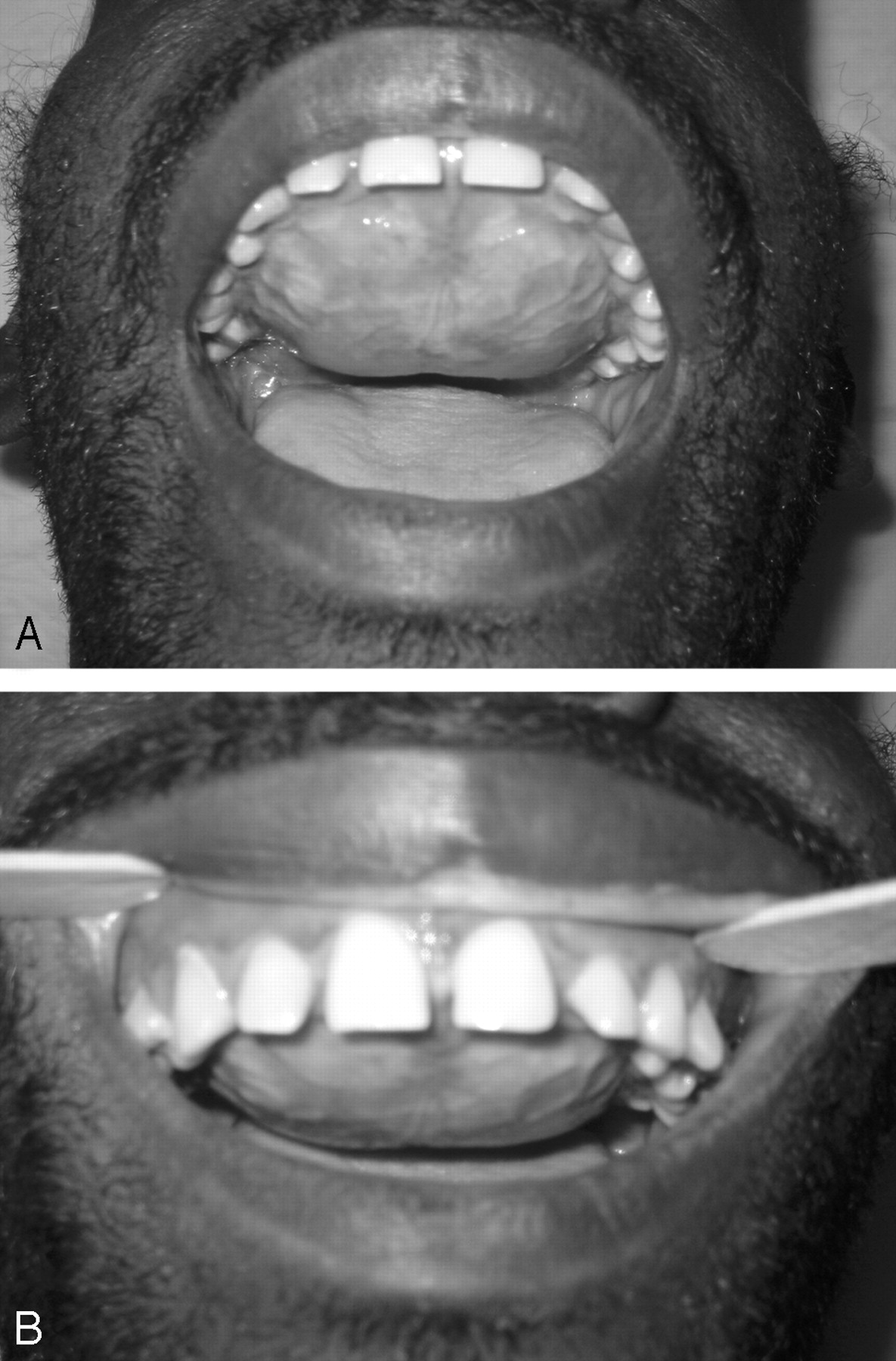
A 37-year-old man with a history of end stage renal disease who was on hemodialysis for 5 years presented with an expanding palatal lesion of 6-months’ duration, which caused dysarthria and oral dysphagia. He denied any other regional symptoms. Physical examination revealed a firm enlarged nontender hard palate that had descended to the occlusal plane. Combined with extensive maxillary hypertrophy bilaterally, the palatal changes caused the maxillary teeth to splay outwards (Fig 1).

Clinical photographs (A and B) show maxillary hypertrophy with convex palate and splayed dentition.
A noncontrast CT scan of his sinuses showed diffuse bony thickening of the hard palate with low-attenuation serpentine “tunneling” extending through the bone. Similar but less-pronounced changes were present in his mandible (Fig 2). The affected areas lacked clearly defined cortical bone and, thus, had no corticomedullary distinction. A Panorex film showed decreased cortical bone and loss of the lamina dura around the teeth (Fig 3). Punch biopsy of the palatal mass revealed a fibro-osseous lesion composed of irregular curved individual spicules of bone within fibrous tissue. No normal bone was identified. The specimen was pathologically interpreted to be consistent with fibrous dysplasia.

CT without contrast. Axial (A and B) and coronal (C and D) views. There is diffuse hypertrophy of the palate with extensive serpentine channels present throughout the entire maxilla bone and the anterior body and the symphysis of the mandible. The mandibular rami have a ground-glass appearance. There is virtually no cortical bone identified in the palate.

A Panorex film shows loss of lamina dura and eroded cortical bone.
However, the absence of good corticomedullary differentiation on conventional films and CT imaging, a typical finding in fibrous dysplasia, along with the overall clinical picture, suggested that the skeletal changes represented secondary hyperparathyroidism rather than fibrous dysplasia. Further work-up revealed a markedly elevated parathyroid hormone level (4115 pg/mL), an elevated phosphorus level (7.5 mg/dL), an elevated alkaline phosphatase level (810 U/L), and a normal calcium level (10 mg/dL). Additional work-up showed a normal 25-hydroxy (OH) vitamin D level (33 ng/mL) with a depressed 1,25(OH)2 vitamin D level (13 pg/mL). He underwent a 4-gland parathyroidectomy and responded well postoperatively, with a decrease in the parathyroid hormone level to 83 pg/mL, a decrease in the calcium level to 7.9 mg/dL, and a decrease in the phosphorous level to 2.2 mg/dL. The patient was discharged home after calcium levels stabilized. The patient did not return for any follow-up.
Discussion
Chronic renal failure alters bone metabolism by multiple mechanisms. Phosphate retention and decreased vitamin D conversion leads to hypocalcemia, which, in turn, stimulates the parathyroid chief cells to produce more parathyroid hormone. Long-term dialysis also inhibits healthy bone homeostasis, with aluminum deposition that interferes with bone mineralization. Subsequently, there are 2 major types of metabolic bone disease: 1) high-turnover osteodystrophy (increased bone resorption and formation) and 2) low-turnover or aplastic disease (adynamic bone and osteomalacia).1
Presently, the skeletal sequelae of hyperparathyroidism are considered to be mainly of historic interest. Whether primary, secondary, or tertiary, hyperparathyroidism is usually diagnosed by laboratory tests long before abnormal calcium metabolism results in macroscopic skeletal changes. The various skeletal changes have been well documented in the literature and include generalized demineralization, subperiosteal resorption, bone cysts, and pathologic fractures.2 Pathognomic radiologic findings include the loss of the dental lamina, thinning of cortical bone in multiple locations (distal phalanges, distal clavicles, distal ulna, inferior margin of femoral neck and pubis, and medial proximal tibia), a coarsened trabecular pattern, and a “salt-and-pepper” appearance of bone, particularly the skull.1–3 Less commonly encountered are the facial skeletal changes associated with hyperparathyroidism. These vary according to the clinical state of the disease (active or healed), and they have been given a variety of names in the literature, making it difficult to accurately know the number of such reported cases. It is estimated that fewer than 50 cases have been reported, most in the dental and oral surgical literature.4–9 The purpose of this report was to illustrate, with CT scans, an unusual presentation of hyperparathyroidism affecting the facial bones, especially the hard palate.
Facial skeletal changes associated with hyperparathyroidism are infrequently encountered and assume 3 radiographic patterns.5 The classic form is termed “osteitis fibrosa cystica” and presents with a combination of increased bone cell activity, peritrabecular fibrosis, and cystic brown tumors.2 Radiographically, this appears as a constellation of cortical thinning of multiple bones, coarsened trabecular patterns, osteolytic lesions, and “salt-and-pepper” appearance of the skull, which is the result of mixed osteolytic and sclerotic bone.1–3 The second form resembles fibrous dysplasia, with a classic ground-glass pattern on both conventional films and CT. Unlike true fibrous dysplasia, these findings can be diffuse and generalized, with poor corticomedullary distinction, an imaging finding not present in fibrous dysplasia. The 3rd and the rarest form is uremic leontiasis ossea as seen in our patient. Uremic leontiasis ossea is characterized by significant hypertrophy of the jaws with serpiginous “tunneling” or channeling within the bone and poor visualization of the cortical bone. The cause of this unusual structure is not known; no specific patterns of microscopic changes explain the radiographic findings.
The present case emphasizes the need to evaluate pathology specimens in the appropriate clinical context. If the final diagnosis was based only on pathology, an erroneous diagnosis of fibrous dysplasia could have been accepted; however, the clinical and imaging findings suggested an alternate disease process. Although the clinical presentation of this case is rare, once hyperparathyroidism is suspected, it can be readily confirmed by routine metabolic panel screening. The unique imaging findings should also strongly suggest the diagnosis.
- Received June 28, 2006.
- Accepted after revision July 27, 2006.
- Copyright © American Society of Neuroradiology
In this issue


Jump to section
Related Articles
Cited By...
- No citing articles found.