
Article Figures & Data
Figures
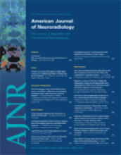
- Fig 1.
Agreement in detection of residual flow at the neck of an aneurysm of the anterior cerebral artery after treatment with detachable coils seen on DSA (A, black arrow). Unenhanced 3D TOF MRA (B) and MIP reconstruction (C) showing the residual flow (white arrows) in agreement with DSA.
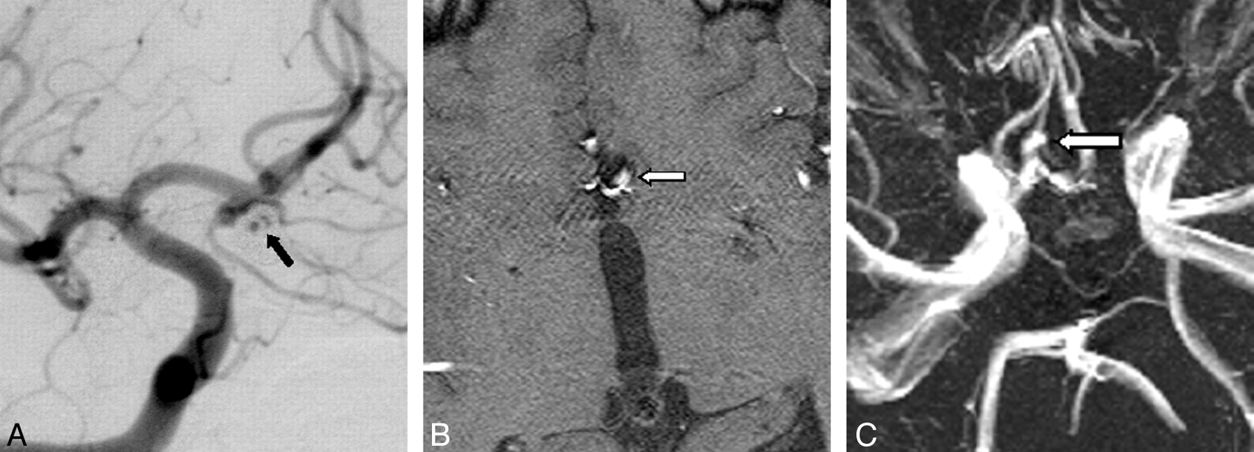
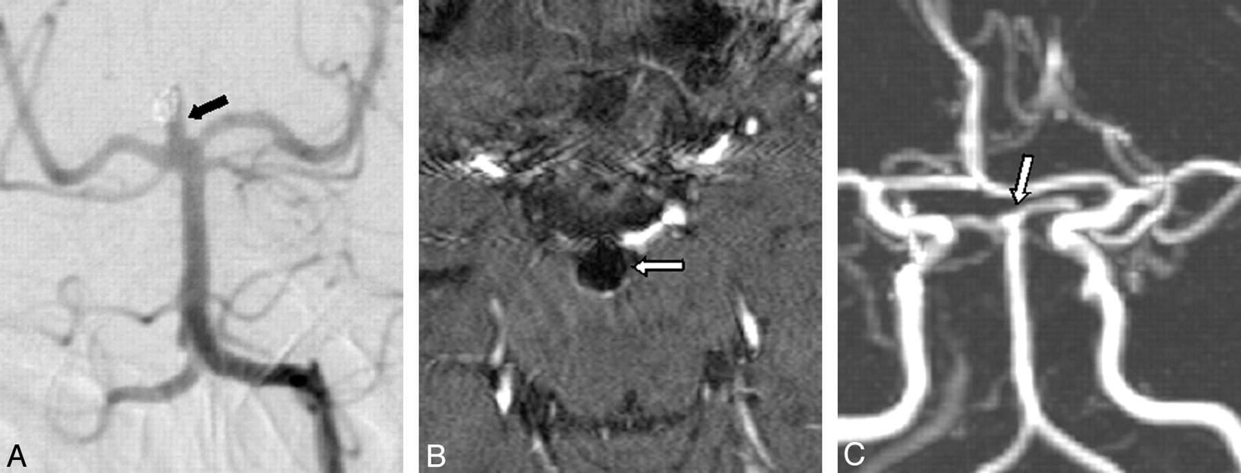
- Fig 2.
Overestimation of a completely occluded aneurysm of the anterior communicating artery seen on DSA (A, black arrow). Unenhanced 3D TOF MR angiography (B) and MIP reconstruction (C) showing a small residual perfusion at the neck of the aneurysm (white arrows).
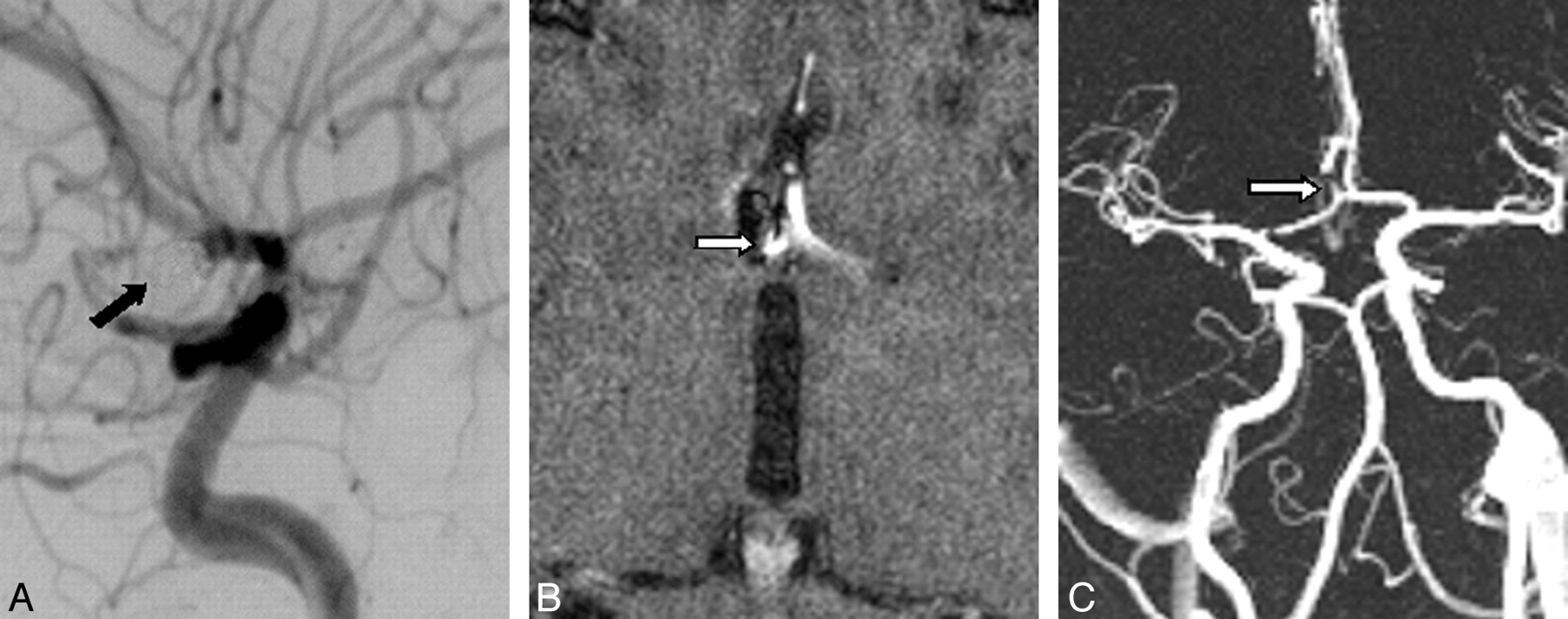
- Fig 3.
Overestimation of a small residual perfusion of a coiled aneurysm of the middle cerebral artery seen on DSA (A, black arrow). Contrast-enhanced 3D TOF MRA (B) and MIP reconstruction (C) showing a large residual neck (white arrows).
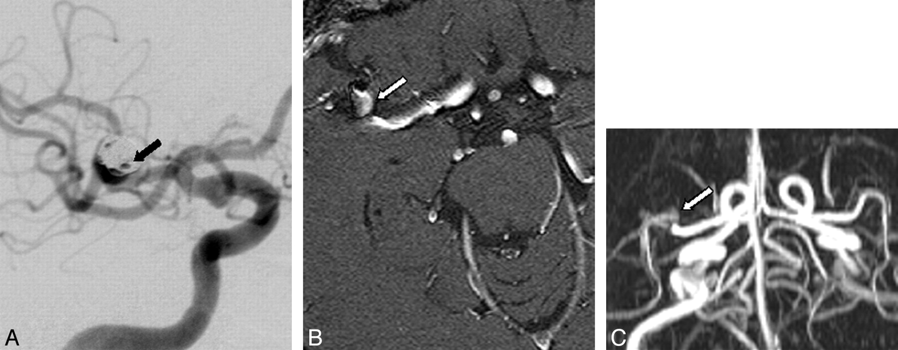
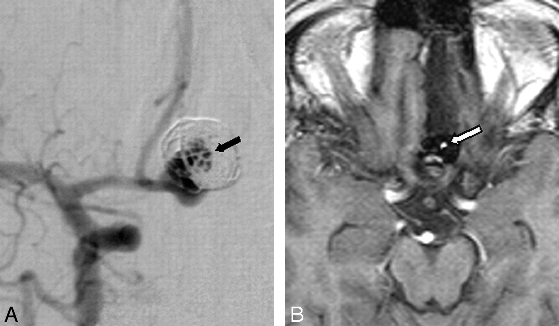
- Fig 4.
Underestimation of a residual perfusion of the neck of a basilaris tip aneurysm seen on DSA (A, black arrow). Contrast-enhanced TOF MRA (B) and MIP reconstruction (C) showing complete occlusion (white arrows).
- Fig 5.
Underestimation of a large residual neck of an aneurysm of the anterior communicating artery seen on DSA (A, black arrow). Unenhanced TOF MRA (B) shows a small residual perfusion of the aneurysm neck (white arrow).


Tables
MRA Finding DSA Finding Large Residual Neck Small Residual Neck Complete Occlusion Not Assessable Total Large residual neck 2 1 1 0 4 Small residual neck 1 50 8 0 59 Complete occlusion 0 7 118 0 125 Not assessable 0 2 3 0 5 Total 3 60 130 0 193 Note:—MRA indicates magnetic resonance angiography; DSA, digital subtraction angiography.
MRA Findings DSA Findings Residual Neck Complete Occlusion ICA Residual neck 12 1 Complete occlusion 1 26 Percentage 92.3 (68–99) 96 (80–99) AcomA Residual neck 18 6 Complete occlusion 1 40 Percentage 94.7 (74–99) 87 (74–95) ACA Residual neck 1 0 Complete occlusion 0 6 Percentage 100 (3–100) 100 (5–100) MCA Residual neck 7 1 Complete occlusion 0 18 Percentage 100 (54–100) 95 (75–99) BA Residual neck 10 1 Complete occlusion 2 10 Percentage 83.3 (52–98) 90.9 (59–99) PcomA Residual neck 5 0 Complete occlusion 3 15 Percentage 62.5 (18–90) 100 (79–100) PICA Residual neck 1 0 Complete occlusion 0 2 Percentage 100 (3–100) 100 (16–100) VA Residual neck 0 0 Complete occlusion 0 1 Percentage 100 (3–100) Total Residual neck 54 9 Complete occlusion 7 118 Percentage 88.5 (78–95) 92.9 (87–97) Note:—MRA indicates magnetic resonance angiography; DSA, digital subtraction angiography; ICA, internal carotid artery; AcomA, anterior communicating artery; ACA, anterior cerebral artery; MCA, middle cerebral artery; BA, basilar artery; PcomA, posterior communicating artery; PICA, posterior inferior cerebellar artery; VA, vertebral artery. Percentages represent sensitivity (left column) and specificity (right column); figures in parentheses are 95% confidence intervals.
Location N (%) Size (mean, mm) Overestimation (Size) Underestimation (Size) ICA 40 (21.3) 10.4 Vessel overlap (9 mm) Pulsation-induced artifacts (10 mm) AcomA 65 (34.6) 5.7 Vessel overlap (8 mm) Susceptibility at bone/air interface (2.5 mm) Vessel overlap and small size (2 mm, 3 mm) Partially thrombosed aneurysm (10 mm) Pulsation-induced artifacts (2 mm, 12 mm) ACA 7 (3.7) 9.1 MCA 26 (13.8) 7.1 Vessel overlap (11 mm) BA 23 (12.2) 7.6 Pulsation-induced artifacts (10 mm) Slow flow and small size (2 mm) Saturation effects and small size (3 mm) PcomA 23 (12.2) 7.3 Vessel overlap (10 mm) Signal void due to slow flow (5 mm) Susceptibility at bone/air interface (3 mm) PICA 3 (1.7) 6.7 VA 1 (0.5) 4.0 Total 188 (100) 7.5 Note:—MRA indicates magnetic resonance angiography; DSA, digital subtraction angiography; ICA, internal carotid artery; AcomA, anterior communicating artery; ACA, anterior cerebral artery; MCA, middle cerebral artery; BA, basilar artery; PcomA, posterior communicating artery; PICA, posterior inferior cerebellar artery; VA, vertebral artery.
MRA Finding DSA Finding Residual Neck Complete Occlusion Aneurysms ≤ 3 mm Residual neck 7 3 Complete occlusion 4 19 Percentage 63.6 (31–89) 86.4 (65–97) Aneurysms >3 mm Residual neck 47 6 Complete occlusion 3 99 Percentage 94 (84–99) 94.3 (88–98) Residual neck 47 6 Aneurysms ≤ 5 mm Residual neck 13 3 Complete occlusion 5 52 Percentage 72.2 (62–82) 94.5 (89–100) Aneurysms > 5 mm Residual neck 41 6 Complete occlusion 2 66 Percentage 95.3 (84–99) 91.7 (83–97) Note:—TOF indicates time of flight; MRA, magnetic resonance angiography; DSA, digital subtraction angiography. Percentages represent sensitivity (left column) and specificity (right column); figures in parentheses are 95% confidence intervals.
MRA Finding DSA Finding Residual Neck Complete Occlusion Unenhanced MRA Residual neck 24 4 Complete occlusion 3 46 Percentage 88.9 (70.1–97.6) 92 (80.8–97.7) Contrast-enhanced MRA Residual neck 30 5 Complete occlusion 4 72 Percentage 88.2 (72.6–96.7) 93.5 (85.5–97.7) Note:—TOF indicates time of flight; MRA, magnetic resonance angiography; DSA, digital subtraction angiography. Percentages represent sensitivity (left column) and specificity (right column); figures in parentheses are 95% confidence intervals.
In this issue


Jump to section
Related Articles
Cited By...
- Differential Subsampling with Cartesian Ordering-MRA for Classifying Residual Treated Aneurysms
- MRA versus DSA for the follow-up imaging of intracranial aneurysms treated using endovascular techniques: a meta-analysis
- Voxel based analysis of recurrence dynamics in intracranial aneurysms after coiling
- MRA Versus DSA for Follow-Up of Coiled Intracranial Aneurysms: A Meta-Analysis
- 3D Computerized Occlusion Rating of Embolized Experimental Aneurysms Using Noninvasive 1.5T MR Imaging
- Residual Flow After Cerebral Aneurysm Coil Occlusion: Diagnostic Accuracy of MR Angiography
- A Prospective Trial of 3T and 1.5T Time-of-Flight and Contrast-Enhanced MR Angiography in the Follow-Up of Coiled Intracranial Aneurysms
- MR Angiographic Follow-Up of Intracranial Aneurysms Treated with Detachable Coils: Evaluation of a Blood-Pool Contrast Medium