Advertisement

Main menu

  • Home
  • Content
    • Current Issue
    • Accepted Manuscripts
    • Article Preview
    • Past Issue Archive
    • AJNR Case Collection
    • Case of the Week Archive
    • Classic Case Archive
    • Case of the Month Archive
  • Special Collections
    • Spinal CSF Leak Articles (Jan 2020-June 2024)
    • 2024 AJNR Journal Awards
    • Most Impactful AJNR Articles
  • Multimedia
    • AJNR Podcast
    • AJNR Scantastics
    • Video Articles
  • For Authors
    • Submit a Manuscript
    • Author Policies
    • Fast publishing of Accepted Manuscripts
    • Graphical Abstract Preparation
    • Manuscript Submission Guidelines
    • Imaging Protocol Submission
    • Submit a Case for the Case Collection
  • About Us
    • About AJNR
    • Editorial Board
  • More
    • Become a Reviewer/Academy of Reviewers
    • Subscribers
    • Permissions
    • Alerts
    • Feedback
    • Advertisers
    • ASNR Home
  • Other Publications
    • ajnr

User menu

  • Alerts
  • Log in

Search

  • Advanced search
American Journal of Neuroradiology

American Journal of Neuroradiology

ASHNR American Society of Functional Neuroradiology ASHNR American Society of Pediatric Neuroradiology ASSR
  • Alerts
  • Log in

Advanced Search

  • Home
  • Content
    • Current Issue
    • Accepted Manuscripts
    • Article Preview
    • Past Issue Archive
    • AJNR Case Collection
    • Case of the Week Archive
    • Classic Case Archive
    • Case of the Month Archive
  • Special Collections
    • Spinal CSF Leak Articles (Jan 2020-June 2024)
    • 2024 AJNR Journal Awards
    • Most Impactful AJNR Articles
  • Multimedia
    • AJNR Podcast
    • AJNR Scantastics
    • Video Articles
  • For Authors
    • Submit a Manuscript
    • Author Policies
    • Fast publishing of Accepted Manuscripts
    • Graphical Abstract Preparation
    • Manuscript Submission Guidelines
    • Imaging Protocol Submission
    • Submit a Case for the Case Collection
  • About Us
    • About AJNR
    • Editorial Board
  • More
    • Become a Reviewer/Academy of Reviewers
    • Subscribers
    • Permissions
    • Alerts
    • Feedback
    • Advertisers
    • ASNR Home
  • Follow AJNR on Twitter
  • Visit AJNR on Facebook
  • Follow AJNR on Instagram
  • Join AJNR on LinkedIn
  • RSS Feeds

Welcome to the new AJNR, Updated Hall of Fame, and more. Read the full announcements.


AJNR is seeking candidates for the position of Associate Section Editor, AJNR Case Collection. Read the full announcement.


Research ArticlePEDIATRICS

Defining Optic Nerve Tortuosity

G.T. Armstrong, A.R. Localio, T. Feygin, L. Bilaniuk, P.C. Phillips, M.J. Fisher, B.L. Strom and R. Zimmerman
American Journal of Neuroradiology April 2007, 28 (4) 666-671;
G.T. Armstrong
  • Find this author on Google Scholar
  • Find this author on PubMed
  • Search for this author on this site
A.R. Localio
  • Find this author on Google Scholar
  • Find this author on PubMed
  • Search for this author on this site
T. Feygin
  • Find this author on Google Scholar
  • Find this author on PubMed
  • Search for this author on this site
L. Bilaniuk
  • Find this author on Google Scholar
  • Find this author on PubMed
  • Search for this author on this site
P.C. Phillips
  • Find this author on Google Scholar
  • Find this author on PubMed
  • Search for this author on this site
M.J. Fisher
  • Find this author on Google Scholar
  • Find this author on PubMed
  • Search for this author on this site
B.L. Strom
  • Find this author on Google Scholar
  • Find this author on PubMed
  • Search for this author on this site
R. Zimmerman
  • Find this author on Google Scholar
  • Find this author on PubMed
  • Search for this author on this site
  • Article
  • Figures & Data
  • Info & Metrics
  • Responses
  • References
  • PDF
Loading

Article Figures & Data

Figures

  • Tables
  • Fig 1.
    • Download figure
    • Open in new tab
    • Download powerpoint
    Fig 1.

    Optic glioma.

    A, Sagittal spin-echo image (TR, 643 ms; TE, 12 ms) shows markedly enlarged optic nerve (arrow).

    B, Axial fast spin-echo (TR, 6000 ms; TE, 84 ms) image shows bilateral enlargement, along with tortuosity of intraorbital optic nerves (arrow).
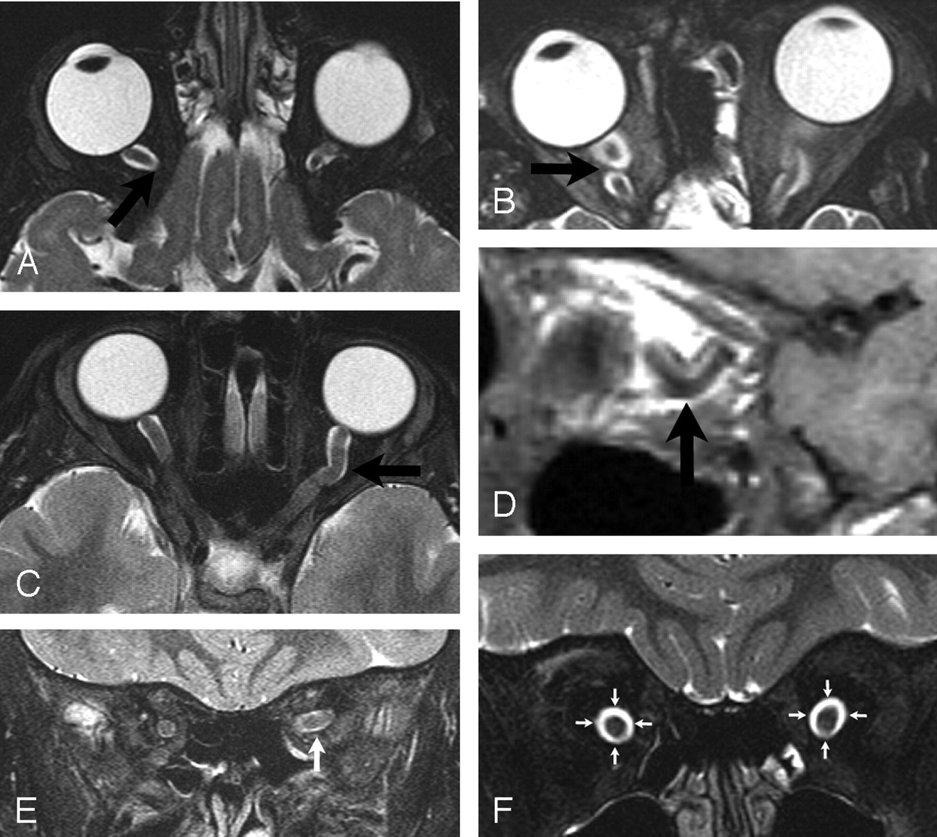
  • Fig 2.
    • Download figure
    • Open in new tab
    • Download powerpoint
    Fig 2.

    Tortuosity of optic nerves

    A, Axial fast spin-echo T2-weighted image showing factor 1: interruption of the optic nerve out of the axial plane (tip of arrow) without return.

    B, Axial fast spin-echo T2-weighted image showing factor 2: interruption of the optic nerve out of the axial plane (tip of arrow) with return of the nerve into the axial plane.

    C, Axial fast spin-echo T2-weighted image showing factor 3: deviation of the optic nerve within the axial plane (arrow).

    D, Sagittal T1-weighted image showing factor 4: increased curvature (tip of arrow) in the sagittal plane.

    E, Coronal fast spin-echo T2-weighted image showing factor 5: lack of congruity of the optic nerves (arrow) in the coronal plane.

    F, Coronal fast spin-echo T2-weighted image showing factor 6: dilation of the subarachnoid space (encircled by arrows) surrounding anterior portion of optic nerves.

  • Figure3
    • Download figure
    • Open in new tab
    • Download powerpoint

Tables

  • Figures
    • View popup
    Table 1:

    5 item global assessment results by case number for 3 readers

    Case No.Definitely Not TortuousProbably Not TortuousToss-UpProbably TortuousDefinitely Tortuous
    1•••○○○○○○○○○○○○
    2○○•••○○○○○○○○○○
    3○○○○○○○○○•○○○••
    4○○○○○○○○○○○○•••
    5•••○○○○○○○○○○○○
    6○○○○○○○○○○○○•••
    7•○○○○○○○○○○•○•○
    8•○•○○○○•○○○○○○○
    9•••○○○○○○○○○○○○
    10•••○○○○○○○○○○○○
    11○○○○○○○○○○○○•••
    12○○○○○○○○○○○•••○
    13○○○○○○○○○○○○•••
    14○○○•○○○○○○○•○•○
    15•••○○○○○○○○○○○○
    16○○○○○○○○○••○○○•
    17○○○○○○○○○○○○•••
    18○○○○○○○○○•○•○•○
    19•○○○○•○○○○○○○•○
    20○○•○○○○•○○○○•○○
    21•••○○○○○○○○○○○○
    22•••○○○○○○○○○○○○
    23••○○○•○○○○○○○○○
    24○○○•○•○○○○•○○○○
    25○○○○○○•○○○••○○○
    26○○○○○○○○•○○○••○
    27•••○○○○○○○○○○○○
    28•••○○○○○○○○○○○○
    Column Perfect Agreement9000514/28 = 50%
    Column Majority Agreement11203823/28 = 82%
    Dichotomous Agreement12921/28 = 75%
    • Note:—• represents, for a given case, the response on the Likert scale that best describes the presence of tortuosity as read by 1 of 3 individual radiologists.

    • View popup
    Table 2:

    Individual predictors of tortuosity

    Case No.Radiographic Factor
    1 Out Axial Plane2 Out/In Axial Plane3 Deviation within Axial Plane4 Curvature Sagittal Plane5 Lack of Coronal Congruity6 Dilation Subarachnoid SpaceMean Total Score
    1○○○○○○○○○○○○n/a○○○
    2○○•••○○○••••○○○○○○2.3
    3○○○○•••••○••••••••4.3
    4••○○○••••○••••••••4.7
    5○○○○○○○○○○○○○○○○○○0
    6••○○○••••••••○••○•4.3
    7○○○○••○•○○••○•○○••2.7
    8○○○○○○○••○○○n/a○○○
    9○○○○○○○○○○○○○○○○○○0
    10○○○○○○○○○○○○n/a○○•
    11○○○○••••••••••••••4.7
    12○○○○••••••••••••••4.7
    13○•○•○•••••••••••••5.0
    14○•○○○•○•••••○•○••○3.3
    15○○○○○○○○○○○○○○○•••1.0
    16○○○••••○○••••○••••4.0
    17○○○○•••••••○••••••4.3
    18○○○•••••○••••••••○4.3
    19○○○•••○○○○••○○○○•○2.0
    20•••○○○•••○○○○•○○○○2.3
    21•••○○○○○○○○•○○○○○○1.3
    22•••○○○○○○n/a○○○•○○
    23○••○○•○○○•••○○○○○○2.0
    24○○○○○••••○○•○•○•○○2.3
    25○○○○••••••○•○••○○○3.0
    26○○○••••○○•••••••○○3.7
    27○○○○○○○○○○○○○○○○○○0
    28•••○○○○○○○○○○○○•○○1.3
    Column Perfect Agree22/28 = 79%15/28 = 54%21/28 = 75%19/27 = 70%18/25 = 72%18/28 = 64%
    • Note:—Radiologist decision: ○, factor absent; •, factor present; n/a, not available for review.

    • View popup
    Table 3:

    Univariate analysis of radiographic factors for predicting a diagnosis of tortuosity

    VariableUnivariate OR for Prediction of Tortuosity
    1.67 (CI .22–2.1)
    24.6 (CI 1.7–12.7)
    314.4 (CI 4.2–49-8)
    47.9 (CI 2.3–26.6)
    523.6 (CI 6.5–85.4)
    628.6 (CI 7.1–114.7)
    • Note:—OR indicates odds ratio; CI, 95% confidence interval.

    • View popup
    Table 4:

    Radiographic factor combinations that predict a diagnosis of tortuosity

    Radiographic Factor CombinationConcordance StatisticSensitivitySpecificity
    Factors 5 & 6.90389 (CI .79–1)93 (CI .86–1)
    Factors 3, 5, & 6.91185 (CI .66–1)93 (CI .86–1)
    Factors 3 & 6.89885 (CI .66–1)91 (CI .83–.98)
    Factor 5.82693 (CI .83–1)80 (CI .64–.94)
    Factor 6.83293 (CI .83–1)76 (CI .58–.91)
    Factor 3.78089 (CI .71–1)69 (CI .46–.86)
    • Note:—CI indicates 95% confidence interval.

    • View popup
    Table 5:

    Likelihood ratios of key radiographic factor combinations

    Radiographic Factor CombinationLikelihood Ratio
    Factors Present
        5 & 613.3 (CI 6.4–46.1)
        3, 5, & 612.7 (CI 6.0–44)
        3 & 69.6 (CI 4.8–34.2)
        54.6 (CI 2.3–11.5)
        63.8 (CI 2.2–9.7)
        32.8 (CI 1.6–7.1)
    Factors Absent
        3.16 (CI 0–.49)
        6.10 (CI 0–.26)
        5.09 (CI 0–2.4)
        3 & 6.07 (CI 0–.27)
        5 & 6.06 (CI 0–.22)
        3, 5, & 60*
    • Note:—CI indicates 95% confidence interval.

    • * Unable to calculate confidence interval.

PreviousNext
Back to top

In this issue

American Journal of Neuroradiology: 28 (4)
American Journal of Neuroradiology
Vol. 28, Issue 4
April 2007
  • Table of Contents
  • Index by author
Advertisement
Print
Download PDF
Email Article

Thank you for your interest in spreading the word on American Journal of Neuroradiology.

NOTE: We only request your email address so that the person you are recommending the page to knows that you wanted them to see it, and that it is not junk mail. We do not capture any email address.

Enter multiple addresses on separate lines or separate them with commas.
Defining Optic Nerve Tortuosity
(Your Name) has sent you a message from American Journal of Neuroradiology
(Your Name) thought you would like to see the American Journal of Neuroradiology web site.
CAPTCHA
This question is for testing whether or not you are a human visitor and to prevent automated spam submissions.
Cite this article
G.T. Armstrong, A.R. Localio, T. Feygin, L. Bilaniuk, P.C. Phillips, M.J. Fisher, B.L. Strom, R. Zimmerman
Defining Optic Nerve Tortuosity
American Journal of Neuroradiology Apr 2007, 28 (4) 666-671;

Citation Manager Formats

  • BibTeX
  • Bookends
  • EasyBib
  • EndNote (tagged)
  • EndNote 8 (xml)
  • Medlars
  • Mendeley
  • Papers
  • RefWorks Tagged
  • Ref Manager
  • RIS
  • Zotero
0 Responses
Respond to this article
Share
Bookmark this article
Defining Optic Nerve Tortuosity
G.T. Armstrong, A.R. Localio, T. Feygin, L. Bilaniuk, P.C. Phillips, M.J. Fisher, B.L. Strom, R. Zimmerman
American Journal of Neuroradiology Apr 2007, 28 (4) 666-671;
del.icio.us logo Twitter logo Facebook logo Mendeley logo
  • Tweet Widget
  • Facebook Like
  • Google Plus One

Jump to section

  • Article
    • Abstract
    • Materials and Methods
    • Results
    • Discussion
    • Conclusion
    • Footnotes
    • References
  • Figures & Data
  • Info & Metrics
  • Responses
  • References
  • PDF

Related Articles

  • No related articles found.
  • PubMed
  • Google Scholar

Cited By...

  • Quantitative MRI criteria for optic pathway enlargement in neurofibromatosis type 1
  • Risk of optic pathway glioma in children with neurofibromatosis type 1 and optic nerve tortuosity or nerve sheath thickening
  • Crossref
  • Google Scholar

This article has not yet been cited by articles in journals that are participating in Crossref Cited-by Linking.

More in this TOC Section

  • SyMRI & MR Fingerprinting in Brainstem Myelination
  • Comparison of Image Quality and Radiation Dose in Pediatric Temporal Bone CT Using Photon-Counting Detector CT and Energy-Integrating Detector CT
  • Pons&Vermis Localization on Fetal MRI Using U-Net
Show more Pediatrics

Similar Articles

Advertisement

Indexed Content

  • Current Issue
  • Accepted Manuscripts
  • Article Preview
  • Past Issues
  • Editorials
  • Editors Choice
  • Fellow Journal Club
  • Letters to the Editor

Cases

  • Case Collection
  • Archive - Case of the Week
  • Archive - Case of the Month
  • Archive - Classic Case

Special Collections

  • Special Collections

Resources

  • News and Updates
  • Turn around Times
  • Submit a Manuscript
  • Author Policies
  • Manuscript Submission Guidelines
  • Evidence-Based Medicine Level Guide
  • Publishing Checklists
  • Graphical Abstract Preparation
  • Imaging Protocol Submission
  • Submit a Case
  • Become a Reviewer/Academy of Reviewers
  • Get Peer Review Credit from Publons

Multimedia

  • AJNR Podcast
  • AJNR SCANtastic
  • Video Articles

About Us

  • About AJNR
  • Editorial Board
  • Not an AJNR Subscriber? Join Now
  • Alerts
  • Feedback
  • Advertise with us
  • Librarian Resources
  • Permissions
  • Terms and Conditions

American Society of Neuroradiology

  • Not an ASNR Member? Join Now

© 2025 by the American Society of Neuroradiology All rights, including for text and data mining, AI training, and similar technologies, are reserved.
Print ISSN: 0195-6108 Online ISSN: 1936-959X

Powered by HighWire