
Article Figures & Data
Figures
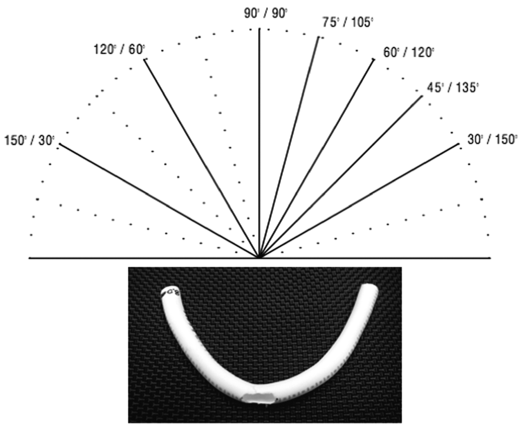
- Fig 1.
A, Angles of bending from 180° (0°) to 30° (inner angle of model curve).
B, PTFE tube, 4-mm diameter with 8-mm opening as used for stent deployment and bending.
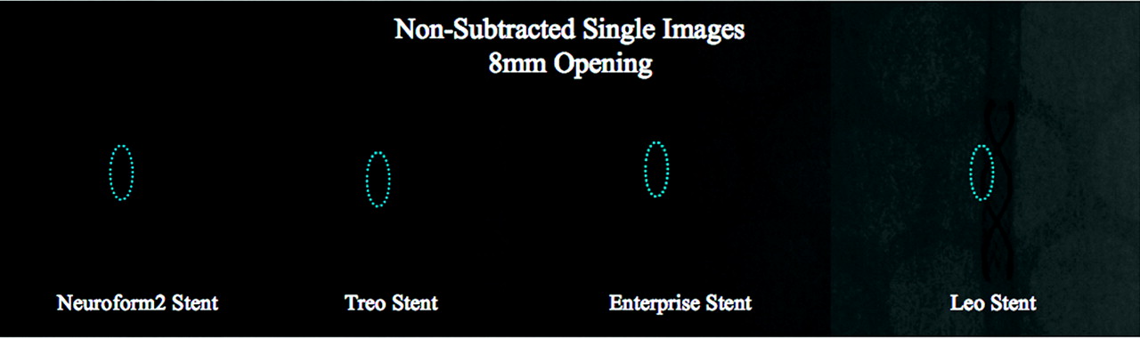
- Fig 2.
Nonsubtracted digital radiography (DR) images of the stents. The DRs of the Neuroform2 and Neuroform2-Treo stents visualize the proximal and distal platinum markers clearly; however, the structure of the stent cells and struts is poorly defined. The Neuroform2 image shows only faintly the prolapse of the struts at the site of the 8-mm opening (dotted blue ellipse). This phenomenon seems to occur to a lesser degree with the Neuroform-Treo stent because of the one extra connector along the circumference of the stent, reducing the area of open cells by about 39%. The Enterprise stent is only visualized by its proximal and distal platinum markers. The stent cells and struts are not defined because of the small size of the stent struts (50 × 50 μm); therefore, the structural details of stent are not visible. The LEO stent is seemingly better visualized throughout its entire length, outlined by 2 platinum wires intertwined along the surface of the stent serving as markers; the stent cells and struts, however, are not identifiable.
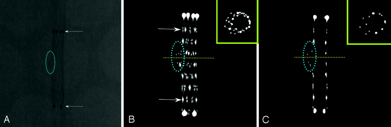
- Fig 3.
Neuroform2, DR and FPCT (MIPs). To increase flexibility and conformability, this stent has an open-cell design with 2 connectors per circumference. The DR shows the proximal and distal markers well (A, dotted arrows); however, the stent cells and struts are poorly defined. FPCT using multiplanar reconstructions or MIPs is superior in displaying the stent architecture in detail, allowing for differentiation between struts, connectors (seen as 2 moderately bright spots per circumference, C, arrows) as well as the platinum markers. Thin sections allow an “in-stent” view and clearly show the protrusion of stent struts through a 5-mm hole (“aneurysm neck,” dotted blue ellipse). This prolapse is difficult if not impossible to identify when using DR or fluoroscopy. FPCT makes it also possible to obtain cross sections of the stent lumen at the level of the opening, creating a double lumen figure (C, miniature yellow box).
- Fig 4.
Neuroform2 Stent: constant opening, increasing bend. MIPs, using section thickness of 1.0 mm (right column) and 5.0 mm (left column), bending with 120°, 90°, and 30° angles, over a simulated “aneurysm neck” (8-mm hole in the PTFE tube, dotted blue ellipse in the left column). The MIPs demonstrate an asymmetric deployment of the stent at the site of the hole. As the angle of the bend decreases, there is an increased opening of the stent cells, along with a tendency of the stent struts to protrude more into the aneurysm cavity (arrowheads). Using 1.0-mm section MIPs, starting at the 90° bend, a minor inward prolapse of the stent struts at the concavity (arrows) occurs. These findings are not detectable using current conventional radiographic techniques such as fluoroscopy or DR.
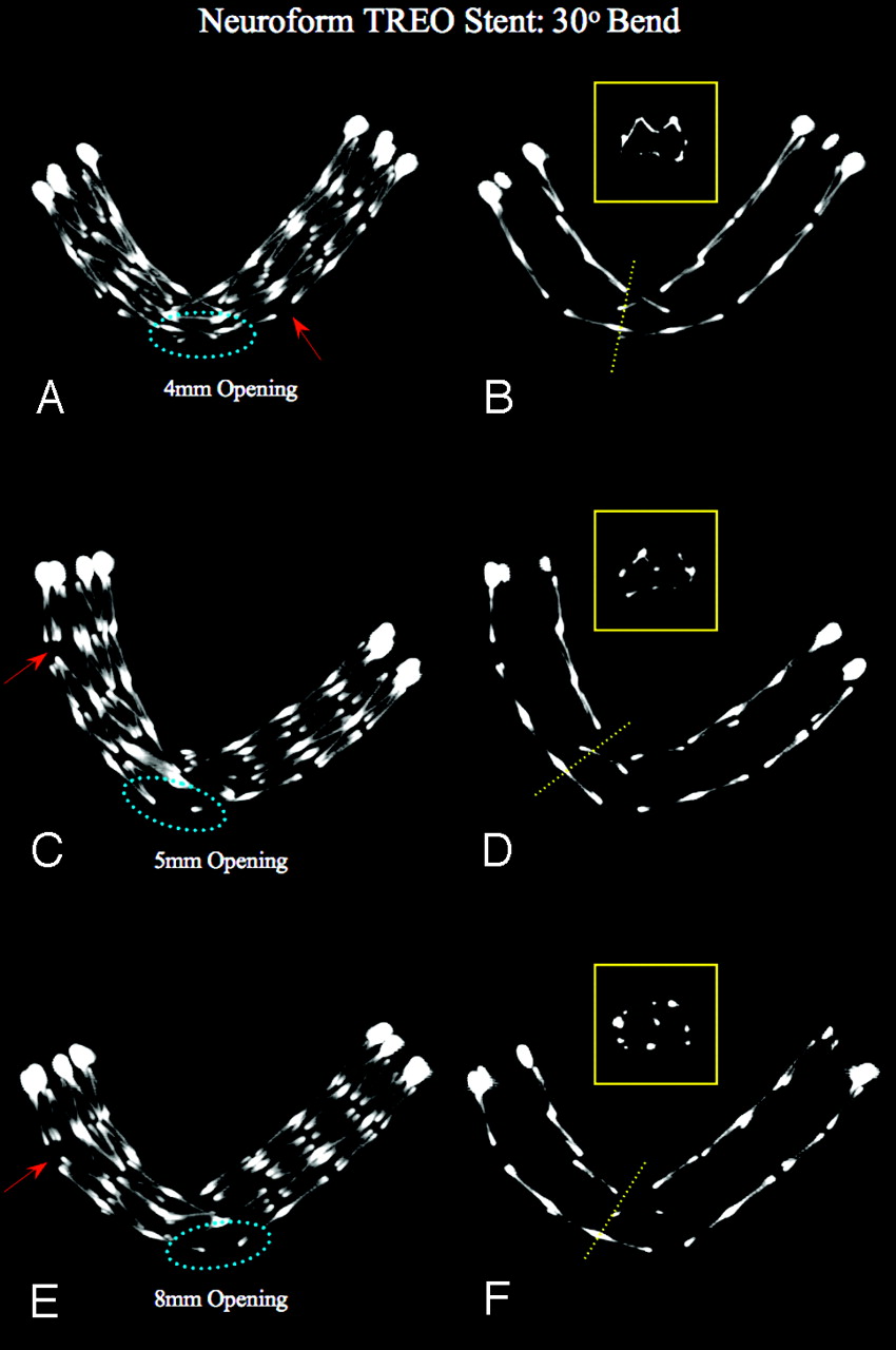
- Fig 5.
Neuroform-Treo Stent: constant bend 30°, increasing size of openings (“aneurysm necks”). MIPs using section thickness of 5.0 mm (left column) and 1.0 mm (right column), bending with 30° angle over 4-, 5-, and 8-mm holes or simulated “aneurysm necks” (left column, dotted blue ellipse). As compared with the NF2 stent, the NF3 shows less increase in cell opening with increased bend, probably because of the presence of one or more connectors per circumference. Some minor increase in opening can be seen remote from the curvature (arrows). The thin-section MIPs show an increased inward prolapse of struts into the stent lumen as the diameter of the holes increases. This seems more pronounced compared with the NF2. However, whether this phenomenon occurs also depends on the positioning of the stent relative to the location of the “aneurysm neck.” In the absence of a connector across the opening, the stent struts still protrude into the “aneurysm,” though to a lesser degree than seen with NF2. Miniatures (yellow boxes, left column): Cross-sectional view of the inner stent lumen at its midsection.
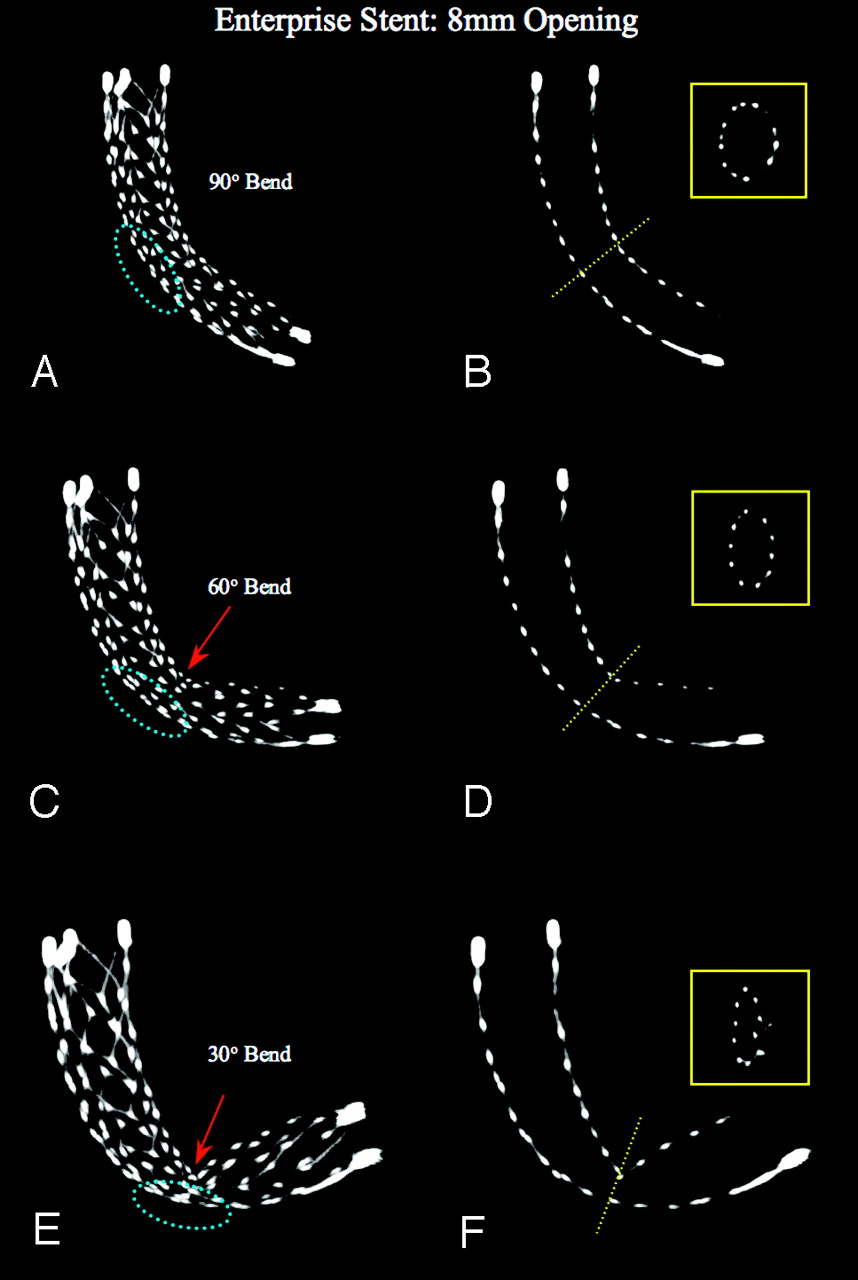
- Fig 6.
Enterprise Stent: constant 8-mm opening, increasing bend. MIPs, using section thickness of 5.0 mm (left column) and 1.0 mm (right column), bending with 90°, 60°, and 30° angles, over a 8-mm hole simulated “aneurysm neck” (dotted blue ellipse). It is evident that starting from a 90° bend, the Enterprise stent shows initially a minor flattening at its midsection that increases with 60° and becomes a true kinking at 30°. It also documents that the stent tends not to completely collapse, even though the midsection of the stent significantly changes its shape from round to oval and the diameter becomes relatively small (miniatures).
- Fig 7.
LEO Stent: constant bend, increasing size of openings (“aneurysm necks”). MIPs using section thicknesses of 5.0 and 1.0 mm, bending with 30° angle, in 4-, 5-, and 8-mm simulated aneurysm necks. The MIPs show that as the size of the holes (“aneurysm necks,” dotted blue ellipse) increases, the stent tends to flatten more at its midsection (arrows), whereas the degree of bending is unchanged. This phenomenon seems more pronounced if the platinum markers do not cross the aneurysm neck, hence the nitinol wires are thinner and softer. The MIPs further demonstrate an inward crimping of the proximal and distal ends, significantly narrowing the lumen of the stent at these points (double arrows).


Tables
Stent Material Design Diameter (mm) Length (mm) Manufacturer Neuroform2 Nitinol Open cell 4.0 25.0 Boston Scientific Neuroform-Treo Nitinol Open cell 3.5 20.0 Boston Scientific Enterprise Nitinol Closed cell 4.0 ± 0.5 22.0 Cordis LEO Nitinol Braided wires 4.5 ± 0.5 25.0 ± 2.0 Balt Extrusion Deployment Configuration Strut Characteristics Neuroform2 Asymmetric Increased opening of stent cells with decreasing angle Inward and outward prolapse of the stent struts starting at 110° curve Neuroform-Treo Asymmetric Increased cell opening of the stent with decreasing angle (less than NF2) Less outward prolapse but more inward prolapse of the struts than NF2 with decreasing angles, starting at 75° Loss of normal strut configuration starting at 110° curve Enterprise Symmetric Change of shape from round to oval Increasing trend to flatten and to kink at the concavity with increasing size of the “aneurysm neck” Reduction of the stent diameter at the midsection starting at 90° LEO Symmetric Flattening of the midsection of the stent starting at 90° Less tendency to kink and flatten than Enterprise Flattening and narrowing of the distal and proximal ends starting at 115° Inward crimping of proximal and distal ends with lumen narrowing
In this issue


Jump to section
Related Articles
Cited By...
- Safety of Non-invasive Brain Stimulation in Patients with Implants: A Computational Study
- Propensity Score Analysis of Flow Diverters Placed in Scaffolding Stents
- Early experience treating intracranial aneurysms using Accero: a novel, fully visible, low profile braided stent with platinum-nitinol composite wire technology
- Curative cerebrovascular reconstruction with the Pipeline embolization device: the emergence of definitive endovascular therapy for intracranial aneurysms
- Stent-assisted coiling of cerebrovascular aneurysms: experience at a large tertiary care center with a focus on predictors of recurrence
- Clinical Impact of Flat Panel Volume CT Angiography in Evaluating the Accurate Intraoperative Deployment of Flow-Diverter Stents
- Final results of the US humanitarian device exemption study of the low-profile visualized intraluminal support (LVIS) device
- Measurement in the angiography suite: evaluation of vessel sizing techniques
- Urgent off-label use of the pipeline flow diverter stent in selected ischemic cerebrovascular conditions: thrombotic segments and tortuous arteries
- Safety and performance of the Penumbra Liberty stent system in a rabbit aneurysm model
- "Y" and "X" Stent-Assisted Coiling of Complex and Wide-Neck Intracranial Bifurcation Aneurysms
- Effect of Structural Remodeling (Retraction and Recoil) of the Pipeline Embolization Device on Aneurysm Occlusion Rate
- Double-barrel entanglement of intracranial Enterprise stents resulting from undetected incomplete stent apposition
- Reply:
- Delivery technique plays an important role in determining vessel wall apposition of the Enterprise self-expanding intracranial stent
- Parent Vessel Size and Curvature Strongly Influence Risk of Incomplete Stent Apposition in Enterprise Intracranial Aneurysm Stent Coiling
- X-Configured Stent-Assisted Coiling in the Endovascular Treatment of Complex Anterior Communicating Artery Aneurysms: A Novel Reconstructive Technique
- Endovascular rescue of a misshapen intracranial stent: report of two cases
- Contrast-Enhanced Angiographic Cone-Beam CT of Cerebrovascular Stents: Experimental Optimization and Clinical Application
- Angiographic CT after Intravenous Contrast Agent Application: A Noninvasive Follow-Up Tool after Intracranial Angioplasty and Stenting
- Possible Mechanisms for Delayed Migration of the Closed Cell--Designed Enterprise Stent When Used in the Adjunctive Treatment of a Basilar Artery Aneurysm
- Feasibility of Angiographic CT in Peri-Interventional Diagnostic Imaging: A Comparative Study with Multidetector CT
- Balloon assisted treatment of intracranial aneurysms: the conglomerate coil mass technique
- Stent-Assisted Embolization of Wide-Neck Anterior Communicating Artery Aneurysms: Review of 21 Consecutive Cases
- Curative cerebrovascular reconstruction with the Pipeline embolization device: the emergence of definitive endovascular therapy for intracranial aneurysms
- Bailout Stent Deployment during Coil Embolization of Intracranial Aneurysms
- Wall Shear Stress in Intracranial Self-Expanding Stents Studied Using Ultra-High-Resolution 3D Reconstructions