
Article Figures & Data
Figures
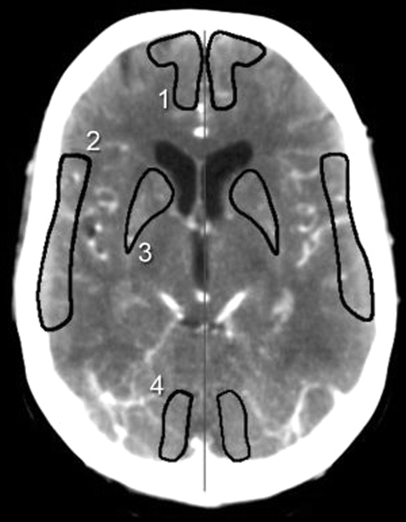
- Fig 1.
Analysis of manual outlined ROIs according to territorial division of Damasio.26 1, ACA territory; 2, MCA territory; 3, basal ganglia; 4, PCA territory.
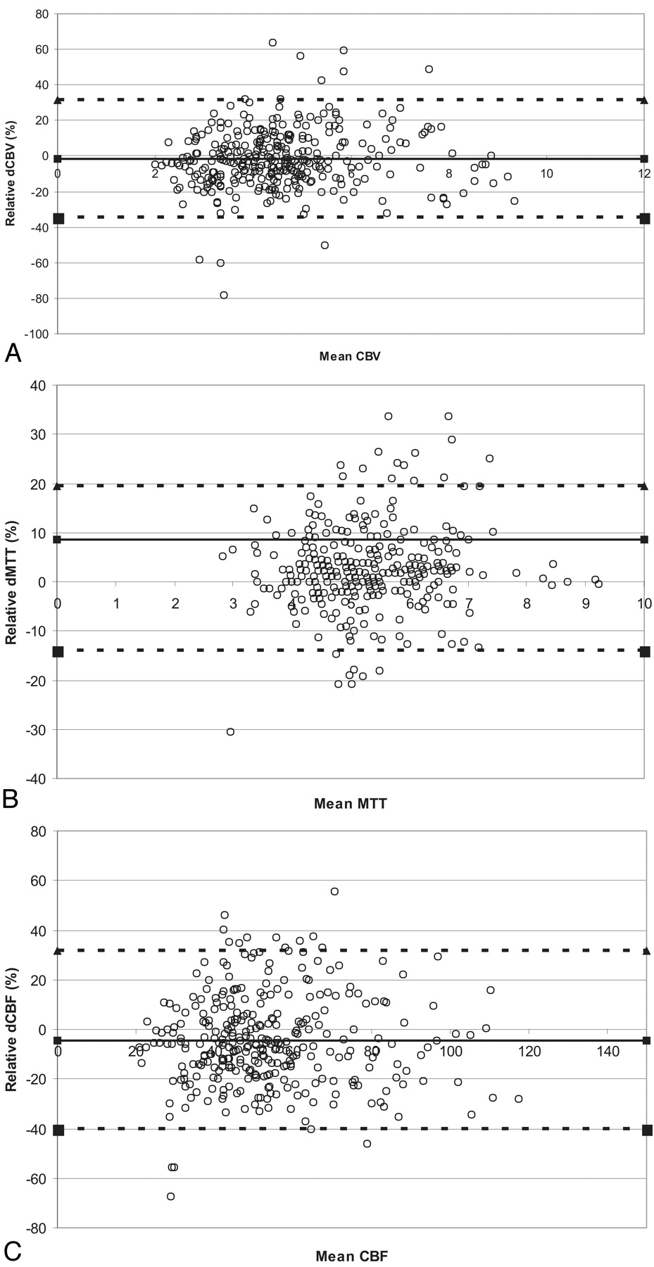
- Fig 2.
Bland-Altman plots of the relative differences (interobserver variability, pooled data) against the mean absolute value for CBF (milliliters per 100 g per minute) (A), CBV (milliliters per 100 g) (B), and MTT (seconds) (C). The relative ΔCBV, ΔCBF, and ΔMTT indicate the difference between 2 observations divided by the mean of those 2 observations, given as a percentage. The thick line represents the mean bias, and the dotted lines indicate the upper and lower limits of agreement. These upper and lower limits of agreement for the relative differences were 37% and −37% for CBV, 38% and −43% for CBF, and 21% and −16% for MTT, respectively.


Tables
Patient demographics Mean age ± SD 68 ± 11.1 years (range, 48–84 years) Male:female 14:6 Presenting event Amaurosis fugax 2 Transient ischemic attack 9 Stroke 9 Carotid arteries Symptomatic side, left: right 10: 10 Mean stenosis ± SD, symptomatic side 88 ± 13 (range, 60% to 99%) Mean stenosis ± SD, asymptomatic side 11 ± 17% (range, 0% to 50%) Cerebral damage (noncontrast CT) No infarct 11 Small infarct (small branch area)* 5 Medium size infarct (major branch area)* 4 * Infarct size was classified according to Lodder et al.36
Territory CBV (ml/100 g) CBF (ml/100 g/min) MTT (seconds) CT perfusion values—symptomatic hemisphere ACA 4.1 ± 1.4 50.4 ± 18.8 5.0 ± 0.7 MCA 5.3 ± 1.4 56.1 ± 17.8 5.9 ± 1.3 Basal ganglia 4.3 ± 1.3 49.3 ± 17.6 5.5 ± 1.2 PCA 4.9 ± 1.4 50.9 ± 15.4 5.8 ± 0.7 CT perfusion values—asymptomatic hemisphere ACA 3.9 ± 1.3 50.9 ± 18.1 4.7 ± 0.7 MCA 5.3 ± 1.6 70.7 ± 22.3 4.5 ± 0.6 Basal ganglia 4.3 ± 1.3 57.2 ± 20.5 4.6 ± 0.9 PCA 5.0 ± 1.6 53.7 ± 16.9 5.6 ± 0.9 CT perfusion ratios (symptomatic/asymptomatic side) ACA 1.05 ± 0.14 1.01 ± 0.13 1.06 ± 0.10 MCA 1.01 ± 0.12 0.81 ± 0.14 1.28 ± 0.17 Basal ganglia 1.03 ± 0.09 0.88 ± 0.08 1.18 ± 0.12 PCA 1.05 ± 0.22 1.00 ± 0.17 1.06 ± 0.13 Note:—CBV indicates cerebral blood volume; CBF, cerebral blood flow; MTT, mean transit time; ACA, anterior cerebral artery; MCA, middle cerebral artery; PCA, posterior cerebral artery. Perfusion characteristics in our study group of 20 patients with symptomatic carotid artery stenosis. CT perfusion values and ratios (mean ± SD) are given for the various vascular flow territories. Note that numbers were calculated from mean values of 3 observations. For CT perfusion values, 80 data points (20 patients × 2 slabs × 2 hemispheres) were used for each territory. For CT perfusion ratios, 40 data points (20 patients × 2 slabs) were used for each territory.
- Table 3:
Comparison of intraobserver and interobserver variability for CT perfusion values and CT perfusion ratios
Variability SDDrel (Range of Relative Differences) CBV CBF MTT Intraobserver CT perfusion values 16% (−62%, 54%) 18% (−59%, 63%) 5% (−35%, 21%) CT perfusion ratios 11% (−51%, 37%) 10% (−42%, 33%) 6% (−30%, 23%) P* .000 .000 .048 Interobserver CT perfusion values 17% (−78%, 63%) 18% (−67%, 55%) 9% (−30%, 34%) CT perfusion ratios 16% (−44%, 71%) 13% (−49%, 51%) 9% (−36%, 30%) P* .135 .000 .295 Note:—SDDrel indicates SDs of relative differences; CBV, cerebral blood volume; CBF, cerebral blood flow; MTT, mean transit time. Pooled data from all flow territories. Variability is expressed as the SD of the relative differences for each pair of observations (SDDrel) and is thus expressed as a percentage. The range of relative differences is given in parentheses. Note that both intraobserver and interobserver variability are significantly lower for CBF ratios than for absolute CBF values, whereas for CBV, this effect was seen only in intraobserver variability. For MTT, the difference was small and significant for intraobserver variability only.
* F test.
- Table 4:
Comparison of intraobserver and interobserver variability for CT perfusion values separately for the symptomatic and asymptomatic hemisphere
Variability SDDrel (%) Symptomatic Side Asymptomatic Side CBV CBF MTT CBV CBF MTT Intraobserver ACA 22 22 5 20 21 5 MCA 18 20 4 19 22 5 Basal ganglia 18 20 4 19 20 7* PCA 20 22 8 20 20 4 Interobserver ACA 15 19 9 20* 22 8 MCA 12 17 6 14 19 8 Basal ganglia 15 15 8 21 18 9 PCA 22 16 11 14 13 6 Note:—SDDrel indicates SDs of relative differences; CBV, cerebral blood volume; CBF, cerebral blood flow; MTT, mean transit time; ACA, anterior cerebral artery; MCA, middle cerebral artery; PCA, posterior cerebral artery.
* Difference between asymptomatic and symptomatic hemisphere significantly different (P < .05).
- Table 5.
Comparison of intraobserver and interobserver variability (SDDrel) in the four flow territories
Variability SDDrel (%) CBV CBF MTT CBV ratio CBF ratio MTT ratio Intraobserver ACA 17 18 5 12 12 4 MCA 15 17 5 5 6 5 Basal ganglia 16 17 6 8 8 7 PCA 18 18 6 16 12 8 P* .730 .884 .210 .249 .029 .090 Interobserver ACA 16 19 8 15 16 8 MCA 11 15 6 11 9 6 Basal ganglia 17 15 8 13 8 8 PCA 18 15 9 23 17 10 P* .005 .005 .179 .000 .001 .001 Note:—SDDrel indicates SDs of relative differences; CBV, cerebral blood volume; CBF, cerebral blood flow; MTT, mean transit time; ACA, anterior cerebral artery; MCA, middle cerebral artery; PCA, posterior cerebral artery. The test of Levine for homogeneity of variance was used to check whether variability differed between flow territories. The least variability was commonly seen for the MCA territory. For CTP ratios, the largest variability was usually found for the PCA territory.
* Test of Levine for homogeneity of variance
In this issue


Jump to section
Related Articles
Cited By...
- Can Iterative Reconstruction Improve Imaging Quality for Lower Radiation CT Perfusion? Initial Experience
- Evaluation of CT Perfusion in the Setting of Cerebral Ischemia: Patterns and Pitfalls
- Intra- and Interobserver Agreement and Impact of Arterial Input Selection in Perfusion CT Measurements Performed in Squamous Cell Carcinoma of the Upper Aerodigestive Tract
- The Acetazolamide Challenge: Techniques and Applications in the Evaluation of Chronic Cerebral Ischemia