
Article Figures & Data
Figures
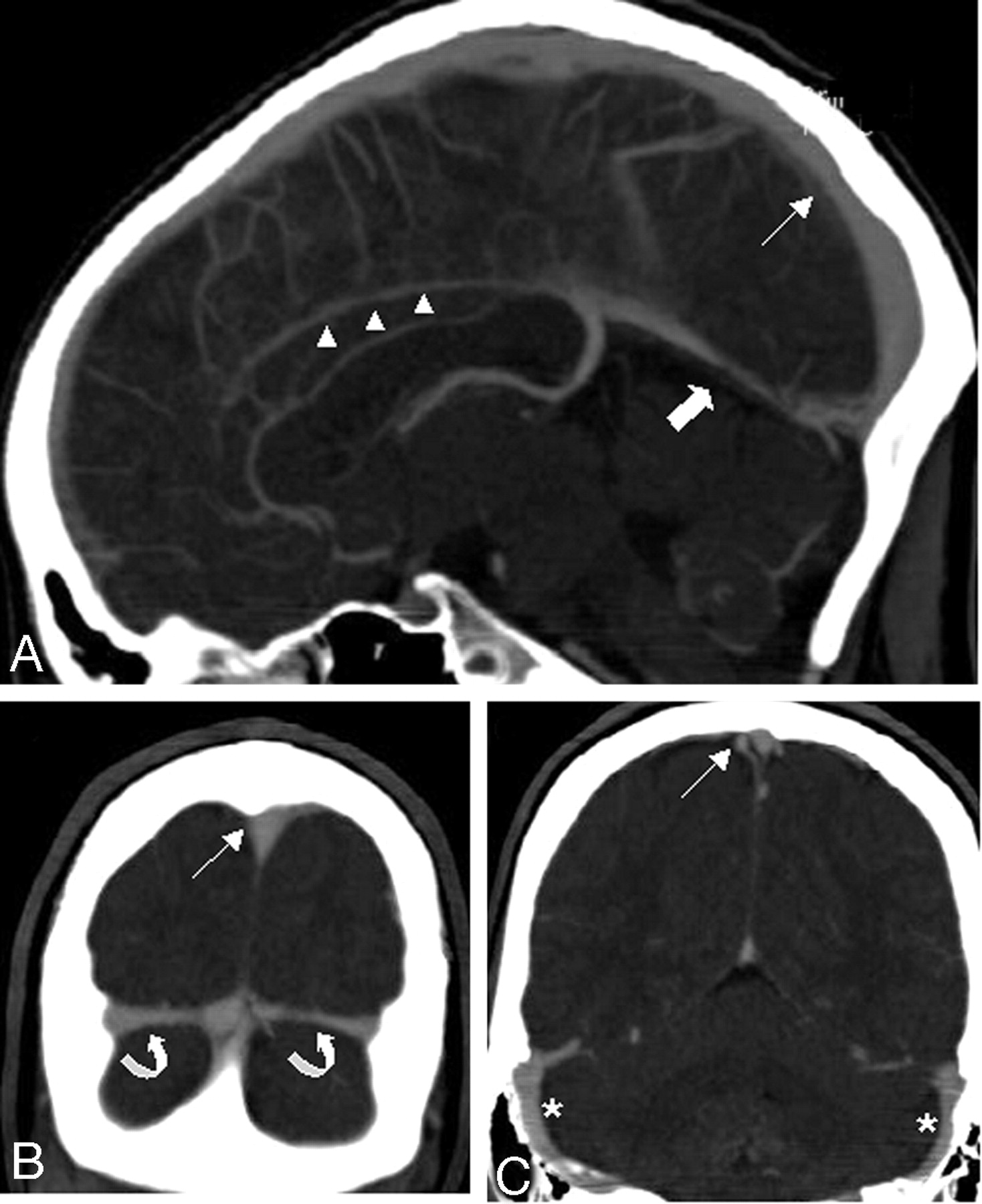
- Fig 1.
Sagittal (A) and coronal (B, C) sections of MIP reformations in a 22-year-old woman (patient 11) demonstrate the normal venous anatomy: superior sagittal sinus (thin arrow), the inferior sagittal sinus (arrowheads), the straight sinus (thick arrow), and the transverse sinuses (curved arrows). No venous pathologic condition was present in this patient. The MDCTA was performed on a 64-row-detector system.
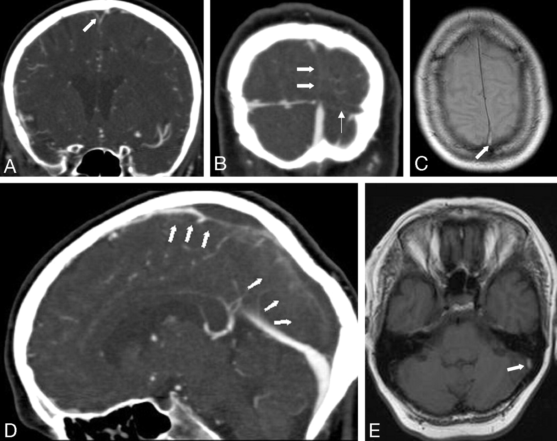
- Fig 2.
Coronal (A, B) and sagittal (D) sections of MIP reformations of a MDCTA performed on a 4-row-detector system in a 54-year old woman (patient 10) with an acute thrombosis of the superior sagittal sinus (thick arrows) and the left transverse sinus (thin arrow) show filling defects in the respective sinuses. Axial T1- (C) and PD-weighted (E) MR images demonstrate hyperintense signal intensity in the thrombosed left transverse sinus (C, arrow) and the superior sagittal sinus (E, arrow).
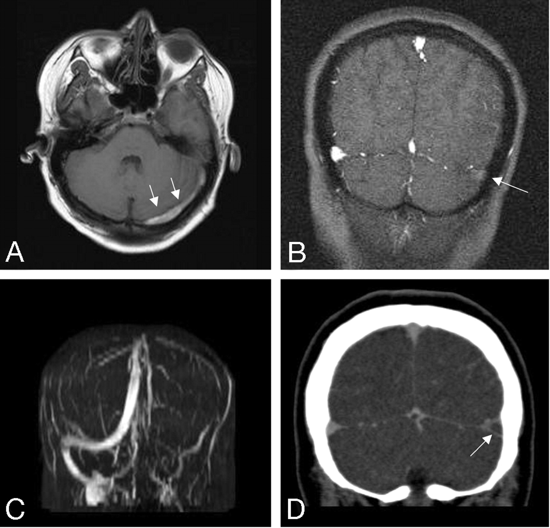
- Fig 3.
A 23-year-old male patient presenting with headache, amnestic aphasia, and visual disturbances (patient 4). MR imaging and MDCTA, performed on a 4-row-detector system, demonstrate a thrombosis of the left transverse (arrows) and sigmoid sinuses.
A, Axial T1-weighted MR depicts hyperintense, thrombotic material in the left transverse sinus.
B, C, 2D time-of-flight MR venography shows no flow void in the left transverse and sigmoid sinuses.
D, Coronal sections of MIP reformations demonstrate a filling defect in the left transverse sinus.
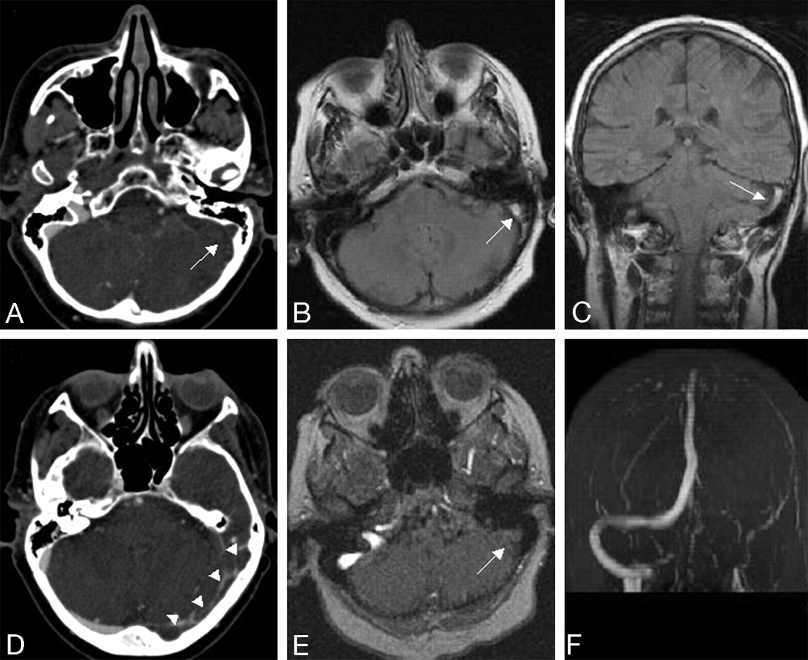
- Fig 4.
MDCTA and MR images of a 29-year-old female patient (patient 7) presenting with headache and seizures. Transversal sections of MIP reformations of the MDCTA (A, D) demonstrate filling defects in the left transverse (arrowheads) and sigmoid sinuses (arrow). Axial PD-weighted (B) and coronal fluid-attenuated inversion recovery (FLAIR) images (C) depict hyperintense, thrombotic material in the left sigmoid sinus.
E, F, 2D time-of-flight MR venography shows no flow void in the left transverse and sigmoid sinuses.


Tables
Patient Age/Sex Clinical Symptoms Diagnosis Site of Thrombosis Venous Edema or Intracerebral Hemorrhage 1 37/f Hemiparesis and sensory symptoms (l) Infarction right middle cerebri artery 2* 83/f Headache Sinus thrombosis LTS; LSS 3 47/f Headache Sinus thrombosis; drowsiness SSS; RTS 4 23/m Headache; visual disturbances; aphasia Sinus thrombosis LTS; LSS Edema 5 63/f Headache; aphasia; hemiparesis (r) Sinus thrombosis; postoperative state; cerebral amyloid angiopathy SSS Edema 6 42/m Headache Normal 7 29/f Headache Sinus thrombosis; seizures LTS; LSS Edema; ICH 8 43/m Headache Normal 9 35/f Headache; drowsiness Normal 10 54/f Headache; seizures Sinus thrombosis SSS; LTS 11 22/f Drowsiness; aphasia Normal; seizures 12 81/f Headache; drowsiness Postoperative state 13 76/f Headache; aphasia; drowsiness Normal 14 50/m Seizures; headache Sinus thrombosis SSS; SS; RTS; LTS Edema 15 74/f Confusion; seizures Normal 16 52/m Mental-status disorder Sinus thrombosis SSS; RTS; RSS 17 62/m Headache; sensory symptoms (l) Sinus thrombosis RTS 18 62/f Seizure Infarction right middle cerebri artery 19 36/f Headache Sinus thrombosis SSS Note:—f indicates female; m, male; l, left-sided; r, right-sided; SSS, superior sagittal sinus; SS, straight sinus; RTS, right transverse sinus; LTS, left transverse sinus; RSS, right sigmoid sinus; LTS, left sigmoid sinus; ICH, intracerebral hemorrhage.
* Excluded from analysis due to significant motion artifacts.
- Table 2:
Identification of venous sinus and cerebral veins in MSCTA and initial diagnosis of thrombosis made by the readers
Sinus or Vein Identified and Evaluated (n) Thrombosis Diagnosed (n) n Positive (%) True-Positive Total (R1/2/3) False-PositiveTotal (R1/2/3) True-NegativeTotal (R1/2/3) False-Negative Total (R1/2/3) Superior sagittal sinus 54 100 18 (6/6/6) 0 (0/0/0) 36 (12/12/12) 0 (0/0/0) Inferior sagittal sinus 54 100 0 (0/0/0) 0 (0/0/0) 54 (18/18/18) 0 (0/0/0) Straight sinus 54 100 2 (0/1/1) 0 (0/0/0) 51 (17/17/17) 1 (1/0/0) Left transverse sinus 54 100 10 (3/4/3) 3 (1/2/0) 39 (13/12/14) 2 (1/0/1) Right transverse sinus 54 100 10 (3/4/3) 0 (0/0/0) 42 (14/14/14) 2 (1/0/1) Left sigmoid sinus 52 96.3 3 (1/2/0) 1 (1/0/0) 45 (14/16/15) 3 (1/0/2) Right sigmoid sinus 53 98.1 2 (1/0/1) 0 (0/0/0) 50 (17/17/16) 1 (0/1/0) Vein of Galen 53 98.1 0 (0/0/0) 0 (0/0/0) 53 (18/18/17) 0 (0/0/0) Internal cerebral veins 50 92.6 0 (0/0/0) 0 (0/0/0) 50 (18/17/15) 0 (0/0/0) Left basal vein of Rosenthal 49 90.7 0 (0/0/0) 0 (0/0/0) 49 (18/15/16) 0 (0/0/0) Right basal vein of Rosenthal 48 88.9 0 (0/0/0) 0 (0/0/0) 48 (18/14/16) 0 (0/0/0) Left vein of Labbé 44 81.5 0 (0/0/0) 0 (0/0/0) 44 (15/14/15) 0 (0/0/0) Right vein of Labbé 43 79.6 0 (0/0/0) 0 (0/0/0) 43 (12/14/17) 0 (0/0/0) Note:—n indicates number of readings; R, reader.
- Table 3:
Presence of venous edema, intracerebral hemorrhage, and prominent Pacchioni granulations and diagnostic confidence
Venous edema and/or ICH diagnosed (n readings) Prominent Pachioni Granulations Found (in n of n readings) Diagnostic Confidence* (R 1/2/3) Additional Imaging Wanted** (R 1/2/3) Patient True-Pos. False-Pos. True-Neg. False-Neg. 1 0 0 3 0 Yes (2/3) 5/4/3 –/–/– 2† 3 3 0 0 0 Yes (2/3) 5/4/5 –/–/– 4 2 0 0 1 No 5/5/4 –/–/– 5 0 0 0 3 No 4/5/4 –/–/– 6 0 0 3 0 Yes (2/3) 5/4/4 –/–/– 7 2 0 0 1 No 5/5/2 –/–/MRI 8 0 0 3 0 Yes (2/3) 5/5/4 –/–/– 9 0 0 3 0 Yes (2/3) 5/5/5 –/–/– 10 0 0 3 0 No 5/3/4 –/–/– 11 0 0 3 0 No 5/3/4 –/MRI/– 12 0 0 3 0 Yes (1/3) 5/5/4 –/–/– 13 0 0 3 0 Yes (1/3) 5/3/4 –/–/MRI 14 0 0 0 3 No 3/5/4 MRI/–/MRI 15 0 0 3 0 Yes (2/3) 5/5/4 –/–/– 16 3 0 0 0 Yes (1/3) 5/5/5 –/–/– 17 0 0 3 0 No 3/5/3 MRI/–/cCT 18 0 0 3 0 No 5/4/5 –/MRI/– 19 0 0 3 0 Yes (1/3) 4/4/4 –/–/– Note:—n indicates number; R, reader; cCT native cranial CT.
* Rated on a 5-point scale (1, absolutely; 2, very; 3, intermediate; 4, not very certain; 5, uncertain).
** No additional imaging wanted.
† Excluded from analysis due to significant motion artifacts.
Patient False-Positive Readings False-Negative Readings R1 R2 R3 R1 R2 R3 1 2* 3 4 LSS 5 LTS, LSS LTS 6 7 LSS LSS 8 9 10 LTS 11 12 13 14 SS, RTS RTS, LTS 15 16 17 LTS RSS 18 19 Note:—No entry indicates no false-positive or -negative readings; R, reader; SS, straight sinus; RTS, right transverse sinus; LTS, left transverse sinus; RSS, right sigmoid sinus; LSS, left sigmoid sinus.
* Excluded from analysis due to significant motion artifacts.
In this issue


Jump to section
Related Articles
Cited By...
- Internal cerebral vein asymmetry is an independent predictor of poor functional outcome in endovascular thrombectomy
- Cerebral venous thrombosis: a practical guide
- Current endovascular strategies for cerebral venous thrombosis: report of the SNIS Standards and Guidelines Committee
- Management of Brain Arteriovenous Malformations: A Scientific Statement for Healthcare Professionals From the American Heart Association/American Stroke Association
- Early Detection and Quantification of Cerebral Venous Thrombosis by Magnetic Resonance Black-Blood Thrombus Imaging
- Cerebral Venous Thrombosis
- Diagnosis and Management of Cerebral Venous Thrombosis: A Statement for Healthcare Professionals From the American Heart Association/American Stroke Association
- Isolated Acute Nontraumatic Cortical Subarachnoid Hemorrhage
- Diagnostic Accuracy and Yield of Multidetector CT Angiography in the Evaluation of Spontaneous Intraparenchymal Cerebral Hemorrhage
- Noncontrast CT in Deep Cerebral Venous Thrombosis and Sinus Thrombosis: Comparison of its Diagnostic Value for Both Entities