Advertisement

Main menu

  • Home
  • Content
    • Current Issue
    • Accepted Manuscripts
    • Article Preview
    • Past Issue Archive
    • AJNR Case Collection
    • Case of the Week Archive
    • Classic Case Archive
    • Case of the Month Archive
  • Special Collections
    • Spinal CSF Leak Articles (Jan 2020-June 2024)
    • 2024 AJNR Journal Awards
    • Most Impactful AJNR Articles
  • Multimedia
    • AJNR Podcast
    • AJNR Scantastics
    • Video Articles
  • For Authors
    • Submit a Manuscript
    • Author Policies
    • Fast publishing of Accepted Manuscripts
    • Graphical Abstract Preparation
    • Manuscript Submission Guidelines
    • Imaging Protocol Submission
    • Submit a Case for the Case Collection
  • About Us
    • About AJNR
    • Editorial Board
  • More
    • Become a Reviewer/Academy of Reviewers
    • Subscribers
    • Permissions
    • Alerts
    • Feedback
    • Advertisers
    • ASNR Home
  • Other Publications
    • ajnr

User menu

  • Alerts
  • Log in

Search

  • Advanced search
American Journal of Neuroradiology

American Journal of Neuroradiology

ASHNR American Society of Functional Neuroradiology ASHNR American Society of Pediatric Neuroradiology ASSR
  • Alerts
  • Log in

Advanced Search

  • Home
  • Content
    • Current Issue
    • Accepted Manuscripts
    • Article Preview
    • Past Issue Archive
    • AJNR Case Collection
    • Case of the Week Archive
    • Classic Case Archive
    • Case of the Month Archive
  • Special Collections
    • Spinal CSF Leak Articles (Jan 2020-June 2024)
    • 2024 AJNR Journal Awards
    • Most Impactful AJNR Articles
  • Multimedia
    • AJNR Podcast
    • AJNR Scantastics
    • Video Articles
  • For Authors
    • Submit a Manuscript
    • Author Policies
    • Fast publishing of Accepted Manuscripts
    • Graphical Abstract Preparation
    • Manuscript Submission Guidelines
    • Imaging Protocol Submission
    • Submit a Case for the Case Collection
  • About Us
    • About AJNR
    • Editorial Board
  • More
    • Become a Reviewer/Academy of Reviewers
    • Subscribers
    • Permissions
    • Alerts
    • Feedback
    • Advertisers
    • ASNR Home
  • Follow AJNR on Twitter
  • Visit AJNR on Facebook
  • Follow AJNR on Instagram
  • Join AJNR on LinkedIn
  • RSS Feeds

Welcome to the new AJNR, Updated Hall of Fame, and more. Read the full announcements.


AJNR is seeking candidates for the position of Associate Section Editor, AJNR Case Collection. Read the full announcement.


Research ArticleBRAIN

MR Imaging of Metronidazole-Induced Encephalopathy: Lesion Distribution and Diffusion-Weighted Imaging Findings

E. Kim, D.G. Na, E.Y. Kim, J.H. Kim, K.R. Son and K.H. Chang
American Journal of Neuroradiology October 2007, 28 (9) 1652-1658; DOI: https://doi.org/10.3174/ajnr.A0655
E. Kim
  • Find this author on Google Scholar
  • Find this author on PubMed
  • Search for this author on this site
D.G. Na
  • Find this author on Google Scholar
  • Find this author on PubMed
  • Search for this author on this site
E.Y. Kim
  • Find this author on Google Scholar
  • Find this author on PubMed
  • Search for this author on this site
J.H. Kim
  • Find this author on Google Scholar
  • Find this author on PubMed
  • Search for this author on this site
K.R. Son
  • Find this author on Google Scholar
  • Find this author on PubMed
  • Search for this author on this site
K.H. Chang
  • Find this author on Google Scholar
  • Find this author on PubMed
  • Search for this author on this site
  • Article
  • Figures & Data
  • Info & Metrics
  • Responses
  • References
  • PDF
Loading

Article Figures & Data

Figures

  • Tables
  • Fig 1.
    • Download figure
    • Open in new tab
    • Download powerpoint
    Fig 1.

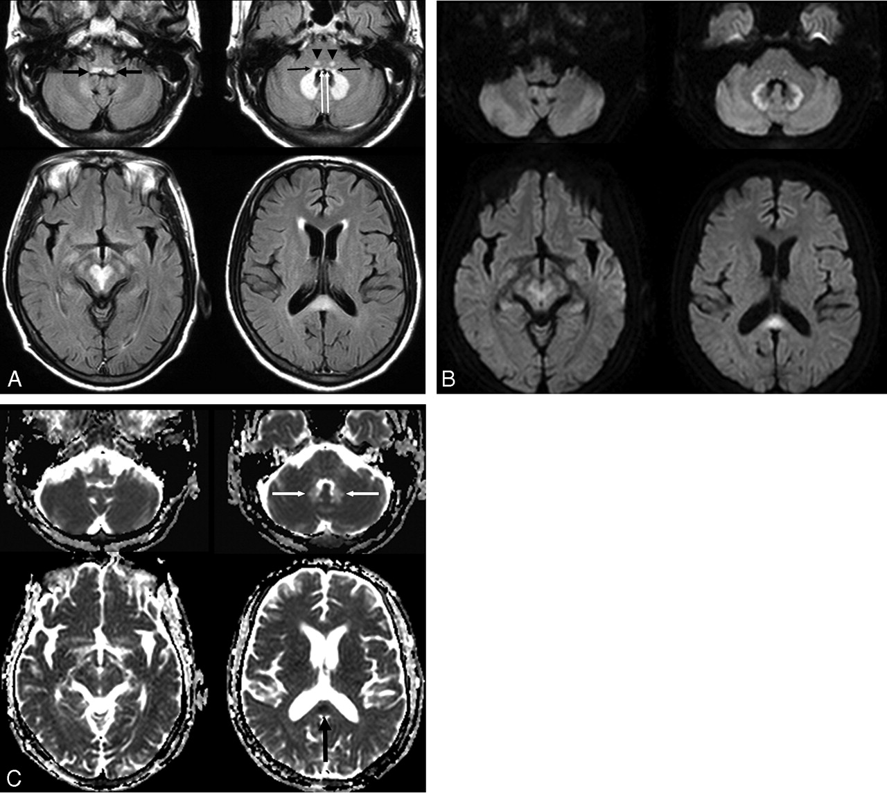
    A 54-year-old man (patient 4) with spontaneous bacterial peritonitis. A, Axial FLAIR (TR/TE/TI = 6000/120/2000 ms) images demonstrate bilateral symmetric hyperintense lesions in the dorsal medulla (black thick arrows), vestibular (black thin arrows), abducens (white arrows), and a focal tegmental lesion of the superior olivary nuclei (arrowheads) of the dorsal pons, dentate nuclei of the cerebellum, red nuclei and tegmentum of the midbrain, and the splenium of the corpus callosum. B, DWIs (TR/TE = 3396/60) show bright signal intensity at the peripheral part of the cerebellar dentate nuclei and central part of the splenium. The lesions of the low pons and tegmentum of the midbrain are slightly hyperintense, and the lesions of dorsal medulla and central part of dentate nuclei are isointense on DWI. C, ADC maps show a focal area of low ADC in the splenium of the corpus callosum (black arrow) and a high ADC area in most areas of the dentate nuclei (white arrows).

  • Fig 2.
    • Download figure
    • Open in new tab
    • Download powerpoint
    Fig 2.

    A 55-year-old man with ischemic colitis (patient 6). A, Axial T2-weighted (TR/ TE = 5000/110) images demonstrate bilateral symmetric hyperintense lesions in the dorsal medulla, vestibular, abducens, and focal tegmental lesions of the superior olivary nuclei of the dorsal pons, dentate nuclei of the cerebellum, the tectum of the midbrain, and the splenium of the corpus callosum. B, DWI (TR/TE = 4000/73) and ADC maps show mild hyperintensity and slightly high ADC of the dentate nuclei and obvious hyperintensity and very low ADC of the splenium of the corpus callosum. C, Follow-up MR images obtained 15 days after drug discontinuation. T2-weighted images show that the hyperintense lesions of the dentate nucleus and pons have disappeared, but a residual hyperintense lesion is seen in the splenium of corpus callosum. Note the near normalization of ADC and residual hyperintensity of the residual splenium lesion on DWI.

  • Fig 3.
    • Download figure
    • Open in new tab
    • Download powerpoint
    Fig 3.

    A 64-year-old man with an intra-abdominal abscess (patient 3). A, Axial FLAIR (TR/TE/TI = 10,000/122/2000) images show bilateral symmetric hyperintense lesions in the inferior olivary nuclei (arrows) and dorsal medulla, dorsal pons, cerebellar dentate nuclei, splenium and genu of corpus callosum, and subcortical white matter of both cerebral hemispheres. B, Follow-up FLAIR images obtained 17 days after drug discontinuation show complete reversal of all lesions.

Tables

  • Figures
    • View popup
    Table 1:

    Demographic and clinical data of seven patients with MIE

    PatientAge/SexUnderlying DiseaseDrug Dosage (g)/Duration (days)SymptomsMedication Duration Before Symptom Development (days)Time to Clinical Improvement after Drug Discontinuation (days)Duration Between Symptom Onset and Initial MRI (days)Duration Between Drug Discontinuation and Follow-Up MRI (days)a
    149/MCrohn disease135/90Weakness of extremities, dysarthria, gait disturbance521038–
    270/MBrain abscess, LC57/38Weakness of extremities, dysarthria227714
    364/MIntraabdominal abscess37.5/25Dysarthria, gait disturbance, visual blurring1771717
    454/MSpontaneous bacterial peritonitis, LC49.5/33Dysarthria, confusion15521–
    571/MDM foot, CRF66/44Dysarthria, gait disturbance374334
    655/MIschemic colitis, LC21/14Dysarthria, gait disturbance, tingling sensation of both extremities117315
    761/FPseudo-membranous colitis40.5/27Dysarthria, gait disturbance2475–
    • Note:—LC indicates liver cirrhosis; CRF, chronic renal failure; DM, diabetes mellitus.

    • a Follow-up MR imaging was not studied in 3 patients (patients 1, 4, and 7).

    • View popup
    Table 2:

    Lesion distribution on T2-weighted and FLAIR MR images in the seven patients with MIE

    PatientCerebellum (Dentate Nuclei)MedullaPonsMidbrainCorpus CallosumSubcortical WM
    Dorsal MedullaIONVNANSONTectumTegmentumRN
    1+−−+++−++−−
    2+−−+−+++++−
    3++++−+++−++
    4++−+++−+++−
    5++−++++−−−−
    6++−++++−−+−
    7+−−−−−+−−−−
    • Note:—WM indicates white matter; ION, inferior olivary nucleus; VN, vestibular nucleus; AN, abducens nucleus; SON, superior olivary nucleus; RN, red nucleus; +, presence of lesion at each anatomic location; −, no abnormality on MR at each anatomic location.

    • View popup
    Table 3:

    Signal intensities and ADC values of lesions of DWI on initial MR imaging in five patients and follow-up MR imaging in one patient

    LesionsSignal Intensity on DWI (High/Mixed/Iso)ADC Value (10−6 mm2/s)ADC Ratioa
    Initial patients (n = 5)
        Cerebellar dentate nucleus (n = 5)2/2/1885 ± 671.2 ± 0.06
        Midbrain (n = 5)
        Tectum (n = 3)2/0/1872 ± 591.1 ± 0.10
        Red nucleus (n = 2)1/1/0929 ± 21.3 ± 0.01
        Tegmentum around peri-aqueductal GM (n = 2)2/0/01021 ± 591.5 ± 0.09
        Dorsal pons (n = 4)2/0/2844 ± 751.1 ± 0.16
        Dorsal medulla (n = 3)1/1/1959 ± 1621.3 ± 0.25
        Splenium (n = 2)2/0/0515 ± 1950.7 ± 0.30
    Follow-up patient (n = 1)
        Cerebellar dentate nucleus (n = 1)(0/0/1)7341.0
        Splenium (n = 1)(1/0/0)10971.4
    • Note:—Iso indicates isointense; GM, gray matter.

    • a ADC values and ratios are presented as mean ± SD. ADC ratio is the ratio of the ADC value of a lesion to that of normal subcortical white matter.

PreviousNext
Back to top

In this issue

American Journal of Neuroradiology: 28 (9)
American Journal of Neuroradiology
Vol. 28, Issue 9
October 2007
  • Table of Contents
  • Index by author
Advertisement
Print
Download PDF
Email Article

Thank you for your interest in spreading the word on American Journal of Neuroradiology.

NOTE: We only request your email address so that the person you are recommending the page to knows that you wanted them to see it, and that it is not junk mail. We do not capture any email address.

Enter multiple addresses on separate lines or separate them with commas.
MR Imaging of Metronidazole-Induced Encephalopathy: Lesion Distribution and Diffusion-Weighted Imaging Findings
(Your Name) has sent you a message from American Journal of Neuroradiology
(Your Name) thought you would like to see the American Journal of Neuroradiology web site.
CAPTCHA
This question is for testing whether or not you are a human visitor and to prevent automated spam submissions.
Cite this article
E. Kim, D.G. Na, E.Y. Kim, J.H. Kim, K.R. Son, K.H. Chang
MR Imaging of Metronidazole-Induced Encephalopathy: Lesion Distribution and Diffusion-Weighted Imaging Findings
American Journal of Neuroradiology Oct 2007, 28 (9) 1652-1658; DOI: 10.3174/ajnr.A0655

Citation Manager Formats

  • BibTeX
  • Bookends
  • EasyBib
  • EndNote (tagged)
  • EndNote 8 (xml)
  • Medlars
  • Mendeley
  • Papers
  • RefWorks Tagged
  • Ref Manager
  • RIS
  • Zotero
0 Responses
Respond to this article
Share
Bookmark this article
MR Imaging of Metronidazole-Induced Encephalopathy: Lesion Distribution and Diffusion-Weighted Imaging Findings
E. Kim, D.G. Na, E.Y. Kim, J.H. Kim, K.R. Son, K.H. Chang
American Journal of Neuroradiology Oct 2007, 28 (9) 1652-1658; DOI: 10.3174/ajnr.A0655
del.icio.us logo Twitter logo Facebook logo Mendeley logo
  • Tweet Widget
  • Facebook Like
  • Google Plus One
Purchase

Jump to section

  • Article
    • Abstract
    • Materials and Methods
    • Results
    • Discussion
    • Conclusion
    • References
  • Figures & Data
  • Info & Metrics
  • Responses
  • References
  • PDF

Related Articles

  • No related articles found.
  • PubMed
  • Google Scholar

Cited By...

  • Potentially Reversible and Recognizable Acute Encephalopathic Syndromes: Disease Categorization and MRI Appearances
  • Acute Toxic Leukoencephalopathy: Etiologies, Imaging Findings, and Outcomes in 101 Patients
  • A case of metronidazole-induced neurotoxicity
  • An 82-year-old man with ataxia and dysarthria
  • Mystery Case: Metronidazole-induced encephalopathy
  • Antibiotic-associated encephalopathy
  • MR features of metronidazole-induced encephalopathy
  • Acute cerebellar syndrome associated with metronidazole
  • Metronidazole-induced encephalopathy after prolonged metronidazole course for treatment of C. difficile colitis
  • Metronidazole and Hydroxymetronidazole Central Nervous System Distribution: 1. Microdialysis Assessment of Brain Extracellular Fluid Concentrations in Patients with Acute Brain Injury
  • Metronidazole and Hydroxymetronidazole Central Nervous System Distribution: 2. Cerebrospinal Fluid Concentration Measurements in Patients with External Ventricular Drain
  • Radiographic evolution of a rapidly reversible leukoencephalopathy due to metronidazole
  • Brain MRI evolution of metronidazole intoxication
  • Central Nervous System Involvement in Adults with Epidemic Hemolytic Uremic Syndrome
  • Walking unsteadily: a case of acute cerebellar ataxia
  • Hypertrophic olivary degeneration in children: four new cases and a review of the literature with an emphasis on the MRI findings
  • Painful neuropathy due to skin denervation after metronidazole-induced neurotoxicity
  • MRI of metronidazole induced cerebellar ataxia
  • Reversible cerebellar syndrome caused by metronidazole
  • Heat Stroke: Increased Signal Intensity in the Bilateral Cerebellar Dentate Nuclei and Splenium on Diffusion-Weighted MR Imaging
  • Characterizing the Mesencephalon Using Susceptibility-Weighted Imaging
  • MR Imaging Findings in 56 Patients with Wernicke Encephalopathy: Nonalcoholics May Differ from Alcoholics
  • Crossref (212)
  • Google Scholar

This article has been cited by the following articles in journals that are participating in Crossref Cited-by Linking.

  • Neuroimaging Findings in Acute Wernicke's Encephalopathy: Review of the Literature
    Giulio Zuccoli, Nicolò Pipitone
    American Journal of Roentgenology 2009 192 2
  • Cytotoxic Lesions of the Corpus Callosum That Show Restricted Diffusion: Mechanisms, Causes, and Manifestations
    Jay Starkey, Nobuo Kobayashi, Yuji Numaguchi, Toshio Moritani
    RadioGraphics 2017 37 2
  • Antibiotic-associated encephalopathy
    Shamik Bhattacharyya, R. Ryan Darby, Pooja Raibagkar, L. Nicolas Gonzalez Castro, Aaron L. Berkowitz
    Neurology 2016 86 10
  • MR Imaging Findings in 56 Patients with Wernicke Encephalopathy: Nonalcoholics May Differ from Alcoholics
    G. Zuccoli, D. Santa Cruz, M. Bertolini, A. Rovira, M. Gallucci, C. Carollo, N. Pipitone
    American Journal of Neuroradiology 2009 30 1
  • MR Imaging Findings in Alcoholic and Nonalcoholic Acute Wernicke’s Encephalopathy: A Review
    Gaetana Manzo, Angela De Gennaro, Attilio Cozzolino, Antonietta Serino, Giacomo Fenza, Andrea Manto
    BioMed Research International 2014 2014
  • Toxic and Acquired Metabolic Encephalopathies: MRI Appearance
    Pranshu Sharma, Muneer Eesa, James N. Scott
    American Journal of Roentgenology 2009 193 3
  • Imaging Patterns of Toxic and Metabolic Brain Disorders
    Arthur M. de Oliveira, Matheus V. Paulino, Ana P. F. Vieira, Alexander M. McKinney, Antonio J. da Rocha, Germana T. dos Santos, Claudia da Costa Leite, Luis F. de Souza Godoy, Leandro T. Lucato
    RadioGraphics 2019 39 6
  • Neurotoxicity with Antimicrobials in the Elderly: A Review
    Arun Mattappalil, Kari A. Mergenhagen
    Clinical Therapeutics 2014 36 11
  • Metronidazole-induced encephalopathy: a systematic review
    Caspar Godthaab Sørensen, William Kristian Karlsson, Faisal Mohammad Amin, Mette Lindelof
    Journal of Neurology 2020 267 1
  • Clinical relevance of metronidazole and peripheral neuropathy: a systematic review of the literature
    Tiffany A. Goolsby, Bernadette Jakeman, Robert P. Gaynes
    International Journal of Antimicrobial Agents 2018 51 3

More in this TOC Section

  • Progression of Microstructural Damage in Spinocerebellar Ataxia Type 2: A Longitudinal DTI Study
  • Usefulness of Quantitative Susceptibility Mapping for the Diagnosis of Parkinson Disease
  • Evaluating the Effects of White Matter Multiple Sclerosis Lesions on the Volume Estimation of 6 Brain Tissue Segmentation Methods
Show more Brain

Similar Articles

Advertisement

Indexed Content

  • Current Issue
  • Accepted Manuscripts
  • Article Preview
  • Past Issues
  • Editorials
  • Editors Choice
  • Fellow Journal Club
  • Letters to the Editor

Cases

  • Case Collection
  • Archive - Case of the Week
  • Archive - Case of the Month
  • Archive - Classic Case

Special Collections

  • Special Collections

Resources

  • News and Updates
  • Turn around Times
  • Submit a Manuscript
  • Author Policies
  • Manuscript Submission Guidelines
  • Evidence-Based Medicine Level Guide
  • Publishing Checklists
  • Graphical Abstract Preparation
  • Imaging Protocol Submission
  • Submit a Case
  • Become a Reviewer/Academy of Reviewers
  • Get Peer Review Credit from Publons

Multimedia

  • AJNR Podcast
  • AJNR SCANtastic
  • Video Articles

About Us

  • About AJNR
  • Editorial Board
  • Not an AJNR Subscriber? Join Now
  • Alerts
  • Feedback
  • Advertise with us
  • Librarian Resources
  • Permissions
  • Terms and Conditions

American Society of Neuroradiology

  • Not an ASNR Member? Join Now

© 2025 by the American Society of Neuroradiology All rights, including for text and data mining, AI training, and similar technologies, are reserved.
Print ISSN: 0195-6108 Online ISSN: 1936-959X

Powered by HighWire