
Article Figures & Data
Figures
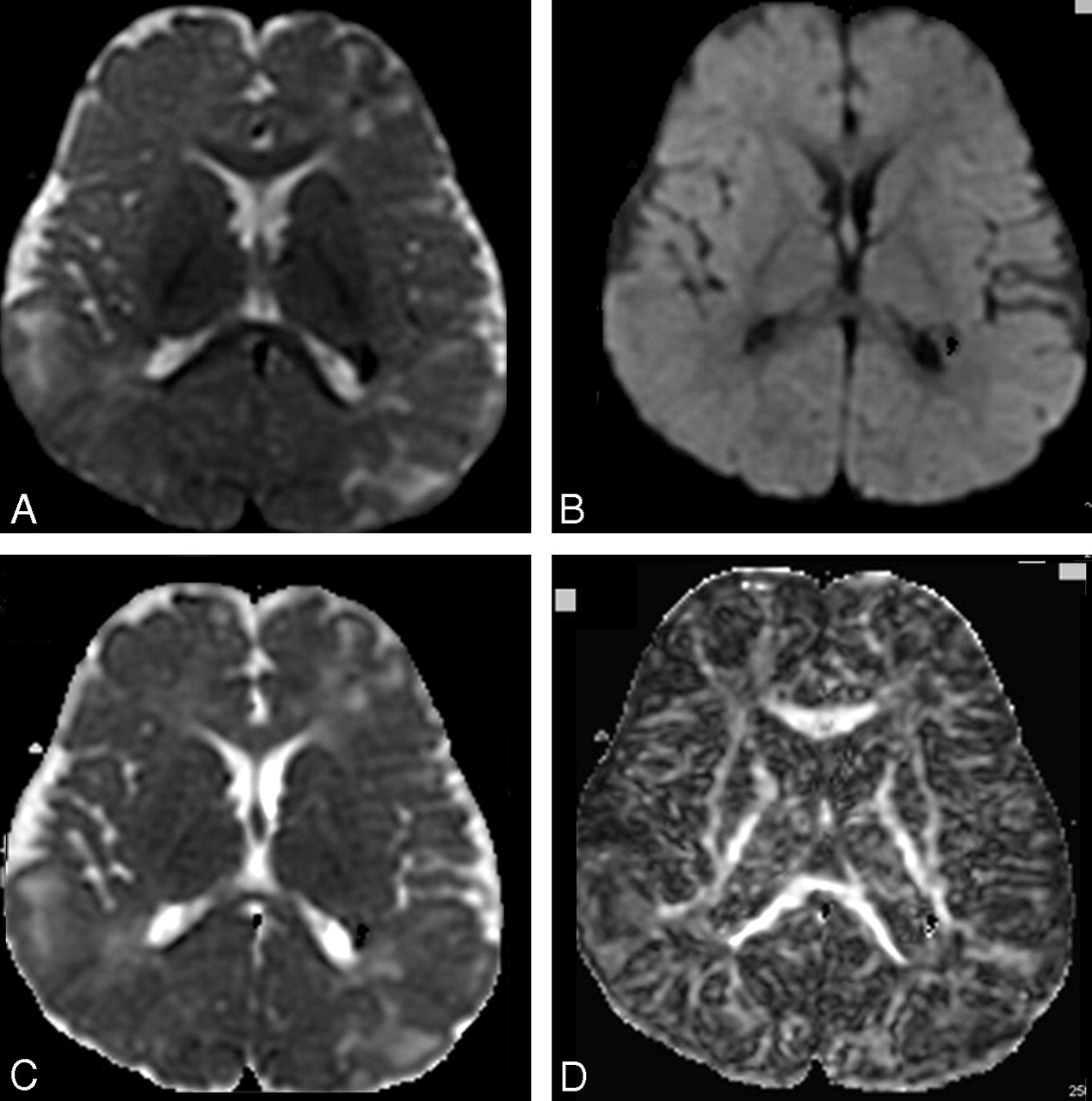
- Fig 1.
A–D, T2-weighted image of the diffusion acquisition (A), diffusion-weighted map as an average of the 6 directions (B), ADC map (C), and FA map (D), processed from DTI acquired on a 1.5T scanner in an 8-year-old patient with TSC (6 diffusion directions, 6 averages, b = 1000 [s/mm2]).
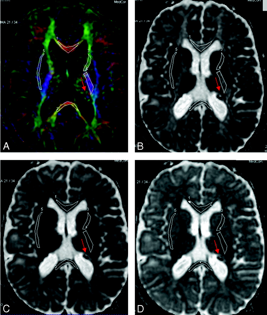
- Fig 2.
A–C, Selected ROIs are carefully drawn over the FA color-encoded images (A) and pasted on the corresponding main eigenvalues maps (B) and ADC map (C). The ROIs are indexed as follows: GCC#1, SCC#2, ALIC#3, PLIC#4, and EC#5. Red arrows indicate some of the TSC lesions.
- Fig 3.
A–D, Selected ROIs on the FA color-encoded image (A) are pasted on the eigenvalues maps (eg, main [B], middle [C], and minor [D] eigenvalues). The ROIs are indexed as follows: GCC#1, SCC#2, ALIC#3, PLIC#4, and EC#5. Red arrows indicate some of the TSC lesions.


Tables
DTI parameter values for NAWM in patients with TSC and the normal control group (NC)a
GCC SCC L-EC R-EC L-PLIC R-PLIC L-ALIC R-ALIC Mean SD Mean SD Mean SD Mean SD Mean SD Mean SD Mean SD Mean SD λ1b TSC 1490 96 1437 171 1011 95 1007 80 1221 72 1220 70 1107 58 1007 80 NC 1344 149 1360 134 963 104 919 81 1153 89 1158 100 963 104 919 81 λ2 TSC 543 125 526 157 653 118 645 111 455 49 483 60 588 69 582 77 NC 414 77 375 91 581 121 588 125 408 35 415 44 510 56 503 59 λ3 TSC 366 98 385 98 452 45 474 59 300 69 306 77 420 90 415 87 NC 275 78 270 104 373 60 373 60 239 25 245 19 321 54 309 51 ADC TSC 800 91 783 126 705 69 709 65 659 56 670 66 705 71 709 70 NC 678 90 668 107 639 67 627 66 600 76 606 72 598 45 577 46 FAc TSC 0.643 0.047 0.627 0.101 0.381 0.011 0.366 0.021 0.639 0.028 0.623 0.031 0.469 0.039 0.434 0.035 NC 0.703 0.051 0.725 0.068 0.437 0.036 0.413 0.035 0.676 0.049 0.670 0.051 0.503 0.035 0.494 0.040 Note:—DTI indicates diffusion tensor imaging; NAWM, normal-appearing white matter; TSC, tuberous sclerosis complex; GCC, genu of corpus callosum; SCC, splenium of corpus callosum; L-EC, left external capsule; R-EC, right external capsule; ADC, apparent diffusion coefficient; FA, fractional anisotropy; L-PLIC, left posterior limb of internal capsule; R-PLIC, right posterior limb of internal capsule; L-ALIC, left anterior limb of internal capsule; R-ALIC, right anterior limb of internal capsule.
a Regions of interest were drawn in the GCC and SCC bilaterally (L and R) over the EC and in both left and right hemispheres over the ALIC and PLIC.
b The 3 eigenvalues, λ1, λ2, and λ3, and ADC are expressed in 10−6 [mm2/s].
c The FA, the relative anisotropy, and the anisotropy index are dimensionless.
In this issue


Jump to section
Related Articles
Cited By...
- Tuberous sclerosis complex: Five new things
- DTI of tuber and perituberal tissue can predict epileptogenicity in tuberous sclerosis complex
- Tubers are neither static nor discrete: Evidence from serial diffusion tensor imaging
- Cerebral Diffusion Tensor MR Tractography in Tuberous Sclerosis Complex: Correlation with Neurologic Severity and Tract-Based Spatial Statistical Analysis
- Everolimus alters white matter diffusion in tuberous sclerosis complex
- Diffusion Tensor Imaging of Commissural and Projection White Matter in Tuberous Sclerosis Complex and Correlation with Tuber Load