Advertisement

Main menu

  • Home
  • Content
    • Current Issue
    • Accepted Manuscripts
    • Article Preview
    • Past Issue Archive
    • AJNR Case Collection
    • Case of the Week Archive
    • Classic Case Archive
    • Case of the Month Archive
  • Special Collections
    • Spinal CSF Leak Articles (Jan 2020-June 2024)
    • 2024 AJNR Journal Awards
    • Most Impactful AJNR Articles
  • Multimedia
    • AJNR Podcast
    • AJNR Scantastics
    • Video Articles
  • For Authors
    • Submit a Manuscript
    • Author Policies
    • Fast publishing of Accepted Manuscripts
    • Graphical Abstract Preparation
    • Manuscript Submission Guidelines
    • Imaging Protocol Submission
    • Submit a Case for the Case Collection
  • About Us
    • About AJNR
    • Editorial Board
  • More
    • Become a Reviewer/Academy of Reviewers
    • Subscribers
    • Permissions
    • Alerts
    • Feedback
    • Advertisers
    • ASNR Home
  • Other Publications
    • ajnr

User menu

  • Alerts
  • Log in

Search

  • Advanced search
American Journal of Neuroradiology

American Journal of Neuroradiology

ASHNR American Society of Functional Neuroradiology ASHNR American Society of Pediatric Neuroradiology ASSR
  • Alerts
  • Log in

Advanced Search

  • Home
  • Content
    • Current Issue
    • Accepted Manuscripts
    • Article Preview
    • Past Issue Archive
    • AJNR Case Collection
    • Case of the Week Archive
    • Classic Case Archive
    • Case of the Month Archive
  • Special Collections
    • Spinal CSF Leak Articles (Jan 2020-June 2024)
    • 2024 AJNR Journal Awards
    • Most Impactful AJNR Articles
  • Multimedia
    • AJNR Podcast
    • AJNR Scantastics
    • Video Articles
  • For Authors
    • Submit a Manuscript
    • Author Policies
    • Fast publishing of Accepted Manuscripts
    • Graphical Abstract Preparation
    • Manuscript Submission Guidelines
    • Imaging Protocol Submission
    • Submit a Case for the Case Collection
  • About Us
    • About AJNR
    • Editorial Board
  • More
    • Become a Reviewer/Academy of Reviewers
    • Subscribers
    • Permissions
    • Alerts
    • Feedback
    • Advertisers
    • ASNR Home
  • Follow AJNR on Twitter
  • Visit AJNR on Facebook
  • Follow AJNR on Instagram
  • Join AJNR on LinkedIn
  • RSS Feeds

Welcome to the new AJNR, Updated Hall of Fame, and more. Read the full announcements.


AJNR is seeking candidates for the position of Associate Section Editor, AJNR Case Collection. Read the full announcement.


OtherBRAIN

3D Rotational Digital Subtraction Angiography May Underestimate Intracranial Aneurysms: Findings from Two Basilar Aneurysms

L.-D. Jou, A. Mohamed, D.H. Lee and M.E. Mawad
American Journal of Neuroradiology October 2007, 28 (9) 1690-1692; DOI: https://doi.org/10.3174/ajnr.A0652
L.-D. Jou
  • Find this author on Google Scholar
  • Find this author on PubMed
  • Search for this author on this site
A. Mohamed
  • Find this author on Google Scholar
  • Find this author on PubMed
  • Search for this author on this site
D.H. Lee
  • Find this author on Google Scholar
  • Find this author on PubMed
  • Search for this author on this site
M.E. Mawad
  • Find this author on Google Scholar
  • Find this author on PubMed
  • Search for this author on this site
  • Article
  • Figures & Data
  • Info & Metrics
  • Responses
  • References
  • PDF
Loading

Abstract

SUMMARY: 3D digital subtraction angiography (DSA) allows clinicians to review intracranial aneurysms and other vascular lesions. We report 2 basilar aneurysms that were imaged by both 3D DSA and DynaCT. These 2 techniques produced very different aneurysm appearances. Anterior portions of the aneurysms were invisible on 3D DSA but were revealed by DynaCT. These aneurysms appeared to have been flattened by image artifacts in 3D DSA. Pulsation and gravity are 2 possible causes of aneurysm underestimation.

Digital subtraction angiography (DSA) has been the “gold standard” for many vascular and cerebrovascular imaging studies. 3D rotational DSA enables clinicians to review intracranial aneurysms and other vascular lesions thoroughly in an angiography study or during an interventional procedure. 3D rotational DSA was shown to perform better than either MR or CT angiography in 1 experimental study1 in which time-of-flight MR angiography underestimated aneurysm volumes, whereas CT angiography overestimated. Nevertheless, 3D DSA still is affected by image artifacts that may potentially influence diagnostic accuracy and impact clinical decisions.2 The requirement of uniformly distributed contrast agent across the region of interest often cannot be met in some vascular regions, such as at a lateral saccular aneurysm with a narrow neck.3 Aneurysms with certain orientation also may not be accessible by contrast injection. This is especially true during endovascular procedures when coils are already inside the aneurysm. Moreover, insufficient contrast influx can introduce image artifacts in 3D DSA.3 For example, parent vessels near an aneurysm may appear stenotic.4 In this report, we describe 2 cases of basilar aneurysms for which there are considerable differences in aneurysm volume and morphology between 3D DSA and DynaCT (Siemens Medical Solutions, Erlangen, Germany).5

Case Report

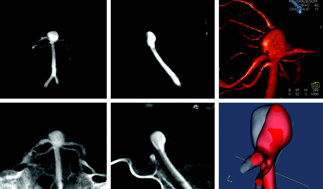
The first case was a 79-year-old woman who had a history of hypertension, diabetes, and hyperlipidemia. She was diagnosed with a basilar tip aneurysm after an episode of fainting. The aneurysm was treated with coil embolization without any incidents. Figure 1 shows the lateral and oblique view of the basilar tip aneurysm from 3D DSA and DynaCT. Because both images have the same resolution, a thick section in the middle of the aneurysm was selected for comparison and is presented in Fig 1.

Figure 1.
  • Download figure
  • Open in new tab
  • Download powerpoint
Figure 1.

Top row shows the front (left) and lateral views (middle) of 3D DSA and a rendering image (right). Bottom row shows the front (left) and lateral views (middle) of DynaCT images and overlap of basilar tip aneurysm models from DSA and DynaCT images (right). Aneurysm from 3D DSA is in red and aneurysm from DynaCT is in light gray. A large portion of aneurysm from DynaCT is missing in 3D DSA. 3D DSA indicates 3D digital subtraction angiography.

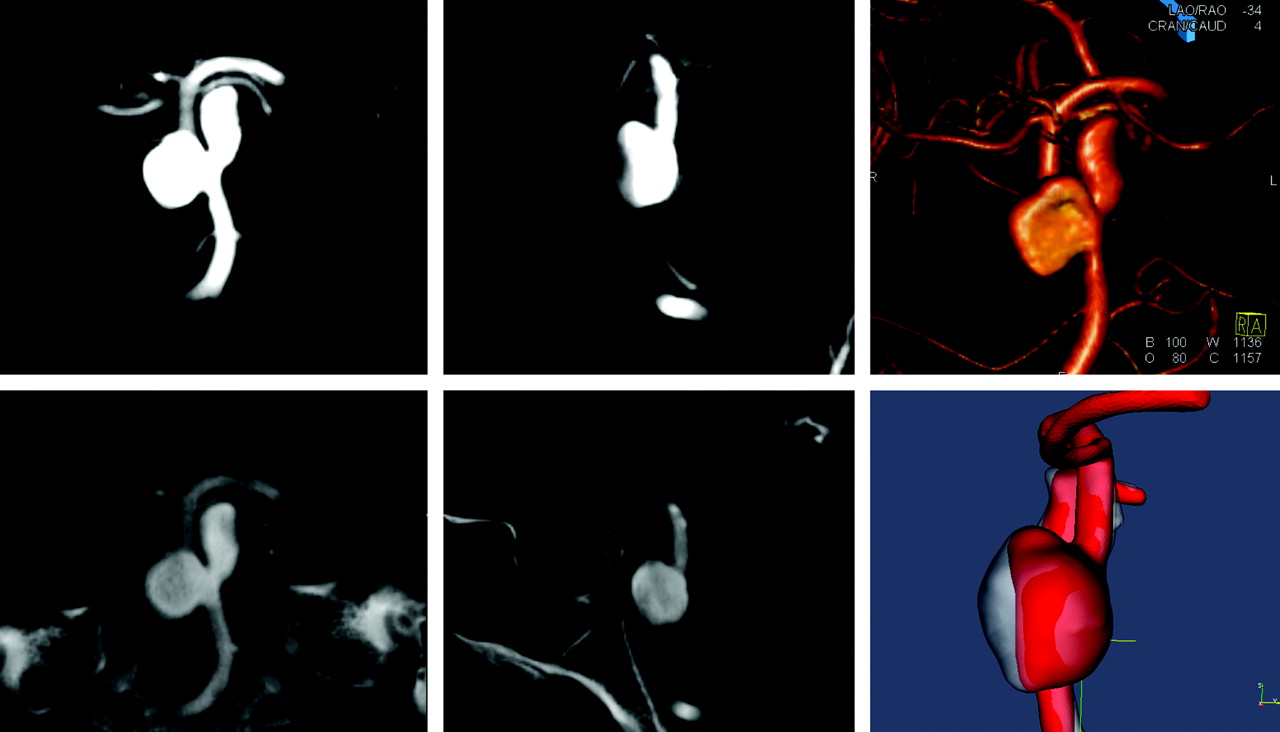
The second case was a 48-year-old man with asthma in whom a basilar trunk aneurysm was diagnosed. His bilobed basilar aneurysm was measured at 9 × 10 mm on the right and 10 × 10 mm on the left. The patient underwent stent placement and coiling for the basilar aneurysm. Both 3D DSA and DynaCT images of the aneurysm are shown in Figure 2. The anterior portion of his right-lobed aneurysm was not visible on 3D DSA.

Figure 2.
  • Download figure
  • Open in new tab
  • Download powerpoint
Figure 2.

Top row shows the front (left) and lateral views (middle) of 3D DSA and a rendering image (right). Bottom row shows the front (left) and lateral views (middle) of DynaCT images and overlap of basilar trunk aneurysm models from DSA and DynaCT images (right). Aneurysm from 3D DSA is in red, and aneurysm from DynaCT is in light gray. A portion of aneurysm in DynaCT is missing in 3D DSA. 3D DSA indicates 3D digital subtraction angiography.

Both patients were imaged with 3D DSA and DynaCT protocols on an AXIOM Artis dBA biplane flat detector system with DynaCT capability (Siemens Medical Solutions). For 3D DSA, 30 mL of contrast agent (Omnipaque 300; GE Healthcare, Princeton, NJ) was injected at 2.5 mL/s for 12 seconds, and 275 projections were acquired in 220° within 10 seconds to reconstruct 3D images. There was a 2-second delay for contrast filling. In DynaCT, 22 mL of same contrast agent diluted by the saline (25%) was injected at 1 mL/s, and 543 projections within 220° were acquired for reconstruction. Both procedures had a 2-second x-ray delay and produced 3D images with a 0.4-mm isotropic resolution after reconstruction. The difference in the appearance of aneurysms in DynaCT and 3D DSA after coregistration is presented in Fig 1 for the first case and in Fig 2 for the second. Both cases showed anterior portions of aneurysms in DynaCT, but they were missing from the 3D DSA. In the 3D DSA images, anterior portions of the aneurysms seemed flat on the X-Leonardo workstation (Siemens Medical Solutions); this appearance may be caused by image artifacts.

Discussion

Presence of artifacts may affect the calculation of aneurysm volume and the determination of packing ratio during an endovascular procedure. In turn, this may influence the clinician's perception about the completion of a procedure.6 Aneurysm volumes for these 2 cases were underestimated by approximately 20% in 3D DSA. The basilar trunk aneurysm in case 2 had a narrow neck, and its image appearance may have been affected by contrast deficit during imaging. The basilar tip aneurysm in case 1 had a wider neck and was directly in the flow path for the basilar artery. Therefore, this artifact cannot be explained simply as insufficient flow into the aneurysm.3 The possible mechanisms that resulted in this artifact are the blood flow pulsation and difference in density between blood and contrast medium.

Although contrast medium is injected at a constant rate, blood flow into the aneurysm is pulsatile, resulting in a fluctuation of contrast medium concentration. This can be observed in projective views of unmasked images. Contrast injection also can increase the flow rate considerably in cerebral vessels. The mean flow rate at the basilar artery is approximately 3 mL/s7; thus, an injection of 2.5 mL/s in these cases may have nearly doubled the mean flow rate at the basilar artery. Injection from only one of the vertebral arteries does not produce blood with uniform contrast at the basilar artery, and a portion of the aneurysm consisting of blood mainly from the noninjecting vertebral artery may appear unopacified.

Gravity also may have played an important role in generating this artifact. The contrast medium used for cerebral angiography has a higher density than the blood and often settles at the posterior part of the aneurysm after washout.8 The artifacts in both cases appear at the anterior side of the aneurysms, indicating that it may be difficult for the contrast medium to reach these regions. Given that the specific density of Omnipaque is 1.35, the specific density of blood injected with contrast medium is 1.18 in 3D DSA and 1.02 in DynaCT for the mean flow rate of 2.5 mL/s at the basilar artery. Therefore, the diluted contrast agent used in DynaCT has a density that is closer to that of the blood and may be able to fill the entire aneurysm better than the concentrated contrast medium used in 3D DSA. This may partly explain why the disappearance is always at the anterior side.

In both of these cases, the significant difference between 3D DSA and DynaCT was at the basilar artery. Although the basilar artery is the only artery we have observed so far that is susceptible to this artifact, there is no reason to believe that it is limited to this part of the vasculature. A careful examination of images from 3D DSA and DynaCT at other regions may allow a better understanding of this artifact.

References

  1. ↵
    Piotin M, Gailloud P, Bidaut L, et al. CT angiography, MR angiography and rotational digital subtraction angiography for volumetric assessment of intracranial aneurysms. An experimental study. Neuroradiology 2003;45:404–09
    CrossRefPubMed
  2. ↵
    Bidaut LM, Laurent C, Piotin M, et al. Second-generation three-dimensional reconstruction for rotational three-dimensional angiography. Acad Radiol 1998;5:836–49
    CrossRefPubMed
  3. ↵
    Ernemann UU, Gronewaller E, Duffner FB, et al. Influence of geometric and hemodynamic parameters on aneurysm visualization during three-dimensional rotational angiography: an in vitro study. AJNR Am J Neuroradiol 2003;24:597–603
    Abstract/FREE Full Text
  4. ↵
    Yamura M, Hirai T, Korogi Y, et al. Quantitative evaluation of measurement accuracy for three-dimensional angiography system using various phantoms. Radiat Med 2005;23:175–81
    PubMed
  5. ↵
    Heran NS, Song JK, Namba K, et al. The utility of DynaCT in neuroendovascular procedures. AJNR Am J Neuroradiol 2006;27:330–32
    Abstract/FREE Full Text
  6. ↵
    Sluzewski M, Rooij WJ, Slob MJ, et al. The relationship between aneurysm volume, packing, and compaction in 145 coiled cerebral aneurysms. Radiology 2004;231:653–58
    CrossRefPubMed
  7. ↵
    Ford MD, Alperin N, Lee SH, et al. Characterization of volumetric flow rate waveforms in the normal internal carotid and vertebral arteries. Physiol Meas 2005;26:477–88
    CrossRefPubMed
  8. ↵
    Wang Z-J, Hoffmann KR, Wang Z, et al. Contrast settling in cerebral aneurysm angiography. Phys Med Biol 2005;50:3171–81
    CrossRefPubMed
  • Received January 16, 2007.
  • Accepted after revision March 23, 2007.
  • Copyright © American Society of Neuroradiology
View Abstract
PreviousNext
Back to top

In this issue

American Journal of Neuroradiology: 28 (9)
American Journal of Neuroradiology
Vol. 28, Issue 9
October 2007
  • Table of Contents
  • Index by author
Advertisement
Print
Download PDF
Email Article

Thank you for your interest in spreading the word on American Journal of Neuroradiology.

NOTE: We only request your email address so that the person you are recommending the page to knows that you wanted them to see it, and that it is not junk mail. We do not capture any email address.

Enter multiple addresses on separate lines or separate them with commas.
3D Rotational Digital Subtraction Angiography May Underestimate Intracranial Aneurysms: Findings from Two Basilar Aneurysms
(Your Name) has sent you a message from American Journal of Neuroradiology
(Your Name) thought you would like to see the American Journal of Neuroradiology web site.
CAPTCHA
This question is for testing whether or not you are a human visitor and to prevent automated spam submissions.
Cite this article
L.-D. Jou, A. Mohamed, D.H. Lee, M.E. Mawad
3D Rotational Digital Subtraction Angiography May Underestimate Intracranial Aneurysms: Findings from Two Basilar Aneurysms
American Journal of Neuroradiology Oct 2007, 28 (9) 1690-1692; DOI: 10.3174/ajnr.A0652

Citation Manager Formats

  • BibTeX
  • Bookends
  • EasyBib
  • EndNote (tagged)
  • EndNote 8 (xml)
  • Medlars
  • Mendeley
  • Papers
  • RefWorks Tagged
  • Ref Manager
  • RIS
  • Zotero
0 Responses
Respond to this article
Share
Bookmark this article
3D Rotational Digital Subtraction Angiography May Underestimate Intracranial Aneurysms: Findings from Two Basilar Aneurysms
L.-D. Jou, A. Mohamed, D.H. Lee, M.E. Mawad
American Journal of Neuroradiology Oct 2007, 28 (9) 1690-1692; DOI: 10.3174/ajnr.A0652
del.icio.us logo Twitter logo Facebook logo Mendeley logo
  • Tweet Widget
  • Facebook Like
  • Google Plus One
Purchase

Jump to section

  • Article
    • Abstract
    • Case Report
    • Discussion
    • References
  • Figures & Data
  • Info & Metrics
  • Responses
  • References
  • PDF

Related Articles

  • No related articles found.
  • PubMed
  • Google Scholar

Cited By...

  • Critical role of angiographic acquisition modality and reconstruction on morphometric and haemodynamic analysis of intracranial aneurysms
  • The utility of cone beam volume CT in the evaluation of thrombosed intracranial aneurysms in subarachnoid hemorrhage
  • In vitro and in silico study of intracranial stent treatments for cerebral aneurysms: effects on perforating vessel flows
  • The utility of cone beam volume CT in the evaluation of thrombosed intracranial aneurysms in subarachnoid hemorrhage
  • Feasibility of Intravenous Flat Panel Detector CT Angiography for Intracranial Arterial Stenosis
  • Patient-Specific Computational Hemodynamics of Intracranial Aneurysms from 3D Rotational Angiography and CT Angiography: An In Vivo Reproducibility Study
  • Use of Angiographic CT Imaging in the Cardiac Catheterization Laboratory for Congenital Heart Disease
  • DynaCT evaluation of in-stent restenosis following Wingspan stenting of intracranial stenosis
  • Crossref (33)
  • Google Scholar

This article has been cited by the following articles in journals that are participating in Crossref Cited-by Linking.

  • Use of Angiographic CT Imaging in the Cardiac Catheterization Laboratory for Congenital Heart Disease
    Andrew C. Glatz, Xiaowei Zhu, Matthew J. Gillespie, Brian D. Hanna, Jonathan J. Rome
    JACC: Cardiovascular Imaging 2010 3 11
  • Machine Learning Application for Rupture Risk Assessment in Small-Sized Intracranial Aneurysm
    Heung Cheol Kim, Jong Kook Rhim, Jun Hyong Ahn, Jeong Jin Park, Jong Un Moon, Eun Pyo Hong, Mi Ran Kim, Seung Gyu Kim, Seong Hwan Lee, Jae Hoon Jeong, Sung Won Choi, Jin Pyeong Jeon
    Journal of Clinical Medicine 2019 8 5
  • C-arm flat detector computed tomography: the technique and its applications in interventional neuro-radiology
    Mudassar Kamran, Sanjoy Nagaraja, James V. Byrne
    Neuroradiology 2010 52 4
  • Influence of stent configuration on cerebral aneurysm fluid dynamics
    M. Haithem Babiker, L. Fernando Gonzalez, Justin Ryan, Felipe Albuquerque, Daniel Collins, Arius Elvikis, David H. Frakes
    Journal of Biomechanics 2012 45 3
  • Patient-Specific Computational Hemodynamics of Intracranial Aneurysms from 3D Rotational Angiography and CT Angiography: An In Vivo Reproducibility Study
    A.J. Geers, I. Larrabide, A.G. Radaelli, H. Bogunovic, M. Kim, H.A.F. Gratama van Andel, C.B. Majoie, E. VanBavel, A.F. Frangi
    American Journal of Neuroradiology 2011 32 3
  • Finite element modeling of embolic coil deployment: Multifactor characterization of treatment effects on cerebral aneurysm hemodynamics
    M. Haithem Babiker, Brian Chong, L. Fernando Gonzalez, Sachmanik Cheema, David H. Frakes
    Journal of Biomechanics 2013 46 16
  • Quantitative Effects of Coil Packing Density on Cerebral Aneurysm Fluid Dynamics: An In Vitro Steady Flow Study
    M. Haithem Babiker, L. Fernando Gonzalez, Felipe Albuquerque, Daniel Collins, Arius Elvikis, David H. Frakes
    Annals of Biomedical Engineering 2010 38 7
  • In vitro and in silico study of intracranial stent treatments for cerebral aneurysms: effects on perforating vessel flows
    Breigh Nonte Roszelle, M Haithem Babiker, Walter Hafner, L Fernando Gonzalez, Felipe C Albuquerque, David H Frakes
    Journal of NeuroInterventional Surgery 2013 5 4
  • Multi-View Convolutional Neural Networks in Rupture Risk Assessment of Small, Unruptured Intracranial Aneurysms
    Jun Hyong Ahn, Heung Cheol Kim, Jong Kook Rhim, Jeong Jin Park, Dick Sigmund, Min Chan Park, Jae Hoon Jeong, Jin Pyeong Jeon
    Journal of Personalized Medicine 2021 11 4
  • CT Angiography is State-of-the-Art First Vascular Imaging for Subarachnoid Hemorrhage
    A.J. Fox, S.P. Symons, R.I. Aviv
    American Journal of Neuroradiology 2008 29 6

More in this TOC Section

  • Progression of Microstructural Damage in Spinocerebellar Ataxia Type 2: A Longitudinal DTI Study
  • Usefulness of Quantitative Susceptibility Mapping for the Diagnosis of Parkinson Disease
  • Evaluating the Effects of White Matter Multiple Sclerosis Lesions on the Volume Estimation of 6 Brain Tissue Segmentation Methods
Show more Brain

Similar Articles

Advertisement

Indexed Content

  • Current Issue
  • Accepted Manuscripts
  • Article Preview
  • Past Issues
  • Editorials
  • Editors Choice
  • Fellow Journal Club
  • Letters to the Editor

Cases

  • Case Collection
  • Archive - Case of the Week
  • Archive - Case of the Month
  • Archive - Classic Case

Special Collections

  • Special Collections

Resources

  • News and Updates
  • Turn around Times
  • Submit a Manuscript
  • Author Policies
  • Manuscript Submission Guidelines
  • Evidence-Based Medicine Level Guide
  • Publishing Checklists
  • Graphical Abstract Preparation
  • Imaging Protocol Submission
  • Submit a Case
  • Become a Reviewer/Academy of Reviewers
  • Get Peer Review Credit from Publons

Multimedia

  • AJNR Podcast
  • AJNR SCANtastic
  • Video Articles

About Us

  • About AJNR
  • Editorial Board
  • Not an AJNR Subscriber? Join Now
  • Alerts
  • Feedback
  • Advertise with us
  • Librarian Resources
  • Permissions
  • Terms and Conditions

American Society of Neuroradiology

  • Not an ASNR Member? Join Now

© 2025 by the American Society of Neuroradiology All rights, including for text and data mining, AI training, and similar technologies, are reserved.
Print ISSN: 0195-6108 Online ISSN: 1936-959X

Powered by HighWire