
Article Figures & Data
Figures
- Fig 1.
Case 1. Images of the brain of an 80-year-old man with a history of headache, seizure, and left hemiparesis for 2 years. A, Precontrast CT scan shows patchy area of hypoattenuation in the white matter of the right parietal lobe with a punctate calcification located centrally. B, Axial T2-weighted MR image of the same day shows hyperintense area and cortical atrophy in the right parietal lobe. However, calcification seen on CT image cannot be found on MR images. C–D, Sagittal and coronal postcontrast images show tunnel-shaped enhancement representing inflammatory granuloma. No ipsilateral ventricular dilation is seen. E, Postoperative gross photograph of resected specimen shows a degenerated worm of Spirometra mansoni surrounded by inflammatory granulomatous tissues. F, Photomicrograph of histologic specimen shows a removed degenerated worm (W) surrounded by collagen capsule (C) and peripheral inflammatory cells and gliosis (G) (H&E stain × 40). G–H, Sagittal and coronal postcontrast images 1 year after a craniotomy in the same patient show lesions excised, with edematous area in the right parietal lobe.
- Fig 2.
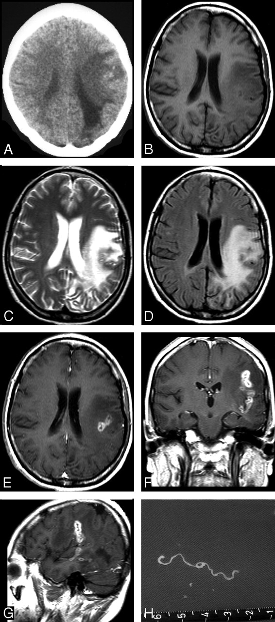
Case 2. Images of the brain of a 45-year-old woman with a 6-year history of severe headache, intermittent seizures, and right hemiparesis. A, Precontrast CT scan reveals unilateral extensive area of low attenuation in the white matter of the left parietal lobe, with ipsilateral ventricular dilation. Small, punctate calcifications are seen in the left parietal lobe. B–D, Axial T1-weighted (A), T2-weighted (B), and FLAIR images (C) of the same section show a wide area of hypointensity on T1-weighted image (B), heterogeneous hyperintensity on T2-weighted (C) and FLAIR images (D), with a small central area of isointensity and slight hyperintensity on T2-weighted image (C), corresponding to isointensity or hypointensity on FLAIR image (D), representing encephalomalacia. E–G, Postcontrast axial (E), coronal (F), and sagittal (G) T1-weighted images show a tunnel about 5 cm in length and 1.5 cm in width, appearing as a hollow tube located in the left temporal and parietal lobe. H, Intraoperative photograph shows a whitish, wrinkled, threadlike live worm approximately 6 cm in length with slow peristalsis.
- Fig 3.
Case 6. Images of the brain of a 14-year-old boy with a 4-year history of seizures and left hemiparesis. A, Precontrast CT scan shows an extensive area of low attenuation in the right basal ganglia with a punctate calcification centrally. B, Axial T2-weighted image of the same section as the CT image in (A) shows hyperintense area in the right basal ganglia. However, calcification seen on CT image cannot be shown clearly on MR image. C–D, Postcontrast axial (C) and coronal (D) T1-weighted images show bead-shaped enhancement in the right basal ganglia. E–F, After 4 months, postcontrast axial (E) and coronal (F) T1-weighted images of the same patient show the tunnel-shaped enhancement in the right parietal lobe with small amounts of residual bead-shaped enhancement in the right basal ganglia, representing the migration of the worm and lesions shifting from the right basal ganglia to the right parietal lobe. Preoperative ELISA on serum and on CSF revealed strongly positive against Spirometra mansoni. A live worm was found at craniotomy.


Tables
- Table 1:
Summery of clinical features, history, methods, and results in 25 patients with cerebral sparganosis
Patient No./Age (y)/Sex Imaging Modalities History Clinical Manifestations Results of ELISA Surgery* Serum CSF Worm Found 1/80/M CT‡, MR§ Raw snake ingestion 40 years previously Headache and seizures for 10 years, left hemiparesis for 2 years + + Degenerated 2/45/F CT‡, MR§ Uncooked frog ingestion 16 years previously Headache, seizures, and right hemiparesis for 6 years + + Live 3/53/F CT‡, MR§ Uncooked frog ingestion 13 years previously Headache, intermittent seizures, and left hemiparesis for 2 years + + Degenerated 4/24/F CT‡, MR§ Contaminated water drinking for 6 years Headache and focal seizures for 7 years + + Degenerated 5/9/M CT†, MR§ Uncertain Headache, intermittent seizures for 2 years + − Live 6/14/M CT†, MR§ Raw snake ingestion 6 years previously Seizures and left hemiparesis for 4 years + + Live 7/39/M MR§ Uncertain Headache and seizures for 5 years + + Degenerated 8/34/M CT† Uncertain Seizures, left hemiparesis for 8 years + + Live 9/37/F CT†, MR§ Flesh of frog applied to the skin wound 5 years previously Headache and seizures for 3 years, right hemiparesis for 8 months + + Degenerated 10/28/F CT† Flesh of snake applied to skin wound 10 years previously Focal seizures for 7 years, right hemiparesis for 13 months + − Degenerated 11/36/F CT†, MR§ Contaminated water drinking for 2 years Headache for 3 years, right hemiparesis for 1 year + + Live 12/38/M MR§ Contaminated water drinking 19 years previously Generalized seizures for 6 years, right hemiparesis for 11 months + + Degenerated 13/47/M MR§ Flesh of frog applied to abscess on back 13 years previously Focal seizures for 8 years, right hemiparesis for 1 year + + Degenerated 14/42/M MR§ Contaminated water drinking 20 years previously Headache, focal seizures for 3 years − + Degenerated worm found 15/83/F MR§ Raw frog ingestion 46 years previously Headache for 30 years, right hemiparesis for 15 months + + Degenerated 16/45/M MR§ Raw snake ingestion 9 years previously Generalized seizures for 6 years − + Degenerated 17/41/F MR§ Raw frog ingestion 17 years previously Seizures and right hemiparesis for 7 years + − Degenerated 18/43/F MR§ Raw snake ingestion 14 years previously Headache, seizures for 9 months + + Degenerated 19/38/M MR§ Raw frog ingestion 9 years previously Headache, seizures for 4 years; + − Degenerated left hemiparesis for 14 months − + Degenerated 20/29/F MR§ Frog ingestion for 7 years Seizures for 3 years + + Degenerated 21/37/M MR§ Raw snake ingestion 11 years previously Headache and seizures for 2 years; + − Degenerated right hemiparesis for 15 months + + Degenerated 22/48/M MR§ Uncooked frog ingestion 7 years previously Headache and seizures for 3 years + None 23/26/F MR§ Uncooked snake ingestion 5 years previously Focal seizures for 2 years + + Degenerated 24/31/F MR§ Contaminated water drinking 11 years previously Headache and seizures for 6 years + − Degenerated 25/20/M MR§ Uncertain Focal seizures for 9 months + + Degenerated Note:—ELISA indicates enzyme-linked immunosorbent assay; +, positive; −, negative.
* In this study, craniotomy was performed in 18 patients and a stereotactic targeting biopsy in 7 patients.
† Unenhanced CT scanning.
‡ Unenhanced, contrast-enhanced CT scanning.
§ Unenhanced, contrast-enhanced MR scanning.
Lesion CT MR Finding Number Finding Number White matter degeneration + 10 + 23 Cortical atrophy + 8 + 23 Ipsilateral ventricle dilation + 9 + 20 Ipsilateral ventricle compression − 0 + 3 Punctate calcification(s) + 6 + 3 Hemorrhage + 4 + 4 Tunnel sign* − 0 + 10 Bead-shaped enhancement† + 1 + 13 Single-ring enhancement + 2 − 0 Nodular enhancement + 1 − 0 Note:—+ indicates positive; −, negative.
* Tunnel sign is commonly seen on coronal and sagittal postcontrast MR images. The tunnel is about 4 cm in length (range, 2–6 cm) and 0.8 cm in width (range, 0.5–1.5 cm) and shows hypointensity on T1-weighted images, hyperintensity on T1-weighted images, and marked enhancement on postcontrast MR images.
† Bead-shaped enhancement is aggregated ringlike enhancement, usually 3 to 6 rings, with a smooth margin; ring wall thickness is 0.1 to 0.2 cm.
In this issue


Jump to section
Related Articles
Cited By...
- Development of a Rapid Diagnostic Kit That Uses an Immunochromatographic Device To Detect Antibodies in Human Sparganosis
- Migration: A Notable Feature of Cerebral Sparganosis on Follow-Up MR Imaging
- Follow-Up MR Imaging for Cerebral Sparganosis
- Letter to the editor: Is there a clear role of imaging in the diagnosis of cerebral sparganosis at present?
- Letter to the editor: Role of imaging in the diagnosis of cerebral sparganosis
- Breast and Scrotal Sparganosis: Sonographic Findings and Pathologic Correlation
- MR spectroscopy and MR perfusion character of cerebral sparganosis: a case report
- Cerebral sparganosis: The wandering lesion