
Article Figures & Data
Figures
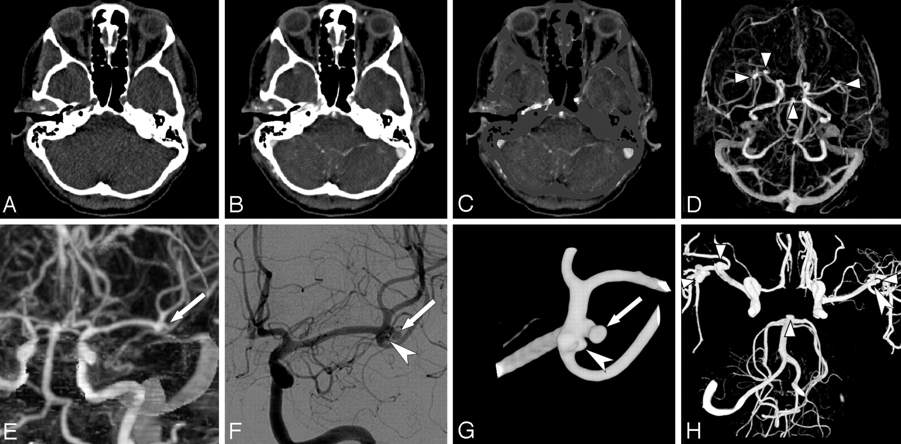
- Fig 1.
Illustration of a MMBE procedure in a 44-year-old woman with a ruptured right middle cerebral artery (MCA) aneurysm. A–C, Axial images of nonenhanced low-dose CT, CTA, and CTA after MMBE. D, Axial MIP obtained after MMBE shows 2 right MCA aneurysms, 1 left MCA aneurysm, and a basilar tip aneurysm (arrowheads). E, Coronal MIP image of the left MCA shows a 2.8-mm MCA aneurysm (arrow) and deceptive thickening of the MCA bifurcation. F, DSA shows the same aneurysm (arrow) as in E, with an additional 1.6-mm MCA aneurysm (large arrowhead). G, 3DRA more clearly shows both MCA aneurysms (arrow and large arrowhead). H, Composite image of three 3DRAs of both ICAs and the right vertebral artery shows all 5 aneurysms (small arrowheads and large arrowhead).
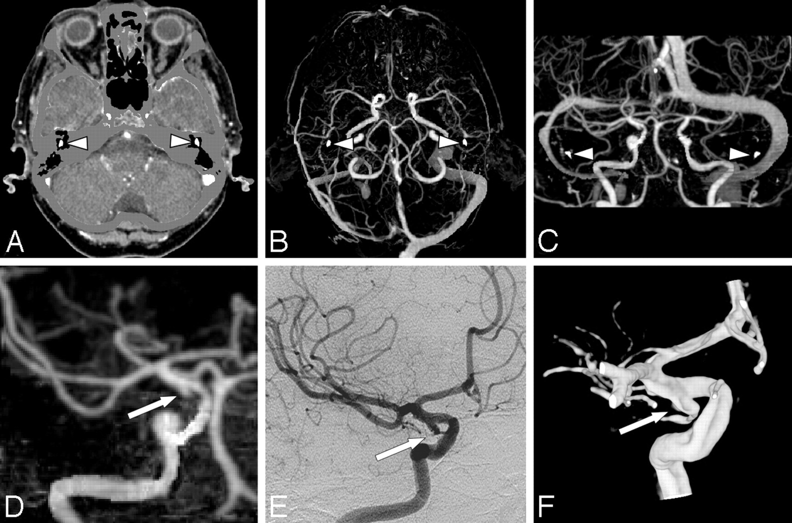
- Fig 2.
A 77-year-old woman with SAH and a false-positive-aneurysm finding on CTA. A, Axial CTA-MMBE image shows near complete bone removal as only the auditory ossicles (arrowheads) are not masked. B and C, Axial MIP and coronal MIP with small bone remnants of auditory ossicles (arrowheads), which do not hinder evaluation. D, Coronal MIP of a volume of interest with a small bulge of the right ICA interpreted as a small aneurysm (arrow). This infundibulum was mistaken for an aneurysm because the posterior communicating artery (PcomA) is not visible. E and F, DSA and 3DRA show the infundibulum of small PcomA (arrow).


Tables
- Table 1:
Number of detected aneurysms on DSA and 3DRA on 27 predefined aneurysm locations in 4 subgroups
Subgroup Location* No. of Aneurysms Anterior cerebral artery Anterior communicating artery 32 Pericallosal artery 4 ICA ICA tip 3 ICA, other locations 8 Posterior communicating artery 20 MCA M1 segment 7 M2 segment 3 Bifurcation 21 Posterior circulation Anterior inferior cerebellar artery 0 Basilar tip 8 Basilar trunk 2 Posterior cerebral artery 1 Posterior inferior cerebellar artery 4 Superior cerebellar artery 3 Vertebral artery 1 Total 117 * All locations were left and right, except for the anterior communicating artery, basilar trunk, and basilar tip, resulting in 27 predefined locations.
Quality Observer A Observer B Overall CTA Good 80 92 Fair 22 11 Moderate 4 4 Poor 2 1 CTA-MMBE Complete 3 5 Near-complete 98 101 Incomplete 7 2 Sensitivity Specificity PPV NPV Per patient Observer A 0.94 (83/88) 0.95 (19/20) 0.99 (83/84) 0.79 (19/24) 95% CI 0.87–0.98 0.75–1.00 0.93–1.00 0.59–0.91 Observer B 0.99 (87/88) 0.90 (18/20) 0.98 (87/89) 0.95 (18/19) 95% CI 0.93–1.00 0.69–0.98 0.92–1.00 0.74–1.00 Consensus 0.99 (87/88) 0.90 (18/20) 0.98 (87/89) 0.95 (18/19) 95% CI 0.93–1.00 0.69–0.98 0.92–1.00 0.74–1.00 Per location Observer A 0.87 (102/117) 1.00 (2070/2071) 0.99 (102/103) 0.99 (2070/2085) 95% CI 0.80–0.92 1.00–1.00 0.94–1.00 0.99–1.00 Observer B 0.88 (103/117) 1.00 (2066/2071) 0.95 (103/108) 0.99 (2066/2080) 95% CI 0.81–0.93 0.99–1.00 0.89–0.98 0.99–1.00 Consensus 0.91 (106/117) 1.00 (2068/2071) 0.97 (106/109) 0.99 (2068/2079) 95% CI 0.84–0.95 1.00–1.00 0.92–0.99 0.99–1.00 * Numbers between parentheses are aneurysms or locations. With DSA, 117 aneurysms were detected in 88 patients on 2188 predefined observed locations.
Location Sensitivity Specificity PPV NPV Anterior cerebral artery 0.97 (35/36) 1.00 (81/81) 1.00 (35/35) 0.99 (81/82) 95% CI 0.81–0.99 0.95–1.00 0.88–1.00 0.93–1.00 ICA 0.81 (25/31) 0.97 (83/86) 0.89 (25/28) 0.93 (83/89) 95% CI 0.63–0.91 0.90–0.99 0.72–0.97 0.86–0,97 MCA 0.90 (28/31) 1.00 (86/86) 1.00 (28/28) 0.97 (86/89) 95% CI 0.74–0.97 0.95–1.00 0.86–1.00 0.90–0.99 Posterior circulation 0.95 (18/19) 1.00 (98/98) 1.00 (18/18) 0.99 (98/99) 95% CI 0.74–1.00 0.95–1.00 0.79–1.00 0.94–1.00 * Numbers between parentheses are aneurysms.
Size Sensitivity PPV <3 mm 0.38 (6/16) 0.86 (6/7) 95% CI 0.18–0.61 0.57–0.99 3–5.9 mm 0.98 (49/50) 0.98 (49/50) 95% CI 0.89–1.00 0.89–1.00 6–10 mm 1.00 (36/36) 0.97 (36/37) 95% CI 0.89–1.00 0.85–1.00 >10 mm 1.00 (15/15) 1.00 (15/15) 95% CI 0.76–1.00 0.76–1.00 * Numbers between parentheses are aneurysms.
Aneurysm Location Size (mm) Visible in Retrospect on CTA Main Reason for Missing Aneurysm Ophthalmic artery 2.5 Yes Very small, in direct contact with bony structure (anterior clinoid process) and evaluated as irregularity of the ICA ICA cavernous segment 2.0 Yes Very small and poor quality of CTA ICA supraclinoid segment 2.0 Yes Very small and evaluated as part of multilobulated aneurysm ICA tip 1.8 No Very small and poor quality of CTA ICA tip 2.2 Yes Very small MCA 1.5 No Very small and presence of vasospasm MCA 2.0 Yes Very small and only visible on 1 MIP image MCA 1.6 Yes Very small and evaluated as thickening of MCA bifurcation Pericallosal artery 2.0 Yes Very small PICA 4.0 Yes Dissecting aneurysm, vasospasm, and incomplete bone removal due to patient movement PcomA 2.0 No Very small and low arterial contrast * All except 1 were <3 mm. One 4-mm aneurysm was undetected due to poor-quality CTA-MMBE.
In this issue


Jump to section
Related Articles
Cited By...
- Diagnostic Impact of Bone-Subtraction CT Angiography for Patients with Acute Subarachnoid Hemorrhage
- Dimensions of the posterior cerebral circulation: an analysis based on advanced non-invasive imaging
- Cerebrovascular geometry in the anterior circulation: an analysis of diameter, length and the vessel taper
- Guidelines for the Management of Aneurysmal Subarachnoid Hemorrhage: A Guideline for Healthcare Professionals From the American Heart Association/American Stroke Association
- Angiographic CT with Intravenous Contrast Injection Compared with Conventional Rotational Angiography in the Diagnostic Work-Up of Cerebral Aneurysms
- Contrast-free MRA at 3.0 T for the detection of intracranial aneurysms
- Patient-Specific Computational Hemodynamics of Intracranial Aneurysms from 3D Rotational Angiography and CT Angiography: An In Vivo Reproducibility Study
- Utility of CT Angiography in the Identification and Characterization of Supraclinoid Internal Carotid Artery Blister Aneurysms