Advertisement

Main menu

  • Home
  • Content
    • Current Issue
    • Accepted Manuscripts
    • Article Preview
    • Past Issue Archive
    • AJNR Case Collection
    • Case of the Week Archive
    • Classic Case Archive
    • Case of the Month Archive
  • Special Collections
    • Spinal CSF Leak Articles (Jan 2020-June 2024)
    • 2024 AJNR Journal Awards
    • Most Impactful AJNR Articles
  • Multimedia
    • AJNR Podcast
    • AJNR Scantastics
    • Video Articles
  • For Authors
    • Submit a Manuscript
    • Author Policies
    • Fast publishing of Accepted Manuscripts
    • Graphical Abstract Preparation
    • Manuscript Submission Guidelines
    • Imaging Protocol Submission
    • Submit a Case for the Case Collection
  • About Us
    • About AJNR
    • Editorial Board
  • More
    • Become a Reviewer/Academy of Reviewers
    • Subscribers
    • Permissions
    • Alerts
    • Feedback
    • Advertisers
    • ASNR Home
  • Other Publications
    • ajnr

User menu

  • Alerts
  • Log in

Search

  • Advanced search
American Journal of Neuroradiology

American Journal of Neuroradiology

ASHNR American Society of Functional Neuroradiology ASHNR American Society of Pediatric Neuroradiology ASSR
  • Alerts
  • Log in

Advanced Search

  • Home
  • Content
    • Current Issue
    • Accepted Manuscripts
    • Article Preview
    • Past Issue Archive
    • AJNR Case Collection
    • Case of the Week Archive
    • Classic Case Archive
    • Case of the Month Archive
  • Special Collections
    • Spinal CSF Leak Articles (Jan 2020-June 2024)
    • 2024 AJNR Journal Awards
    • Most Impactful AJNR Articles
  • Multimedia
    • AJNR Podcast
    • AJNR Scantastics
    • Video Articles
  • For Authors
    • Submit a Manuscript
    • Author Policies
    • Fast publishing of Accepted Manuscripts
    • Graphical Abstract Preparation
    • Manuscript Submission Guidelines
    • Imaging Protocol Submission
    • Submit a Case for the Case Collection
  • About Us
    • About AJNR
    • Editorial Board
  • More
    • Become a Reviewer/Academy of Reviewers
    • Subscribers
    • Permissions
    • Alerts
    • Feedback
    • Advertisers
    • ASNR Home
  • Follow AJNR on Twitter
  • Visit AJNR on Facebook
  • Follow AJNR on Instagram
  • Join AJNR on LinkedIn
  • RSS Feeds

Welcome to the new AJNR, Updated Hall of Fame, and more. Read the full announcements.


AJNR is seeking candidates for the position of Associate Section Editor, AJNR Case Collection. Read the full announcement.


Research ArticleINTERVENTIONAL

Dose Measurement on Both Patients and Operators during Neurointerventional Procedures Using Photoluminescence Glass Dosimeters

T. Moritake, Y. Matsumaru, T. Takigawa, K. Nishizawa, A. Matsumura and K. Tsuboi
American Journal of Neuroradiology November 2008, 29 (10) 1910-1917; DOI: https://doi.org/10.3174/ajnr.A1235
T. Moritake
  • Find this author on Google Scholar
  • Find this author on PubMed
  • Search for this author on this site
Y. Matsumaru
  • Find this author on Google Scholar
  • Find this author on PubMed
  • Search for this author on this site
T. Takigawa
  • Find this author on Google Scholar
  • Find this author on PubMed
  • Search for this author on this site
K. Nishizawa
  • Find this author on Google Scholar
  • Find this author on PubMed
  • Search for this author on this site
A. Matsumura
  • Find this author on Google Scholar
  • Find this author on PubMed
  • Search for this author on this site
K. Tsuboi
  • Find this author on Google Scholar
  • Find this author on PubMed
  • Search for this author on this site
  • Article
  • Figures & Data
  • Supplemental
  • Info & Metrics
  • Responses
  • References
  • PDF
Loading

Article Figures & Data

Figures

  • Tables
  • Fig 1.
    • Download figure
    • Open in new tab
    • Download powerpoint
    Fig 1.

    The small PLD chips used in this study are 12 mm in length and 1.5 mm in diameter. A small square in the background represents 1 mm2.
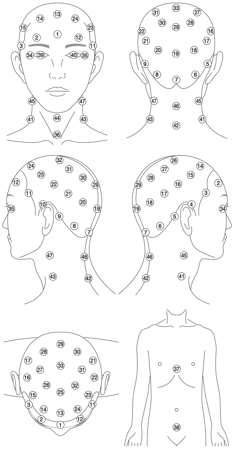
  • Fig 2.
    • Download figure
    • Open in new tab
    • Download powerpoint
    Fig 2.

    Dose-monitoring points for patients. The number in each circle represents the position number for the PLD placement.

  • Fig 3.
    • Download figure
    • Open in new tab
    • Download powerpoint
    Fig 3.

    Geometric distribution of operators’ average ESDs per procedure. ESDs on No. 3 (thyroid) and Nos. 6–10 are measured inside of the lead protector. The number in each circle represents the position number for PLD placement.

  • Fig 4.
    • Download figure
    • Open in new tab
    • Download powerpoint
    Fig 4.

    A, The relative response of PLD to x-rays with different effective energy compared with an ionization chamber. PLD chips are irradiated on the tissue-equivalent phantom8 with (open circle) and without (black circle) a filter for energy compensation. B, Calibration factor converting the PLD readout value to the ESD as a function of the effective energy. All the PLD chips are irradiated without a filter for energy compensation.
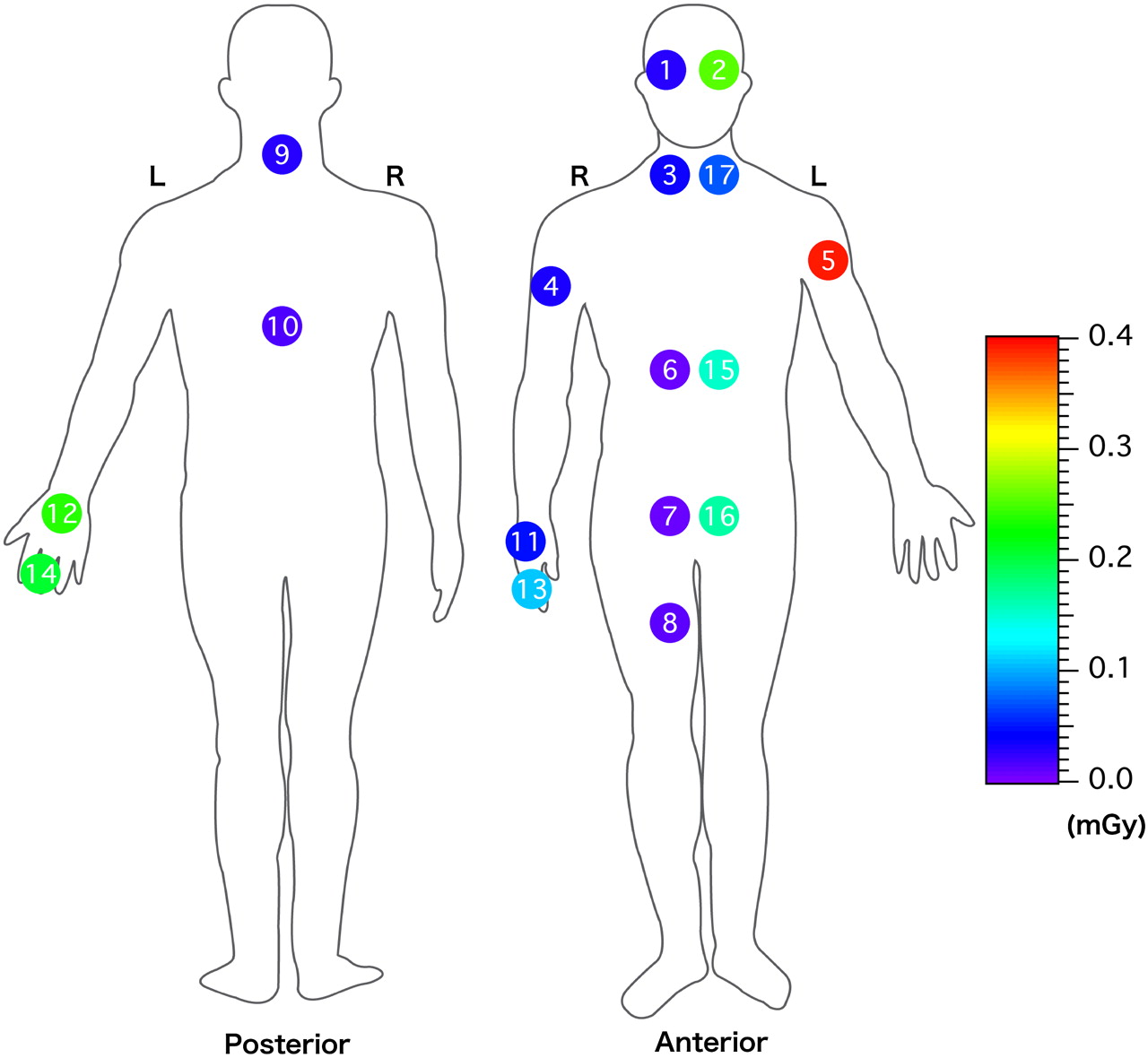
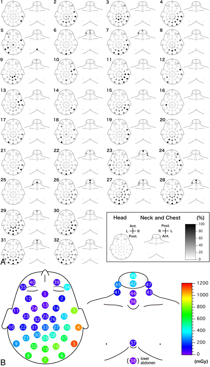
  • Fig 5.
    • Download figure
    • Open in new tab
    • Download powerpoint
    Fig 5.

    A, The relative (percentage) dose distribution to the maximum ESD for each patient. Each number represents a corresponding patient number listed in on-line Table. B, Geometric distribution of the average ESDs per procedure of 28 interventional radiology patients. The number in each circle represents the position number for PLD placement.
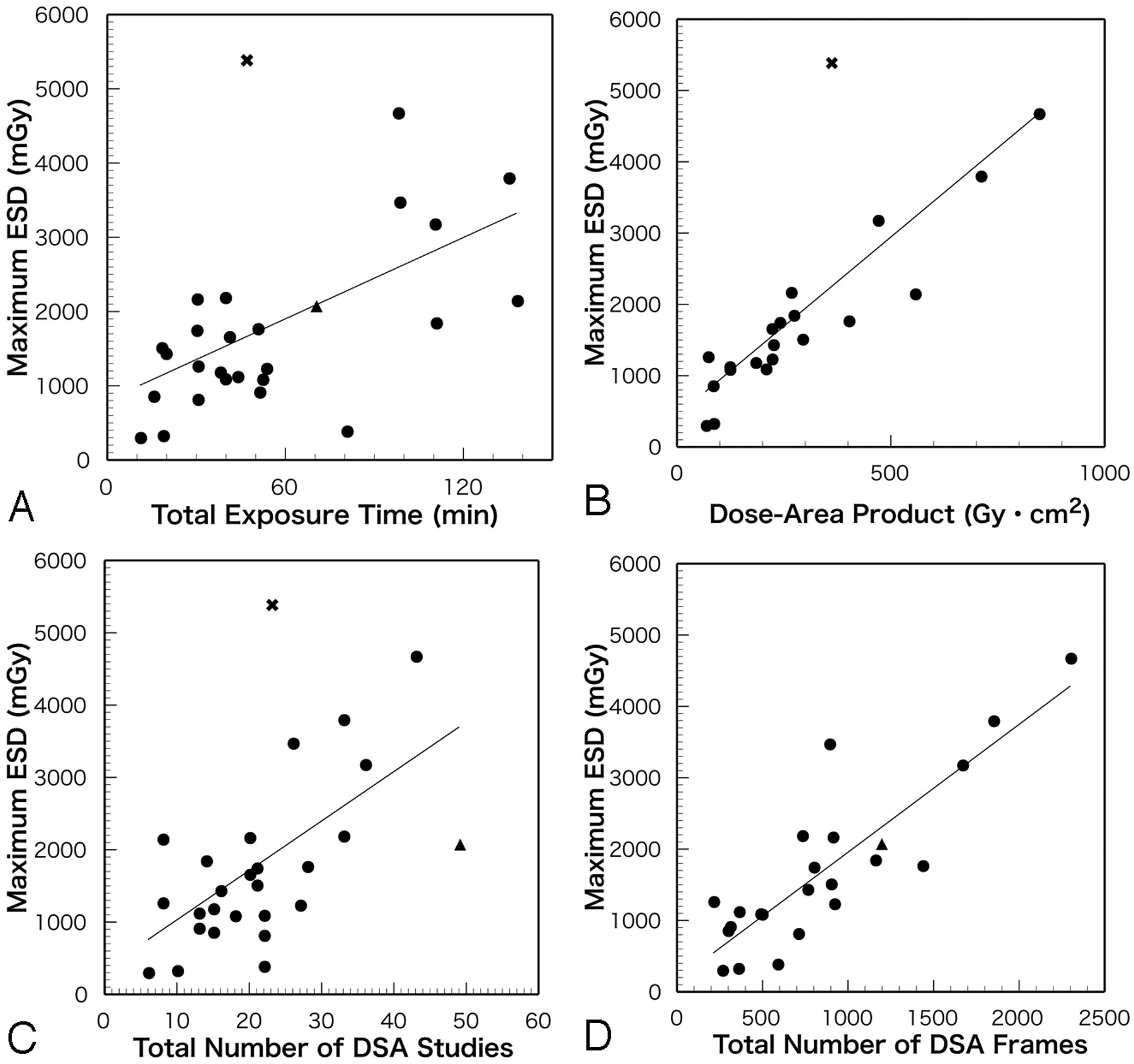
  • Fig 6.
    • Download figure
    • Open in new tab
    • Download powerpoint
    Fig 6.

    Correlations between the maximum ESD and angiographic parameters. Circle indicates a neurointerventional procedure without skin injury; x, patient No. 1 in on-line Table exhibited a depilation; triangle, patient No. 9 in on-line Table exhibited a depilation. A, Correlation between maximum ESD and total exposure time (r = 0.5283, P = .005, n = 27). B, Correlation between maximum ESD and dose-area product (r = 0.7917, P < .001, n = 21). C, Correlation between maximum ESD and total number of DSA studies (r = 0.5636, P = .002, n = 27). D, Correlation between maximum ESD and total number of DSA frames (r = 0.8583, P < .001, n = 23). Lines on the graphs indicate linear regressions.
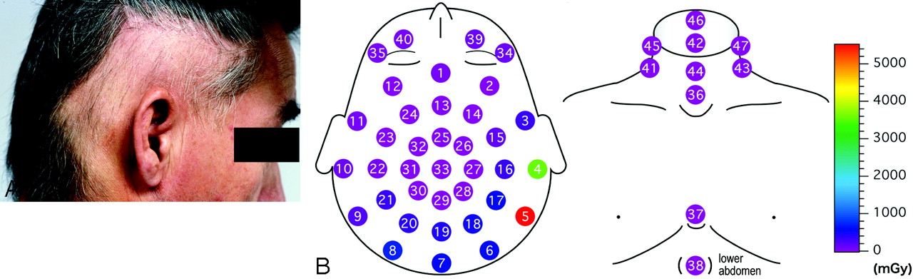
  • Fig 7.
    • Download figure
    • Open in new tab
    • Download powerpoint
    Fig 7.

    A 49-year-old man presents with radiation-induced depilation and erythema after a neurointerventional procedure. A, Square depilation in the right temporal area. B, Distribution of ESDs of this patient. The number in each circle represents the position number for PLD placement.

Tables

  • Figures
    • View popup
    Table 1:

    Average ESDs to the patients for 28 therapeutic interventional radiology procedures

    Point No.AreaAverage (mGy; mean ± SD)Range (mGy)Median (mGy)25–75 Percentiles (mGy)
    5 in Fig 5BRight temporal1124 ± 134965–5373573210–1440
    1 in Fig 5BFrontal33 ± 343–1431913–38
    34 in Fig 5BRight eye380 ± 5937–207910444–445
    35 in Fig 5BLeft eye79 ± 1735–9133218–60
    • View popup
    Table 2:

    Average ESDs to the operators for 25 therapeutic interventional radiology procedures

    Point No.AreaAverage (mGy; mean ± SD)Range (mGy)Median (mGy)25–75 percentiles (mGy)
    1 in Fig 3Right eye0.028 ± 0.0320.006–0.1330.0180.012–0.027
    2 in Fig 3Left eye0.254 ± 0.3380.014–1.2410.1320.076–0.230
    3 in Fig 3Thyroid (underneath protector)0.035 ± 0.0560.002–0.1980.0120.005–0.025
    17 in Fig 3Thyroid (outside protector)0.072 ± 0.0710.008–0.2100.0500.015–0.094
    9 in Fig 3Posterior neck (underneath protector)0.029 ± 0.0420.001–0.1820.0150.005–0.026
    4 in Fig 3Right upper arm0.032 ± 0.0460.005–0.1880.0150.010–0.026
    5 in Fig 3Left upper arm0.390 ± 0.7320.013–3.2960.1680.042–0.271
    11 in Fig 3Back of right hand0.050 ± 0.0700.004–0.2420.0180.014–0.037
    12 in Fig 3Back of left hand0.240 ± 0.4980.009–2.5060.1010.050–0.210
    13 in Fig 3Right finger0.110 ± 0.1560.004–0.5160.0370.020–0.082
    14 in Fig 3Left finger0.208 ± 0.3410.006–1.3330.0680.028–0.171
    6 in Fig 3Chest (underneath protector)0.009 ± 0.0210.000–0.1050.0020.001–0.005
    15 in Fig 3Chest (outside protector)0.152 ± 0.2600.008–1.0990.0660.026–0.109
    10 in Fig 3Back (underneath protector)0.016 ± 0.0300.000–0.1360.0080.002–0.013
    7 in Fig 3Abdomen (underneath protector)0.008 ± 0.0170.000–0.0690.0020.001–0.005
    16 in Fig 3Abdomen (outside protector)0.165 ± 0.3030.003–1.3340.0580.027–0.129
    8 in Fig 3Femur (underneath protector)0.013 ± 0.0240.000–0.0900.0040.001–0.010
PreviousNext
Back to top

In this issue

American Journal of Neuroradiology: 29 (10)
American Journal of Neuroradiology
Vol. 29, Issue 10
November 2008
  • Table of Contents
  • Index by author
Advertisement
Print
Download PDF
Email Article

Thank you for your interest in spreading the word on American Journal of Neuroradiology.

NOTE: We only request your email address so that the person you are recommending the page to knows that you wanted them to see it, and that it is not junk mail. We do not capture any email address.

Enter multiple addresses on separate lines or separate them with commas.
Dose Measurement on Both Patients and Operators during Neurointerventional Procedures Using Photoluminescence Glass Dosimeters
(Your Name) has sent you a message from American Journal of Neuroradiology
(Your Name) thought you would like to see the American Journal of Neuroradiology web site.
CAPTCHA
This question is for testing whether or not you are a human visitor and to prevent automated spam submissions.
Cite this article
T. Moritake, Y. Matsumaru, T. Takigawa, K. Nishizawa, A. Matsumura, K. Tsuboi
Dose Measurement on Both Patients and Operators during Neurointerventional Procedures Using Photoluminescence Glass Dosimeters
American Journal of Neuroradiology Nov 2008, 29 (10) 1910-1917; DOI: 10.3174/ajnr.A1235

Citation Manager Formats

  • BibTeX
  • Bookends
  • EasyBib
  • EndNote (tagged)
  • EndNote 8 (xml)
  • Medlars
  • Mendeley
  • Papers
  • RefWorks Tagged
  • Ref Manager
  • RIS
  • Zotero
0 Responses
Respond to this article
Share
Bookmark this article
Dose Measurement on Both Patients and Operators during Neurointerventional Procedures Using Photoluminescence Glass Dosimeters
T. Moritake, Y. Matsumaru, T. Takigawa, K. Nishizawa, A. Matsumura, K. Tsuboi
American Journal of Neuroradiology Nov 2008, 29 (10) 1910-1917; DOI: 10.3174/ajnr.A1235
del.icio.us logo Twitter logo Facebook logo Mendeley logo
  • Tweet Widget
  • Facebook Like
  • Google Plus One
Purchase

Jump to section

  • Article
    • Abstract
    • Materials and Methods
    • Results
    • Discussion
    • Conclusions
    • Acknowledgments
    • Footnotes
    • References
  • Figures & Data
  • Supplemental
  • Info & Metrics
  • Responses
  • References
  • PDF

Related Articles

  • PubMed
  • Google Scholar

Cited By...

  • The Efficacy of Shielding Systems for Reducing Operator Exposure during Neurointerventional Procedures: A Real-World Prospective Study
  • Radiation Doses in Patient Eye Lenses during Interventional Neuroradiology Procedures
  • Patient Radiation Dose Management in the Follow-Up of Potential Skin Injuries in Neuroradiology
  • A Comparison of Radiation Exposure between Diagnostic CTA and DSA Examinations of Cerebral and Cervicocerebral Vessels
  • Crossref (63)
  • Google Scholar

This article has been cited by the following articles in journals that are participating in Crossref Cited-by Linking.

  • Occupational Radiation Doses to Operators Performing Fluoroscopically-Guided Procedures
    Kwang Pyo Kim, Donald L. Miller, Amy Berrington de Gonzalez, Stephen Balter, Ruth A. Kleinerman, Evgenia Ostroumova, Steven L. Simon, Martha S. Linet
    Health Physics 2012 103 1
  • A Comparison of Radiation Exposure between Diagnostic CTA and DSA Examinations of Cerebral and Cervicocerebral Vessels
    A.-L. Manninen, J.-M. Isokangas, A. Karttunen, T. Siniluoto, M.T. Nieminen
    American Journal of Neuroradiology 2012 33 11
  • Radiation Dose and Radiation Protection for Patients and Physicians During Interventional Procedure
    Koichi CHIDA, Mamoru KATO, Yutaka KAGAYA, Masayuki ZUGUCHI, Haruo SAITO, Tadashi ISHIBASHI, Shoki TAKAHASHI, Shogo YAMADA, Yoshihiro TAKAI
    Journal of Radiation Research 2010 51 2
  • OCCUPATIONAL RADIATION EXPOSURE OF THE EYE IN NEUROVASCULAR INTERVENTIONAL PHYSICIAN
    Mamoru Kato, Koichi Chida, Takato Ishida, Hideto Toyoshima, Yasuyuki Yoshida, Shotaro Yoshioka, Junta Moroi, Toshibumi Kinoshita
    Radiation Protection Dosimetry 2019 185 2
  • What are useful methods to reduce occupational radiation exposure among radiological medical workers, especially for interventional radiology personnel?
    Koichi Chida
    Radiological Physics and Technology 2022 15 2
  • Patient Radiation Dose Management in the Follow-Up of Potential Skin Injuries in Neuroradiology
    E. Vano, J.M. Fernandez, R.M. Sanchez, D. Martinez, L. Lopez Ibor, A. Gil, C. Serna-Candel
    American Journal of Neuroradiology 2013 34 2
  • OCCUPATIONAL RADIATION EXPOSURE DOSE OF THE EYE IN DEPARTMENT OF CARDIAC ARRHYTHMIA PHYSICIAN
    Mamoru Kato, Koichi Chida, Takato Ishida, Fumiaki Sasaki, Hideto Toyoshima, Hajime Oosaka, Ken Terata, Yoshihisa Abe, Toshibumi Kinoshita
    Radiation Protection Dosimetry 2019 187 3
  • Local skin and eye lens equivalent doses in interventional neuroradiology
    Michael Sandborg, Sandro Rossitti, Håkan Pettersson
    European Radiology 2010 20 3
  • Direct measurement of patient&#039;s entrance skin dose during neurointerventional procedure to avoid further radiation-induced skin injuries
    Mikito Hayakawa, Takashi Moritake, Fumikatsu Kataoka, Tomoji Takigawa, Yasuhiro Koguchi, Yuka Miyamoto, Keiichi Akahane, Yuji Matsumaru
    Clinical Neurology and Neurosurgery 2010 112 6
  • Radiation Doses in Patient Eye Lenses during Interventional Neuroradiology Procedures
    R.M. Sánchez, E. Vañó, J.M. Fernández, S. Rosati, L. López-Ibor
    American Journal of Neuroradiology 2016 37 3

More in this TOC Section

  • SAVE vs. Solumbra Techniques for Thrombectomy
  • CT Perfusion&Reperfusion in Acute Ischemic Stroke
  • Delayed Reperfusion Post-Thrombectomy&Thrombolysis
Show more Interventional

Similar Articles

Advertisement

Indexed Content

  • Current Issue
  • Accepted Manuscripts
  • Article Preview
  • Past Issues
  • Editorials
  • Editors Choice
  • Fellow Journal Club
  • Letters to the Editor

Cases

  • Case Collection
  • Archive - Case of the Week
  • Archive - Case of the Month
  • Archive - Classic Case

Special Collections

  • Special Collections

Resources

  • News and Updates
  • Turn around Times
  • Submit a Manuscript
  • Author Policies
  • Manuscript Submission Guidelines
  • Evidence-Based Medicine Level Guide
  • Publishing Checklists
  • Graphical Abstract Preparation
  • Imaging Protocol Submission
  • Submit a Case
  • Become a Reviewer/Academy of Reviewers
  • Get Peer Review Credit from Publons

Multimedia

  • AJNR Podcast
  • AJNR SCANtastic
  • Video Articles

About Us

  • About AJNR
  • Editorial Board
  • Not an AJNR Subscriber? Join Now
  • Alerts
  • Feedback
  • Advertise with us
  • Librarian Resources
  • Permissions
  • Terms and Conditions

American Society of Neuroradiology

  • Not an ASNR Member? Join Now

© 2025 by the American Society of Neuroradiology All rights, including for text and data mining, AI training, and similar technologies, are reserved.
Print ISSN: 0195-6108 Online ISSN: 1936-959X

Powered by HighWire