
Article Figures & Data
Figures
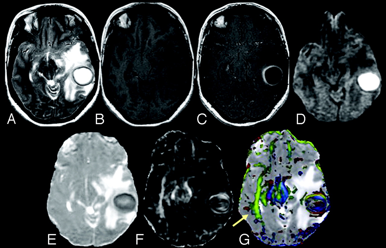
- Fig 1.
A 31-year-old female patient presenting with pyogenic brain abscess in the left temporal lobe. A, Axial T2-weighted image shows a well-defined hyperintense lesion with a hypointense wall. B, The lesion appears hypointense on the axial T1-weighted image with an isointense wall. C, On the postcontrast T1-weighted image, the lesion shows ring enhancement. D, DWI shows homogeneous hyperintensity in the cavity that appears hypointense on the MD map (E). The FA (F) and red-green-blue color-modulated FA map fused with the MD map (G) show that high FA in the abscess cavity is similar to what is observed in the contralateral inferior longitudinal fasciculus and midbrain (arrow).
- Fig 2.
A 25-year-old male patient presenting with pyogenic brain abscess with a heterogeneously hyperintense lesion in the left temporal lobe. A, Axial T2-weighted image shows a hyperintense lesion with a hypointense wall. B, The lesion appears hypointense on the axial T1-weighted image with an isointense wall. C, On the postcontrast T1-weighted image, the lesion shows ring enhancement. D, The lesion appears heterogeneously hyperintense on the DWI in the cavity that appears heterogeneously hypointense on the MD map (E). The FA (F) and red-green-blue color-modulated FA map fused with MD map (G) show that high FA in the abscess cavity is similar to what is observed in the contralateral inferior longitudinal fasciculus and midbrain.
- Fig 3.
A, Plots showing the relationship (A) between FA and IL1-β, TNF-α, and LFA-1. B, FA and sICAM-1 in pus from the abscess cavity. C–F, The relationship between FA and NMs in heat-killed S aureus–treated as well as nontreated cell lines at different time points (1, 24, 48, and 72 hours [hrs]) is depicted in the form of bar plots.
- Fig 4.
A, Coronal images of MD, FA, and color-coded FA maps fused with MD of Jurket cell lines treated with heat-killed S aureus at 1, 24, and 48 hours (hr), respectively, show an increase in FA and MD. B, The corresponding maps for nontreated Jurket cells show very few changes in FA and MD. C, Expression of LFA-1, IL1-β, TNF-α, and GAPDH (housekeeping) genes in nontreated and heat-killed S aureus–treated Jurket cell lines at 1, 24, 48, and 72 hours.
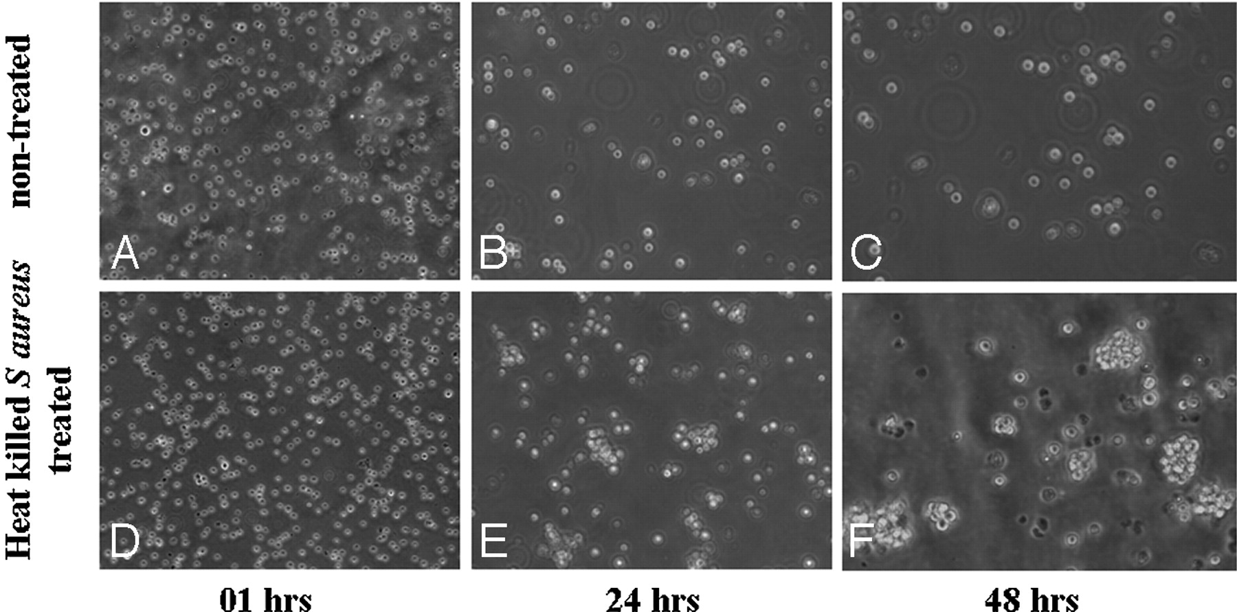
- Fig 5.
Cell aggregation at different time points in nontreated (A–C) and heat-killed S aureus–treated (D–F) cell lines. Photomicrographs of heat-killed S aureus–treated cell line with a time course show an increased degree of cell aggregation compared with that in nontreated cell line. hrs indicates hours.


In this issue


Jump to section
Related Articles
Cited By...
- Differential Gene Expression in Glioblastoma Defined by ADC Histogram Analysis: Relationship to Extracellular Matrix Molecules and Survival
- Differentiation of Tumefactive Demyelinating Lesions from High-Grade Gliomas with the Use of Diffusion Tensor Imaging
- Apparent Diffusion Coefficient with Higher b-Value Correlates Better with Viable Cell Count Quantified from the Cavity of Brain Abscess
- Differentiation of Brain Abscesses from Necrotic Glioblastomas and Cystic Metastatic Brain Tumors with Diffusion Tensor Imaging
- Correlation of Quantitative Diffusion Tensor Tractography with Clinical Grades of Subacute Sclerosing Panencephalitis
- In Vivo Proton MR Spectroscopy Evaluation of Pyogenic Brain Abscesses: A Report of 194 Cases