
Article Figures & Data
Figures
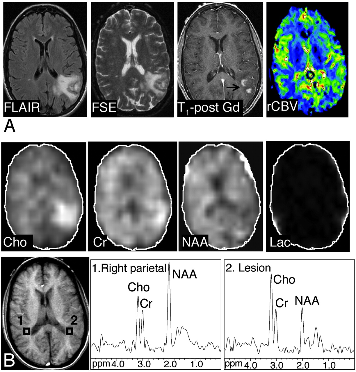
- Fig 1.
Axial FLAIR image, T2-weighted FSE MR image, axial contrast-enhanced T1-weighted SE MR image, and axial CBV map in a 38-year-old woman with primary CNS lymphoma. There is T2 hyperintensity involving the left temporoparietal lobe. There is minimal mass effect; the lesion exhibits patchy heterogeneous enhancement on the postgadolinium images. The CBV map demonstrates a moderately increased blood volume in the lesion compared with the normal contralateral side (rCBV, 1.66, which is higher than the cutoff point of 1.5). A, B, The abnormal signal intensity of the lesion extended over 8 FLAIR sections (5-mm section thickness, no gap; with 5 sections showing the bulk of the lesion). Images shown of the Cho, Cr, NAA, and lactate metabolites were reconstructed from the MRSI section exhibiting the largest metabolic abnormalities (MRSI, section 2). Axial T1-weighted SE localizer image with proton MR spectra of the lesion (voxel 2) and the corresponding control spectrum (voxel 1) are shown. Compared with the contralateral side, the metabolite images and spectra of the lesion show elevated Cho and Cr, and decreased NAA signals, with an NAA/Cho ratio of 0.58 (below the cutoff point of 0.61). Slight contamination of spectra with lipid signals, most likely because of the patient’s head motion, was noted in this examination.
- Fig 2.
Axial FLAIR, T2-weighted FSE, contrast-enhanced T1-weighted SE MR images and axial CBV map in a 27-year-old woman with meningoencephalitis. There is an abnormal high T2 signal intensity in the left frontal lobe with minimal enhancement on the postgadolinium image. The CBV map shows slightly elevated levels of blood volume in the lesion compared with the normal contralateral side, with a rCBV of 1.35. The original diagnosis on the basis of conventional MR imaging favored a neoplasm over an inflammatory cause. At 3 weeks of follow-up (MR spectroscopy was not performed), an overall improved appearance with a decrease in the size of the frontal subcortical and deep white matter T2 signal intensity abnormality was noted. A, B, The abnormal signal intensity of the lesion extended over 5 FSE sections (5-mm section thickness, no gap; with 2 sections showing the bulk of the lesion). One MRSI section (the bottom section shown in the figure) covered the lesion. Images of the Cho, Cr, NAA, and lactate metabolites and proton MR spectra of the lesion (voxel 2) and control region (voxel 1) are shown. The NAA/Cho ratio of 1.15 was above the cutoff point of 0.61, Chonorm was 0.8, and rCBV was slightly below the cutoff point of 1.5. On the basis of MRSI and perfusion MR imaging, the presence of a nonneoplastic lesion was favored. Discriminant function analysis classified this lesion as nonneoplastic on the basis of MRSI data alone and MRSI and perfusion MR imaging data.
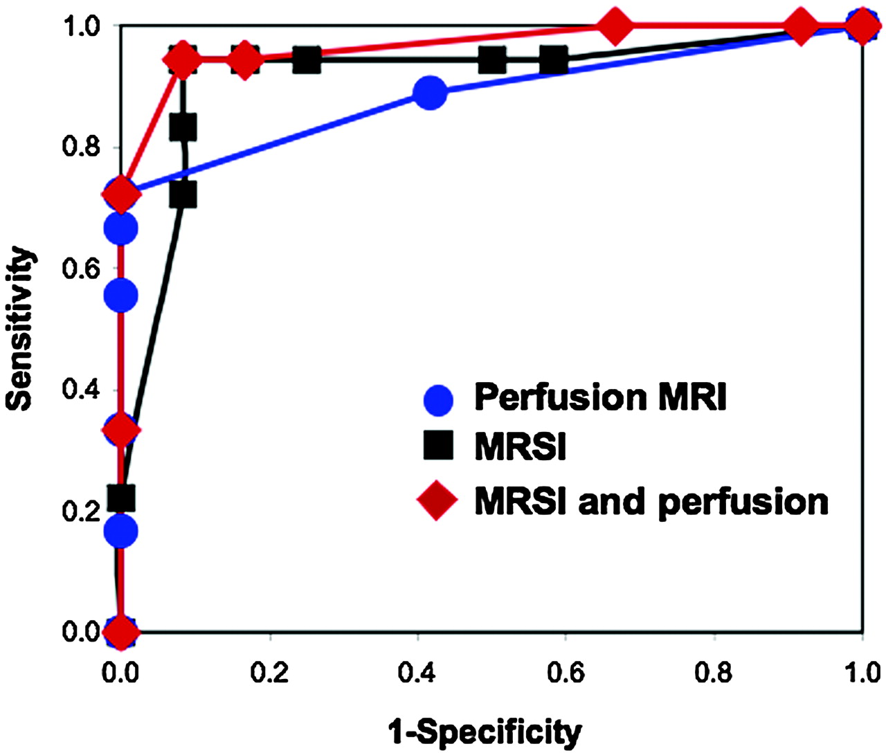
- Fig 3.
ROC curves representing discriminatory capability of perfusion MR imaging, 1H-MRSI and combination of 1H-MRSI and perfusion MR imaging to differentiate between tumors and nonneoplastic lesions. We constructed the curves using data on 30 subjects evaluated with both perfusion MR imaging and MRSI. The calculated areas under the ROC curves were 0.92 for MRSI, 0.89 for perfusion MR imaging, and 0.96 for the analysis on the basis of MRSI and perfusion MR imaging. We found no significant differences when comparing the areas under the curves, indicating that each procedure had similar discriminatory capability for these subjects.


Tables
Tumors and nonneoplastic lesions: metabolite ratios and rCBV*
Metabolites Low-Grade Tumors High-Grade Tumors All Tumors Stroke Demyelination Proved Benign Stable Lesions All Nonneoplastic Lesions P† N = 8 N = 28 N = 36 N = 4 N = 10 N = 10 N = 9 N = 33 NAA/Cho 0.58 ± 0.31 0.34 ± 0.18 0.40 ± 0.23 0.72 ± 0.46 0.95 ± 0.39 1.08 ± 0.41 0.76 ± 0.22 0.91 ± 0.38 .0001 NAA/Cr 1.05 ± 0.51 0.78 ± 0.28 0.84 ± 0.36 1.02 ± 0.70 1.83 ± 1.01 1.06 ± 0.33 1.07 ± 0.40 1.29 ± 0.73 .002 Cho/Cr 2.10 ± 0.98 2.84 ± 2.43 2.68 ± 2.20 1.45 ± 0.55 1.88 ± 0.60 1.03 ± 0.23 1.40 ± 0.23 1.44 ± 0.52 .002 NAAnorm 0.43 ± 0.20 0.35 ± 0.15 0.37 ± 0.16 0.48 ± 0.36 0.64 ± 0.24 0.48 ± 0.17 0.53 ± 0.14 0.54 ± 0.21 .0001 Chonorm 1.63 ± 0.44 2.10 ± 1.06 2.00 ± 0.98 1.05 ± 0.11 1.55 ± 0.56 0.75 ± 0.27 1.33 ± 0.31 1.19 ± 0.49 .0001 Crnorm 0.94 ± 0.35 1.09 ± 0.52 1.06 ± 0.49 0.93 ± 0.25 1.02 ± 0.41 0.92 ± 0.37 1.06 ± 0.24 0.99 ± 0.33 NS rCBV‡ 1.45 ± 1.16 4.87 ± 3.12 4.11 ± 3.14 1.30 1.31 ± 0.49 0.77 ± 0.27 1.07 ± 0.44 1.00 ± 0.39 .002 Note:—NS indicates not significant; rCBV, relative cerebral blood volume; NAA/Cho, ratio of N-acetylaspartate to choline; NAA/Cr, ratio of N-acetylaspartate to creatine; Cho/Cr, ratio of choline to creatine.
* Data are presented as means ± standard deviations.
† t test comparison of tumors and nonneoplastic lesions.
‡ MRI perfusion cases: low-grade tumors (N = 4), high-grade tumors (N = 14), stroke (N = 1), demyelination (N = 2), proved benign (N = 5), stable lesions (N = 4).
In this issue


Jump to section
Related Articles
Cited By...
- Association of Developmental Venous Anomalies with Demyelinating Lesions in Patients with Multiple Sclerosis
- ASFNR Recommendations for Clinical Performance of MR Dynamic Susceptibility Contrast Perfusion Imaging of the Brain
- Proton MR Spectroscopy Improves Discrimination between Tumor and Pseudotumoral Lesion in Solid Brain Masses