Advertisement

Main menu

  • Home
  • Content
    • Current Issue
    • Accepted Manuscripts
    • Article Preview
    • Past Issue Archive
    • AJNR Case Collection
    • Case of the Week Archive
    • Classic Case Archive
    • Case of the Month Archive
  • Special Collections
    • Spinal CSF Leak Articles (Jan 2020-June 2024)
    • 2024 AJNR Journal Awards
    • Most Impactful AJNR Articles
  • Multimedia
    • AJNR Podcast
    • AJNR Scantastics
    • Video Articles
  • For Authors
    • Submit a Manuscript
    • Author Policies
    • Fast publishing of Accepted Manuscripts
    • Graphical Abstract Preparation
    • Manuscript Submission Guidelines
    • Imaging Protocol Submission
    • Submit a Case for the Case Collection
  • About Us
    • About AJNR
    • Editorial Board
  • More
    • Become a Reviewer/Academy of Reviewers
    • Subscribers
    • Permissions
    • Alerts
    • Feedback
    • Advertisers
    • ASNR Home
  • Other Publications
    • ajnr

User menu

  • Alerts
  • Log in

Search

  • Advanced search
American Journal of Neuroradiology

American Journal of Neuroradiology

ASHNR American Society of Functional Neuroradiology ASHNR American Society of Pediatric Neuroradiology ASSR
  • Alerts
  • Log in

Advanced Search

  • Home
  • Content
    • Current Issue
    • Accepted Manuscripts
    • Article Preview
    • Past Issue Archive
    • AJNR Case Collection
    • Case of the Week Archive
    • Classic Case Archive
    • Case of the Month Archive
  • Special Collections
    • Spinal CSF Leak Articles (Jan 2020-June 2024)
    • 2024 AJNR Journal Awards
    • Most Impactful AJNR Articles
  • Multimedia
    • AJNR Podcast
    • AJNR Scantastics
    • Video Articles
  • For Authors
    • Submit a Manuscript
    • Author Policies
    • Fast publishing of Accepted Manuscripts
    • Graphical Abstract Preparation
    • Manuscript Submission Guidelines
    • Imaging Protocol Submission
    • Submit a Case for the Case Collection
  • About Us
    • About AJNR
    • Editorial Board
  • More
    • Become a Reviewer/Academy of Reviewers
    • Subscribers
    • Permissions
    • Alerts
    • Feedback
    • Advertisers
    • ASNR Home
  • Follow AJNR on Twitter
  • Visit AJNR on Facebook
  • Follow AJNR on Instagram
  • Join AJNR on LinkedIn
  • RSS Feeds

Welcome to the new AJNR, Updated Hall of Fame, and more. Read the full announcements.


AJNR is seeking candidates for the position of Associate Section Editor, AJNR Case Collection. Read the full announcement.


Research ArticleBRAIN

Predicting Grade of Cerebral Glioma Using Vascular-Space Occupancy MR Imaging

H. Lu, E. Pollack, R. Young, J.S. Babb, G. Johnson, D. Zagzag, R. Carson, J.H. Jensen, J.A. Helpern and M. Law
American Journal of Neuroradiology February 2008, 29 (2) 373-378; DOI: https://doi.org/10.3174/ajnr.A0794
H. Lu
  • Find this author on Google Scholar
  • Find this author on PubMed
  • Search for this author on this site
E. Pollack
  • Find this author on Google Scholar
  • Find this author on PubMed
  • Search for this author on this site
R. Young
  • Find this author on Google Scholar
  • Find this author on PubMed
  • Search for this author on this site
J.S. Babb
  • Find this author on Google Scholar
  • Find this author on PubMed
  • Search for this author on this site
G. Johnson
  • Find this author on Google Scholar
  • Find this author on PubMed
  • Search for this author on this site
D. Zagzag
  • Find this author on Google Scholar
  • Find this author on PubMed
  • Search for this author on this site
R. Carson
  • Find this author on Google Scholar
  • Find this author on PubMed
  • Search for this author on this site
J.H. Jensen
  • Find this author on Google Scholar
  • Find this author on PubMed
  • Search for this author on this site
J.A. Helpern
  • Find this author on Google Scholar
  • Find this author on PubMed
  • Search for this author on this site
M. Law
  • Find this author on Google Scholar
  • Find this author on PubMed
  • Search for this author on this site
  • Article
  • Figures & Data
  • Info & Metrics
  • Responses
  • References
  • PDF
Loading

Article Figures & Data

Figures

  • Tables
  • Fig 1.
    • Download figure
    • Open in new tab
    • Download powerpoint
    Fig 1.

    Schematic diagram of the VASO scans and the VASO pulse sequence. VASO uses a nonselective inversion recovery sequence, and the TI is chosen to null the precontrast blood signal intensity. Two scans using identical parameters are performed before and after the contrast agent administration, the signal intensity difference of which can be used to calculate absolute CBV.
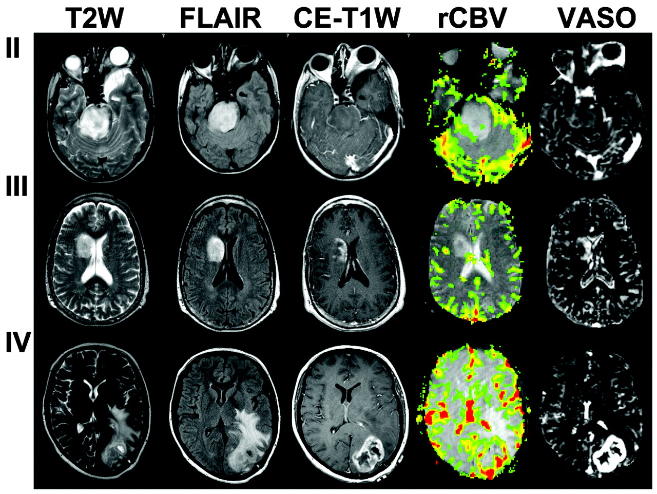
  • Fig 2.
    • Download figure
    • Open in new tab
    • Download powerpoint
    Fig 2.

    T2-weighted, CE-T1-weighted, and FLAIR images, as well as VASO maps for WHO grade II, III, and IV gliomas.

  • Fig 3.
    • Download figure
    • Open in new tab
    • Download powerpoint
    Fig 3.

    Three VASO indices, that is, VASOTumor, VASOContra, and VASORatio, in grade II (n = 9), III (n = 20), and IV (n = 10) gliomas. Error bars indicate SDs. The units for VASOTumor and VASOContra are percentages. The index VASORatio is dimensionless. Significant differences (P < .05) for each pair are indicated by asterisks.
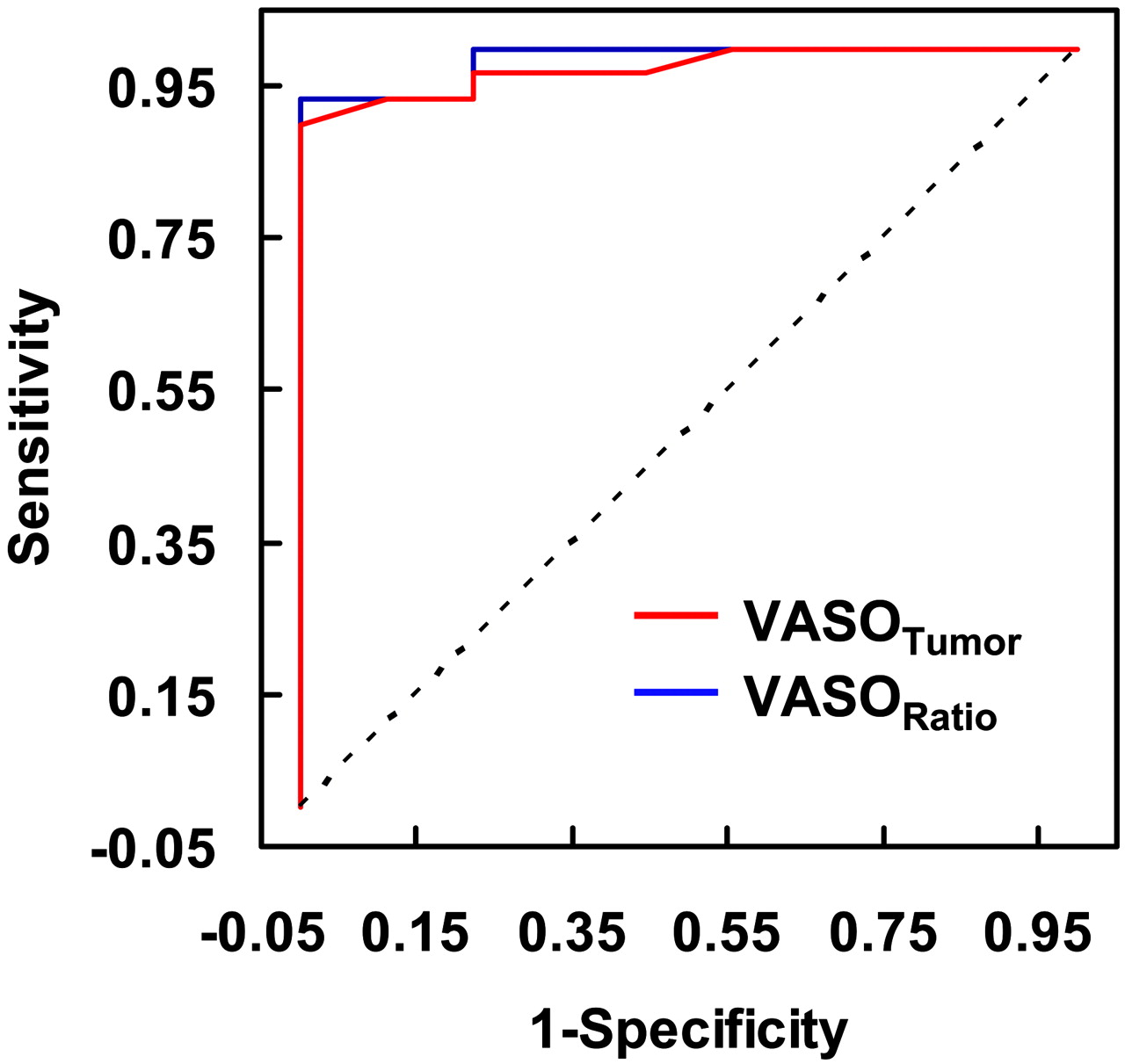
  • Fig 4.
    • Download figure
    • Open in new tab
    • Download powerpoint
    Fig 4.

    ROC curves using VASOTumor or VASORatio. The sensitivity and specificity (in fractions) in terms of distinguishing grade II from higher-grade (grade III and grade IV) tumors are plotted at different thresholds. If the 2 categories have identical distributions of the VASO parameters, the ROC curve would have been a 45° straight line (dotted line). The AUCs are 0.974 and 0.985 for VASOTumor and VASORatio, respectively.

Tables

  • Figures
    • View popup
    Table 1:

    Mean ± SD of each of the VASO and DSC parameters stratified by tumor grade

    GradeVASOTumor, %VASOContra, %VASORatioCBF, mL of Blood per 100 g of Tissue per MinuteCBV, mL of Blood per 100 g of TissuerCBVKTrans, s−1VP, %MTT, s
    II7.5 ± 3.25.4 ± 2.91.5 ± 0.560.1 ± 25.84.2 ± 1.91.4 ± 0.50.011 ± 0.0092.0 ± 1.24.2 ± 0.6
    III41.3 ± 23.66.3 ± 4.28.2 ± 5.5197.8 ± 80.715.3 ± 6.93.8 ± 1.40.019 ± 0.0182.9 ± 2.34.8 ± 1.0
    IV56.1 ± 32.74.8 ± 2.311.6 ± 3.7251.9 ± 160.817.9 ± 9.55.7 ± 2.10.022 ± 0.0373.3 ± 2.74.5 ± 0.8
    • Note:—VASOTumor indicates vascular-space occupancy values in the tumoral regions; VASOContra, vascular-space occupancy signal intensity in regions contralateral to tumor; VASORatio, ratio between tumor side and contralateral side of the vascular-space occupancy; CBF, cerebral blood flow; CBV, cerebral blood volume; rCBV, relative cerebral blood volume; Ktrans, vascular permeability; Vp, fractional plasma volume; MTT, mean transit time.

    • View popup
    Table 2:

    P values from pairwise comparison between different tumor grades using the VASO and DSC parameters

    VariableVASOTumorVASOContraVASORatioCBFCBVrCBVKtransVPMTT
    II vs III0.0003*0.6850.0002*<0.0001*<0.0001*<0.0001*0.2480.4230.170
    II vs IV<0.0001*0.508<0.0001*0.0006*0.0004*<0.0001*0.8580.4000.509
    III vs IV0.1940.3560.0098*0.4420.6500.025*0.3670.7870.522
    • Note:—VASOTumor indicates vascular-space occupancy values in the tumoral regions; VASOContra, vascular-space occupancy signal intensity in regions contralateral to tumor; VASORatio, ratio between tumor side and contralateral side of the vascular-space occupancy; CBF, cerebral blood flow; CBV, cerebral blood volume; rCBV, relative cerebral blood volume; Ktrans, vascular permeability; Vp, fractional plasma volume; MTT, mean transit time.

    • * Significant differences (P < 0.05, not corrected for multiple comparisons).

PreviousNext
Back to top

In this issue

American Journal of Neuroradiology: 29 (2)
American Journal of Neuroradiology
Vol. 29, Issue 2
February 2008
  • Table of Contents
  • Index by author
Advertisement
Print
Download PDF
Email Article

Thank you for your interest in spreading the word on American Journal of Neuroradiology.

NOTE: We only request your email address so that the person you are recommending the page to knows that you wanted them to see it, and that it is not junk mail. We do not capture any email address.

Enter multiple addresses on separate lines or separate them with commas.
Predicting Grade of Cerebral Glioma Using Vascular-Space Occupancy MR Imaging
(Your Name) has sent you a message from American Journal of Neuroradiology
(Your Name) thought you would like to see the American Journal of Neuroradiology web site.
CAPTCHA
This question is for testing whether or not you are a human visitor and to prevent automated spam submissions.
Cite this article
H. Lu, E. Pollack, R. Young, J.S. Babb, G. Johnson, D. Zagzag, R. Carson, J.H. Jensen, J.A. Helpern, M. Law
Predicting Grade of Cerebral Glioma Using Vascular-Space Occupancy MR Imaging
American Journal of Neuroradiology Feb 2008, 29 (2) 373-378; DOI: 10.3174/ajnr.A0794

Citation Manager Formats

  • BibTeX
  • Bookends
  • EasyBib
  • EndNote (tagged)
  • EndNote 8 (xml)
  • Medlars
  • Mendeley
  • Papers
  • RefWorks Tagged
  • Ref Manager
  • RIS
  • Zotero
0 Responses
Respond to this article
Share
Bookmark this article
Predicting Grade of Cerebral Glioma Using Vascular-Space Occupancy MR Imaging
H. Lu, E. Pollack, R. Young, J.S. Babb, G. Johnson, D. Zagzag, R. Carson, J.H. Jensen, J.A. Helpern, M. Law
American Journal of Neuroradiology Feb 2008, 29 (2) 373-378; DOI: 10.3174/ajnr.A0794
del.icio.us logo Twitter logo Facebook logo Mendeley logo
  • Tweet Widget
  • Facebook Like
  • Google Plus One
Purchase

Jump to section

  • Article
    • Abstract
    • Materials and Methods
    • Results
    • Discussion
    • Conclusion
    • Footnotes
    • References
  • Figures & Data
  • Info & Metrics
  • Responses
  • References
  • PDF

Related Articles

  • No related articles found.
  • PubMed
  • Google Scholar

Cited By...

  • Novel MRI Approaches for Assessing Cerebral Hemodynamics in Ischemic Cerebrovascular Disease
  • Imaging biomarkers of angiogenesis and the microvascular environment in cerebral tumours
  • Crossref (27)
  • Google Scholar

This article has been cited by the following articles in journals that are participating in Crossref Cited-by Linking.

  • The diagnostic performance of magnetic resonance spectroscopy in differentiating high-from low-grade gliomas: A systematic review and meta-analysis
    Qun Wang, Hui Zhang, JiaShu Zhang, Chen Wu, WeiJie Zhu, FangYe Li, XiaoLei Chen, BaiNan Xu
    European Radiology 2016 26 8
  • Novel MRI Approaches for Assessing Cerebral Hemodynamics in Ischemic Cerebrovascular Disease
    Manus J. Donahue, Megan K. Strother, Jeroen Hendrikse
    Stroke 2012 43 3
  • PEGylated poly(trimethylene carbonate) nanoparticles loaded with paclitaxel for the treatment of advanced glioma: In vitro and in vivo evaluation
    Xinyi Jiang, Hongliang Xin, Xianyi Sha, Jijin Gu, Ye Jiang, Kitki Law, Yanzuo Chen, Liangcen Chen, Xiao Wang, Xiaoling Fang
    International Journal of Pharmaceutics 2011 420 2
  • In vivo vascular hallmarks of diffuse leukoaraiosis
    Jinsoo Uh, Uma Yezhuvath, Yamei Cheng, Hanzhang Lu
    Journal of Magnetic Resonance Imaging 2010 32 1
  • Imaging biomarkers of angiogenesis and the microvascular environment in cerebral tumours
    G Thompson, S J Mills, D J Coope, J P B O’connor, A Jackson
    The British Journal of Radiology 2011 84 special_issue_2
  • Implementation of vascular‐space‐occupancy MRI at 7T
    Jun Hua, Craig K. Jones, Qin Qin, Peter C. M. van Zijl
    Magnetic Resonance in Medicine 2013 69 4
  • Using magnetic resonance imaging and spectroscopy in cancer diagnostics and monitoring
    Risto A. Kauppinen, Andrew C. Peet
    Cancer Biology & Therapy 2011 12 8
  • Imaging of Brain Tumors: Perfusion/Permeability
    Gerard Thompson, Samantha Jane Mills, Stavros Michael Stivaros, Alan Jackson
    Neuroimaging Clinics of North America 2010 20 3
  • Multiparametric imaging of brain hemodynamics and function using gas-inhalation MRI
    Peiying Liu, Babu G. Welch, Yang Li, Hong Gu, Darlene King, Yihong Yang, Marco Pinho, Hanzhang Lu
    NeuroImage 2017 146
  • Magnetization transfer enhanced vascular‐space‐occupancy (MT‐VASO) functional MRI
    Jun Hua, Manus J. Donahue, Jason M. Zhao, Ksenija Grgac, Alan J. Huang, Jinyuan Zhou, Peter C.M. van Zijl
    Magnetic Resonance in Medicine 2009 61 4

More in this TOC Section

  • Progression of Microstructural Damage in Spinocerebellar Ataxia Type 2: A Longitudinal DTI Study
  • Usefulness of Quantitative Susceptibility Mapping for the Diagnosis of Parkinson Disease
  • Evaluating the Effects of White Matter Multiple Sclerosis Lesions on the Volume Estimation of 6 Brain Tissue Segmentation Methods
Show more Brain

Similar Articles

Advertisement

Indexed Content

  • Current Issue
  • Accepted Manuscripts
  • Article Preview
  • Past Issues
  • Editorials
  • Editors Choice
  • Fellow Journal Club
  • Letters to the Editor

Cases

  • Case Collection
  • Archive - Case of the Week
  • Archive - Case of the Month
  • Archive - Classic Case

Special Collections

  • Special Collections

Resources

  • News and Updates
  • Turn around Times
  • Submit a Manuscript
  • Author Policies
  • Manuscript Submission Guidelines
  • Evidence-Based Medicine Level Guide
  • Publishing Checklists
  • Graphical Abstract Preparation
  • Imaging Protocol Submission
  • Submit a Case
  • Become a Reviewer/Academy of Reviewers
  • Get Peer Review Credit from Publons

Multimedia

  • AJNR Podcast
  • AJNR SCANtastic
  • Video Articles

About Us

  • About AJNR
  • Editorial Board
  • Not an AJNR Subscriber? Join Now
  • Alerts
  • Feedback
  • Advertise with us
  • Librarian Resources
  • Permissions
  • Terms and Conditions

American Society of Neuroradiology

  • Not an ASNR Member? Join Now

© 2025 by the American Society of Neuroradiology All rights, including for text and data mining, AI training, and similar technologies, are reserved.
Print ISSN: 0195-6108 Online ISSN: 1936-959X

Powered by HighWire