
Article Figures & Data
Figures
- Fig 1.
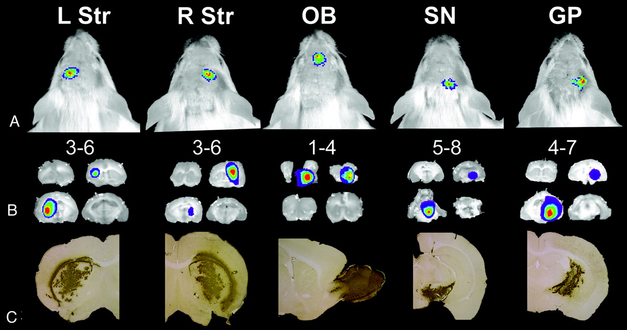
The localization of the bioluminescence imaging signal intensity reflects the anatomic site of injection. A, Bioluminescence imaging scans 14 days after injection of 293T cells transduced with a lentiviral construct encoding enhanced green fluorescent protein (EGFP) and Fluc separated by the internal ribosome entry site (I) of the encephalomyocarditis virus (LV-EGFP-I-Fluc) showing a focus that is located above the injection site: caudal to the eyes and on the left side of the head for the left striatum (L Str), caudal to the eyes and on the right side for the right striatum (R Str), between the eyes for the olfactory bulb (OB), near the caudal edge of the skull for the substantia nigra (SN), and intermediate between R Str and SN for the globus pallidus (GP). These sites correspond to the expected locations on the basis of the injection coordinates. B, Ex vivo bioluminescence images of 1-mm-thick coronal sections show the localization of the signal intensity at the site of injection. The sections are numbered in the anteroposterior direction from the bulbus olfactorius1 to the cerebellum.8 C, Immunohistochemistry for EGFP confirms the site of injection. (Reprinted by permission from Macmillan Publishers: Deroose et al. Mol. Therapy 2006;14:423–31, copyright 2006).
- Fig 2.
Time course of bioluminescence imaging (BLI) signal intensity after lentiviral (LV) transduction of mouse brain. A, Long-term evolution of the BLI signal intensity in a group of mice (n = 10) injected with 17-ng p24 of LV-Fluc and in a group injected with 8.4-ng p24 control vector (LV-enhanced green fluorescent protein [EGFP], n = 4). After a peak at days 8 to 14, the signal intensity declines during the first month to 16% of the maximum value at day 37 and then remains constant at 17.5 ± 2.3% of the maximum value from days 42 to 365. A linear regression line is drawn from days 37 to 365 (R2 = 0.027) for LV-Fluc and for all time points for LV-EGFP (R2 = 0.041). B, BLI of a representative animal shows an initial rise in signal intensity at week (W) 1 followed by a decrease and thereafter a stabilization of the signal intensity. The control animal shown represents the highest signal intensity seen in a control animal. GFP indicates green fluorescent protein; D, day; p, photons. (Reprinted by permission from Macmillan Publishers: Deroose et al. Mol. Therapy 2006;14:423–31, copyright 2006)
- Fig 3.
Brain injury results in activation of TGF-β responsive genes and the Smad-binding element (SBE)-firefly luciferase (luc) reporter. Two SBE-luc mice with similar basal levels of bioluminescence were lesioned with a needle stab to the right hemisphere or were left untreated (control), and bioluminescence was recorded 1 hour later. To highlight the increase in signal intensity in the lesioned mouse, the color scale was adjusted to leave the basal Fluc expression in the control mouse uncolored (<200 photons[p]/s/mm2/sr). (Copyright 2005, The American Association of Immunologists)
- Fig 4.
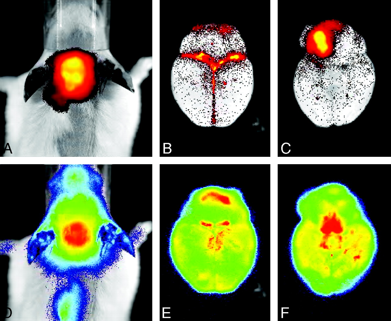
Simultaneous in vivo biphotonic monitoring of pneumococcal meningitis and the accompanying neuronal injury in live transgenic mice. Streptococcus pneumoniae engineered for bioluminescence (lux) was used for direct visualization of disease progression. Host response was monitored in transgenic mice containing an inducible firefly luciferase (Fluc) reporter gene under transcriptional control of the mouse glial fibrillary acidic protein (GFAP) promoter. On the basis of the different spectra of light emission and substrate requirements for Fluc and luc, it is possible to monitor separately the 2 reporters by using a highly sensitive in vivo imaging system. In vivo (A and D) and ex vivo (B, C, E, and F) images of brains from transgenic mice with meningitis were obtained at 19 hours postinfection. A, B, and C show lux imaging and D, E, and F show Fluc imaging. Dorsal and ventral views of an ex vivo brain show the bacterial and GFAP signals individually. Much of the bacterial signal intensity comes from discrete patches, whereas GFAP is induced in the entire brain and there are different intensities of the bioluminescence signal intensity in certain regions of the brain. Note the strong bacterial signal intensity immediately surrounding the inoculation site in the anterior right frontal lobe. (Reprinted by permission from the American Society for Microbiology)


Tables
- Table 1:
Recent examples of applications in molecular neuroimaging using gene marking (of static cells)
Type of Cell Marked Method of Gene Marking Transplant Site Animal Model Imaging Method Application Reference Cancer cells Ex vivo Orthotopic Mice Fluc BLI Evaluation of technical aspects of neuroimaging by testing suitability of Fluc reporter for brain imaging; evaluation of antineoplastic chemotherapy 10 Cancer cells Ex vivo Orthotopic Mice, rats Fluc BLI Evaluation of technical aspects of neuroimaging after lentiviral transduction of various cancer cells 11 Cancer cells Ex vivo Orthotopic Mice Fluc BLI Evaluation of technical aspects of neuroimaging using a herpes simplex virus amplicon vector expressing Fluc from an inducible promoter 12 Cancer cells Ex vivo Orthotopic, heterotopic Mice Fluc BLI Evaluation of technical aspects of neuroimaging by comparing level and time course of light signal from 2 different locations 13 Cancer cells Ex vivo Orthotopic, heterotopic Mice HSV1-tk, PET Evaluation of technical aspects of neuroimaging by testing suitability of a 76Br-labeled uracil analog as a probe in brain imaging 14 Cancer cells Ex vivo Orthotopic Mice Fluc BLI Evaluation of technical aspects of neuroimaging by correlating tumor growth with Fluc BLI and MR imaging; evaluation of antineoplastic chemotherapy 15 Cancer cells Ex vivo Orthotopic Mice Fluc BLI Evaluation of technical aspects of neuroimaging when establishing tumors with varying abilities to disrupt the BBB; evaluation of antineoplastic chemotherapy 16 Cancer cells Ex vivo Orthotopic Mice Fluc BLI Evaluation of technical aspects of neuroimaging by testing hyperspectral/multispectral light analysis as a means of 3D localization in BLI 17 Normal brain In vivo Orthotopic Mice GFP, fluorescence Evaluation of technical aspects of neuroimaging using reflectance fluorescence imaging 18 Normal brain In vivo Orthotopic Rats HSV1-tk, PET Evaluation of technical aspects of neuroimaging in diagnosing early herpes simplex encephalitis 19 Cancer cells Ex vivo Orthotopic Mice Fluc BLI Evaluation of role of activation of G protein-coupled receptor CXCR4 in growth of intracranial tumors; evaluation of antineoplastic chemotherapy 20 Cancer cells Ex vivo Orthotopic Mice Fluc BLI Evaluation of immunotherapy of intracranial tumors 21 Cancer cells Ex vivo Orthotopic Rats Fluc BLI Evaluation of photodynamic therapy of intracranial tumors 22 Cancer cells Ex vivo Orthotopic Mice Fluc BLI Evaluation of tumor angiogenesis by imaging integrin αvβ3receptor expression using fluorescence imaging 23 Note:—BLI indicates bioluminescence imaging; GFP, green fluorescent protein; PET, positron-emission tomography; HSV1-tk, herpes simplex virus–thymidine kinase; BBB, blood-brain barrier; αVβ3, a vitronectin receptor on the cell surface.
- Table 2:
Recent examples of applications in molecular neuroimaging using gene marking (of trafficking cells)
Type of Cell Marked Method of Gene Marking Transplant Site Animal Model Imaging Method Application Reference Viruses Ex vivo Intravenous, or intranasal Mice Fluc BLI Evaluation of effects of interferons on vaccinia viral spread to the brain 26 Viruses Ex vivo Intravenous Mice Fluc BLI Evaluation of factors relating to Sindbis viral spread to the brain 27 Viruses Ex vivo Intravenous Mice Fluc BLI, Rluc BLI Evaluation of effects of valacyclovir on HSV-1 viral spread to the brain and eyes 28 Malaria parasites in RBCs Ex vivo Intravenous Mice, rats Fluc BLI Evaluation of biology of parasite sequestration in cerebral malaria 29 Cancer cells Ex vivo Intravenous Mice Fluc BLI Evaluation of breast cancer metastasis to brain 30 Stem cells Ex vivo Orthotopic Mice Fluc BLI Evaluation of trafficking of stem cells to brain tumors 31 Stem cells Ex vivo Orthotopic Mice Fluc BLI Evaluation of trafficking of stem cells to brain tumors 32 Fluc BLI Evaluation of effect of stem cell-delivered chemotherapy on tumor burden Stem cells Ex vivo Orthotopic Mice Fluc BLI Evaluation of trafficking of stem cells to brain infarcts 33 Stem cells Ex vivo Orthotopic Mice Fluc BLI Evaluation of trafficking of stem cells to ischemic brain in relation to immune status and host immunity 34 Stem cells Ex vivo Orthotopic Mice Fluc BLI Evaluation of trafficking of stem cells to injured spinal cord 35 Note:—BLI indicates bioluminescence imaging; RBC, red blood cells; Rluc, Renilla luciferase.
Type of Vector Location of Imaging Gene, Therapeutic Gene Imaging Gene Linked to Delivery Site of Vector Animal Model Imaging Method Application Ref Adeno-associated viruses In delivery vehicle No In utero Mice Fluc BLI Evaluation of systemic spread of virus, including to brain, for potential to deliver therapeutic genes to ameliorate genetic diseases with perinatal morbidity and mortality 38 Lentivirus In delivery vehicle Yes Intravenous Mice Fluc BLI Evaluation of gene therapy for Fabry disease using gene for α-galactosidase A, including in brain 39 Nonviral Sleeping Beauty transposon In target gliomas No Brain, intratumoral Mice Fluc BLI Evaluation of antiangiogenesis gene therapy delivered in gliomas; tumor burden assessed with Fluc BLI 40 Adenovirus In delivery vehicle Yes Brain, intratumoral Rats Fluc BLI Evaluation of glioma gene therapy using yCD/5-FC 41 Note:—Ref indicates reference; BLI, bioluminescence imaging.
Type of Genetically Engineered Model Promoter Expression Location of Model Animal Method Imaging Application Reference Transgenic Estrogen receptor Ubiquitous + brain Mice Fluc BLI Study of estrogen control of growth, differentiation, and function of many systems; study of implications for estrogen-replacement therapy 44 Transgenic GFAP Brain Mice Fluc BLI Dynamic monitoring of neuronal cell death 45 Transgenic Smad binding element responsive to TGF-β signaling Brain Mice Fluc BLI Study of Smad2/3 activation in traumatic brain injury 46 Transgenic Smad binding element responsive to TGF-β signaling Brain Mice Fluc BLI Study of Smad2activation in neuronal degeneration 47 Transgenic Serum amyloid A protein 1 Ubiquitous + brain Mice Fluc BLI Study of role of SAA1 induction in chronic inflammation associated with amyloid deposition 48 Gene targeting knockin CMV Ubiquitous + brain Mice GFP and RFP fluorescence Study of alternative splicing regulation of FGFR-2 in the brain. 49 Transgenic IκBα Ubiquitous + brain Mice Fluc BLI Study of regulation of IκBαexpression and NF-κB transcriptional activity 50 Transgenic Mouse GFAP Brain Mice Fluc BLI Study of meningitis and accompanying neuronal injury 51 Transgenic c-fos, CMV Ubiquitous + brain Mice Fluc BLI Study of immediate-early genes involved in neural pathways linked to specific behaviors 52 Transgenic Estrogen-responsive elements Ubiquitous + brain Mice Fluc BLI Study of activation of estrogen receptors and kinetics of gene activation by estrogenic compounds 53 Conditional recombinase knockout Pro-opiomelanocortin Pituitary Mice Fluc BLI Study of spontaneous retinoblastoma pathway-dependent pituitary cancer and its response to doxorubicin 54 Transgenic E2F1 Brain Mice Fluc BLI Imaging cell proliferation in gliomas with loss of RB control 55 Note:—BLI indicates bioluminescence imaging; FGFR-2, fibroblast growth factor receptor-2; CMV, cytomegalovirus; GFP, green fluorescent protein; RFP, red fluorescent protein; IκBα, an inhibitor of nuclear transcription factor NF-κB, which regulates the expression of proinflammatory and cytotoxic genes; c-fos, an immediate early gene; RB, retinoblastoma protein.
In this issue


Jump to section
- Article
- Abstract
- Experimental Applications in Molecular Neuroimaging Using Reporter Genes
- Imaging Gene-Marked Cells
- Imaging of Gene Therapies
- Imaging of Transgenic Models of Spontaneous Disease
- Imaging of Molecular Interactions or Events
- Clinical Applications in Molecular Neuroimaging Using Reporter Genes
- Future Outlook
- References
- Figures & Data
- Info & Metrics
- Responses
- References