
Article Figures & Data
Figures
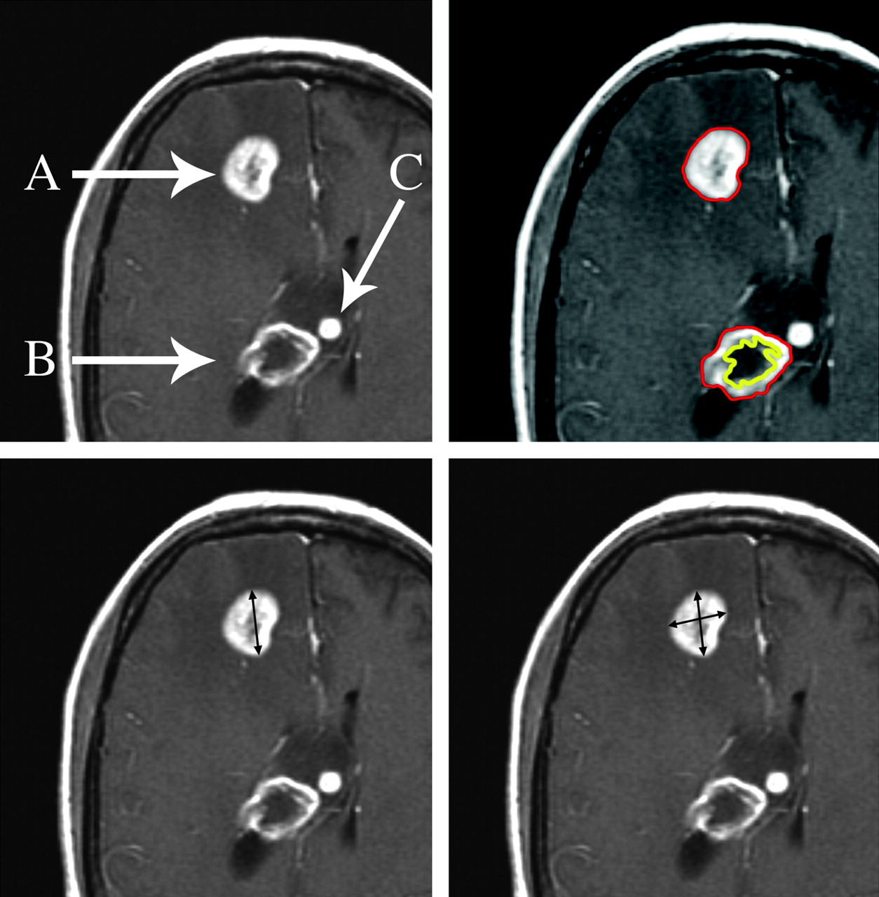
- Fig 1.
Three enhancing foci in a patient with glioblastoma illustrate issues with lesion measurement during clinical trials. Lesion A is homogeneously enhancing and exceeds 10 mm in diameter and thus is ideal for serial measurement by RECIST or 1D (lower left), Macdonald or 2D (lower right), and volumetric (upper right) approaches. Lesion B is predominantly necrotic and is amenable to volumetric measurement (upper right) because the enhancing and nonenhancing components can be segmented. Lesion C is too small in diameter (8 mm) for accurate serial measurement and should be followed as a nonmeasurable lesion (see text). Images are postgadolinium contrast-enhanced axial T1-weighted.
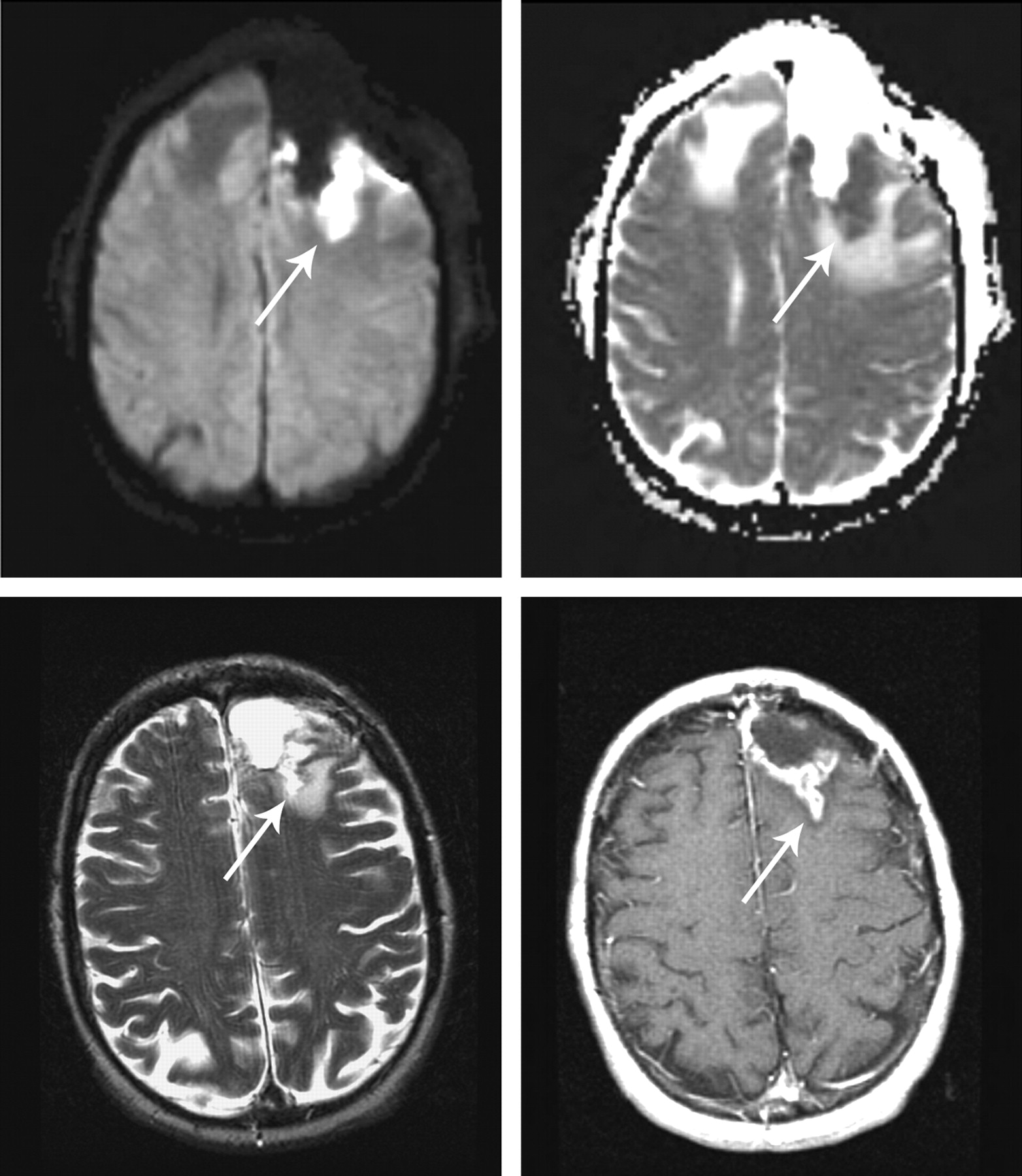
- Fig 2.
Infarcts on immediate postoperative MR images are common. These infarcts often demonstrate nodular gadolinium enhancement on subsequent studies, a finding that could be easily confused with tumor. Immediate postoperative DWI (upper left) and apparent diffusion coefficient (upper right) show restricted diffusion (arrows), followed by a 3-month postoperative T2-weighted (lower left) image and a gadolinium-enhanced T1-weighted (lower right) image showing encephalomalacia and enhancement of the infarct. (Reprinted by permission of Lippincott Williams & Wilkins; Clinical and radiographic features of peritumoral infarction following resection of glioblastoma. Neurology 2006;67:1668–70).
- Fig 3.
Novel therapeutic agents in clinical trials may require use of imaging techniques other than gadolinium-enhancing tumor. Shown here is decreased tumor enhancement but not diameter in a patient with glioblastoma after initiation of a therapy with an inhibitor of VEGF and irinotecan. Note the increase in extent of the infiltrative component of the lesion (lower right). Axial post-gadolinium contrast T1-weighted images (left-hand column) and axial T2-weighted/fluid-attenuated inversion recovery images (right-hand column) were acquired before (upper row) and 7 weeks after (lower row) institution of therapy.


Tables
Comparison of response criteria for different measurement approaches
RECIST (1D)3 Macdonald (2D)4 Volumetric Extrapolated from RECIST*,† Volumetric Extrapolated from Macdonald*,‡ CR Resolution of all enhancing tumor; confirm at 4 weeks Resolution of all enhancing tumor; confirm at 4 weeks Resolution of all enhancing tumor; confirm at 4 weeks Resolution of all enhancing tumor; confirm at 4 weeks PR§ ≥30% decrease in sum of maximal diameters; confirm at 4 weeks ≥50% decrease in product of 2 orthogonal diameters; confirm at 4 weeks ≥66% decrease in volume; confirm at 4 weeks ≥65% decrease in volume; confirm at 4 weeks SD All others All others All others All others PD‖ ≥20% increase in sum of maximal diameters; confirm at 4 weeks ≥25% increase in product of orthogonal diameters; confirm at 4 weeks ≥73% increase in volume; confirm at 4 weeks ≥40% increase in volume; confirm at 4 weeks Comment Single longest diameter of the lesion or sum of longest diameters of multiple measurable lesions (see text) Product of orthogonal diameters on section with largest tumor area; sum of products if multiple measurable lesions Computer-assisted volumetrics using a perimeter methodology; sum of volumes if multiple measurable lesions Use of these values would be equally stringent for PR comparing RECIST and Macdonald criteria but would be more stringent for PD compared with RECIST but comparable with Macdonald criteria Note:—CR indicates complete response; PR, partial response; SD, stable disease; PD, progressive disease.
* “Extrapolated” refers to converting single diameter or orthogonal diameter measurements to a volume assuming a spheric lesion using the formula V = 4/3πr3.
† Volume versus 1D (ie, cube of linear RECIST criteria).
‡ Volume versus 2D.
§ Percentage change from baseline (see text).
‖ Percentage change from nadir (see text).
In this issue


Jump to section
- Article
- Abstract
- Measurement Techniques
- Response Criteria
- Comparison of Diameter and Volumetric Approaches
- Choosing a Measurable Lesion in Clinical Trials
- Technical Considerations
- Timing of Imaging Studies and Confirmation of Response
- Validity of Imaging End Points in Clinical Trials
- Challenges with Novel Therapeutics
- Conclusions
- Footnotes
- References
- Figures & Data
- Info & Metrics
- Responses
- References
Related Articles
Cited By...
- Automated Detection and Segmentation of Brain Metastases in Malignant Melanoma: Evaluation of a Dedicated Deep Learning Model
- "Early Imaging Marker of Progressive Glioblastoma: a window of opportunity"
- Tumor Response Assessment in Diffuse Intrinsic Pontine Glioma: Comparison of Semiautomated Volumetric, Semiautomated Linear, and Manual Linear Tumor Measurement Strategies
- Quantitative Delta T1 (dT1) as a Replacement for Adjudicated Central Reader Analysis of Contrast-Enhancing Tumor Burden: A Subanalysis of the American College of Radiology Imaging Network 6677/Radiation Therapy Oncology Group 0625 Multicenter Brain Tumor Trial
- MR Fingerprinting of Adult Brain Tumors: Initial Experience
- Can an 18F-ALF-NOTA-PRGD2 PET/CT Scan Predict Treatment Sensitivity to Concurrent Chemoradiotherapy in Patients with Newly Diagnosed Glioblastoma?
- A Retrospective Evaluation of Vemurafenib as Treatment for BRAF-Mutant Melanoma Brain Metastases
- Semiautomated Volumetric Measurement on Postcontrast MR Imaging for Analysis of Recurrent and Residual Disease in Glioblastoma Multiforme
- Updated Response Assessment Criteria for High-Grade Gliomas: Response Assessment in Neuro-Oncology Working Group
- End Point Assessment in Gliomas: Novel Treatments Limit Usefulness of Classical Macdonald's Criteria
- Therapeutic Application of Noncytotoxic Molecular Targeted Therapy in Gliomas: Growth Factor Receptors and Angiogenesis Inhibitors