Advertisement

Main menu

  • Home
  • Content
    • Current Issue
    • Accepted Manuscripts
    • Article Preview
    • Past Issue Archive
    • AJNR Case Collection
    • Case of the Week Archive
    • Classic Case Archive
    • Case of the Month Archive
  • Special Collections
    • Spinal CSF Leak Articles (Jan 2020-June 2024)
    • 2024 AJNR Journal Awards
    • Most Impactful AJNR Articles
  • Multimedia
    • AJNR Podcast
    • AJNR Scantastics
    • Video Articles
  • For Authors
    • Submit a Manuscript
    • Author Policies
    • Fast publishing of Accepted Manuscripts
    • Graphical Abstract Preparation
    • Manuscript Submission Guidelines
    • Imaging Protocol Submission
    • Submit a Case for the Case Collection
  • About Us
    • About AJNR
    • Editorial Board
  • More
    • Become a Reviewer/Academy of Reviewers
    • Subscribers
    • Permissions
    • Alerts
    • Feedback
    • Advertisers
    • ASNR Home
  • Other Publications
    • ajnr

User menu

  • Alerts
  • Log in

Search

  • Advanced search
American Journal of Neuroradiology

American Journal of Neuroradiology

ASHNR American Society of Functional Neuroradiology ASHNR American Society of Pediatric Neuroradiology ASSR
  • Alerts
  • Log in

Advanced Search

  • Home
  • Content
    • Current Issue
    • Accepted Manuscripts
    • Article Preview
    • Past Issue Archive
    • AJNR Case Collection
    • Case of the Week Archive
    • Classic Case Archive
    • Case of the Month Archive
  • Special Collections
    • Spinal CSF Leak Articles (Jan 2020-June 2024)
    • 2024 AJNR Journal Awards
    • Most Impactful AJNR Articles
  • Multimedia
    • AJNR Podcast
    • AJNR Scantastics
    • Video Articles
  • For Authors
    • Submit a Manuscript
    • Author Policies
    • Fast publishing of Accepted Manuscripts
    • Graphical Abstract Preparation
    • Manuscript Submission Guidelines
    • Imaging Protocol Submission
    • Submit a Case for the Case Collection
  • About Us
    • About AJNR
    • Editorial Board
  • More
    • Become a Reviewer/Academy of Reviewers
    • Subscribers
    • Permissions
    • Alerts
    • Feedback
    • Advertisers
    • ASNR Home
  • Follow AJNR on Twitter
  • Visit AJNR on Facebook
  • Follow AJNR on Instagram
  • Join AJNR on LinkedIn
  • RSS Feeds

Welcome to the new AJNR, Updated Hall of Fame, and more. Read the full announcements.


AJNR is seeking candidates for the position of Associate Section Editor, AJNR Case Collection. Read the full announcement.


OtherBRAIN

Delayed CSF Enhancement in Posterior Reversible Encephalopathy Syndrome

B.E. Hamilton and G.M. Nesbit
American Journal of Neuroradiology March 2008, 29 (3) 456-457; DOI: https://doi.org/10.3174/ajnr.A0926
B.E. Hamilton
  • Find this author on Google Scholar
  • Find this author on PubMed
  • Search for this author on this site
G.M. Nesbit
  • Find this author on Google Scholar
  • Find this author on PubMed
  • Search for this author on this site
  • Article
  • Figures & Data
  • Info & Metrics
  • Responses
  • References
  • PDF
Loading

Abstract

SUMMARY: We present a case of increased fluid-attenuated inversion recovery signal intensity in the subarachnoid spaces on 6-hour delayed enhanced MR imaging in a patient with posterior reversible encephalopathy syndrome (PRES) in the setting of acute hypertensive encephalopathy. This hyperintensity was believed due to CSF gadolinium enhancement, a finding that would be anticipated in light of the suspected pathogenesis of increased microvascular permeability in PRES.

Posterior reversible encephalopathy syndrome (PRES) is theorized to result from an abnormally leaky blood-brain barrier (BBB) at the level of the microvasculature. MR perfusion studies to date have unsuccessfully clarified the site of abnormally increased vascular permeability as capillary, arteriolar, or venular.1 Increased permeability to small molecules such as water explain the interstitial accumulation of fluid in areas affected by PRES; however, larger molecules such as gadolinium chelates might also cross an abnormally permeable BBB. Because this leak may occur slowly for minutes to hours, it is difficult to capture with dynamic contrast-enhanced perfusion techniques or even immediate postcontrast sequences. We report a case of abnormal fluid-attenuated inversion recovery (FLAIR) hyperintensity in the subarachnoid spaces in a patient with PRES and normal renal function. The hyperintensity was due to delayed CSF gadolinium accumulation, a finding that supports the concept of slow BBB leakage in this syndrome.

Case Report

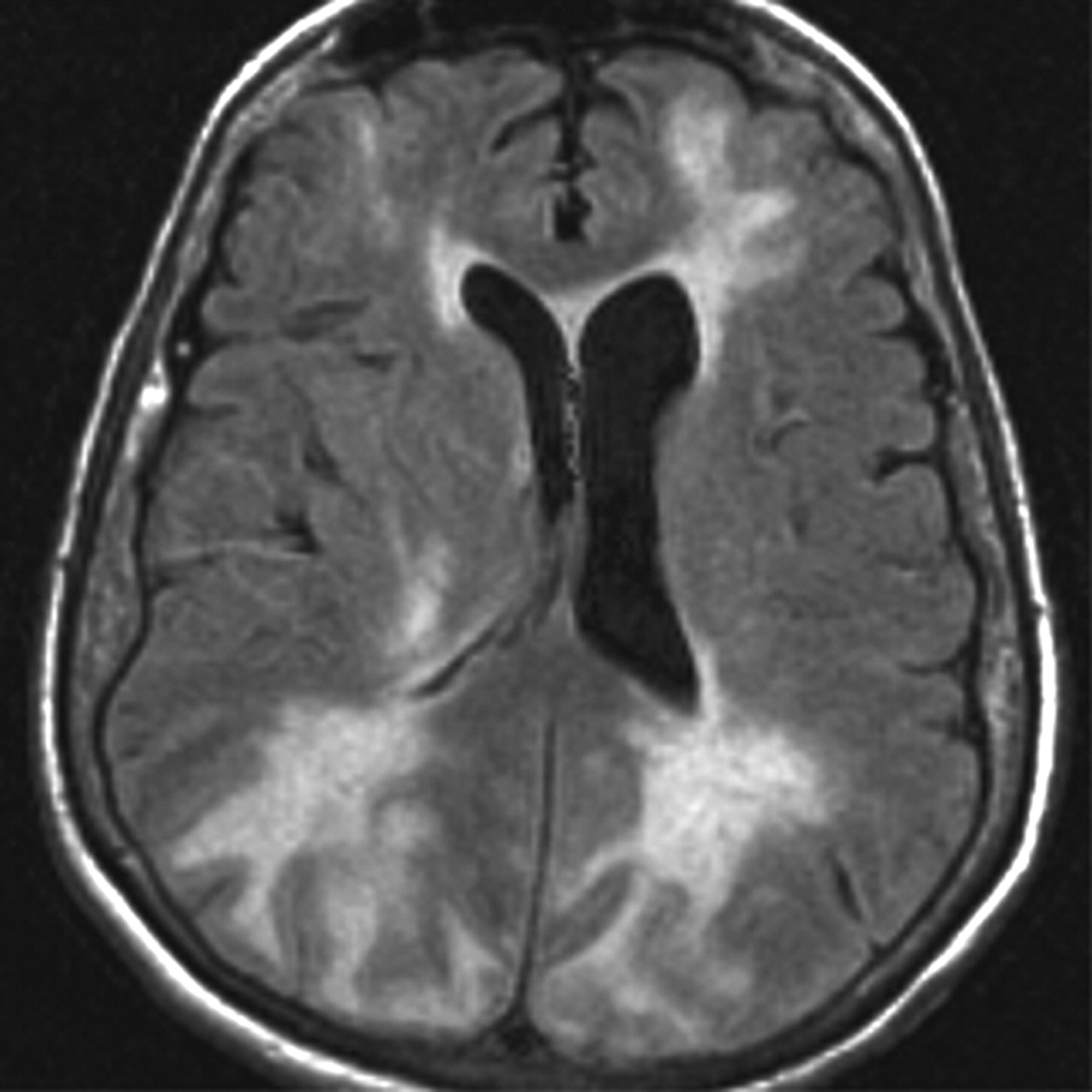
A 77-year-old woman with a medical history significant for chronic hypertension and dementia presented to the emergency department with headache, obtundation, and markedly elevated blood pressures as high as 230/120 mm Hg. Unenhanced FLAIR imaging of the brain performed at this time demonstrated extensive confluent areas of white matter hyperintense signal intensity in both cerebral hemispheres (Fig 1). Diffusion-weighted images and apparent diffusion coefficient maps (not shown) demonstrated increased diffusion in the affected areas and no restricted diffusion. The clinical presentation and MR imaging findings were consistent with PRES in the setting of hypertensive encephalopathy.

Fig 1.
  • Download figure
  • Open in new tab
  • Download powerpoint
Fig 1.

Axial FLAIR image from the patient's first MR imaging examination demonstrates confluent juxtacortical and deep white matter T2 hyperintensity in the bilateral frontal, posterior parietal, and occipital lobes characteristic for PRES. Note the normal hypointense appearance of subarachnoid space fluid.

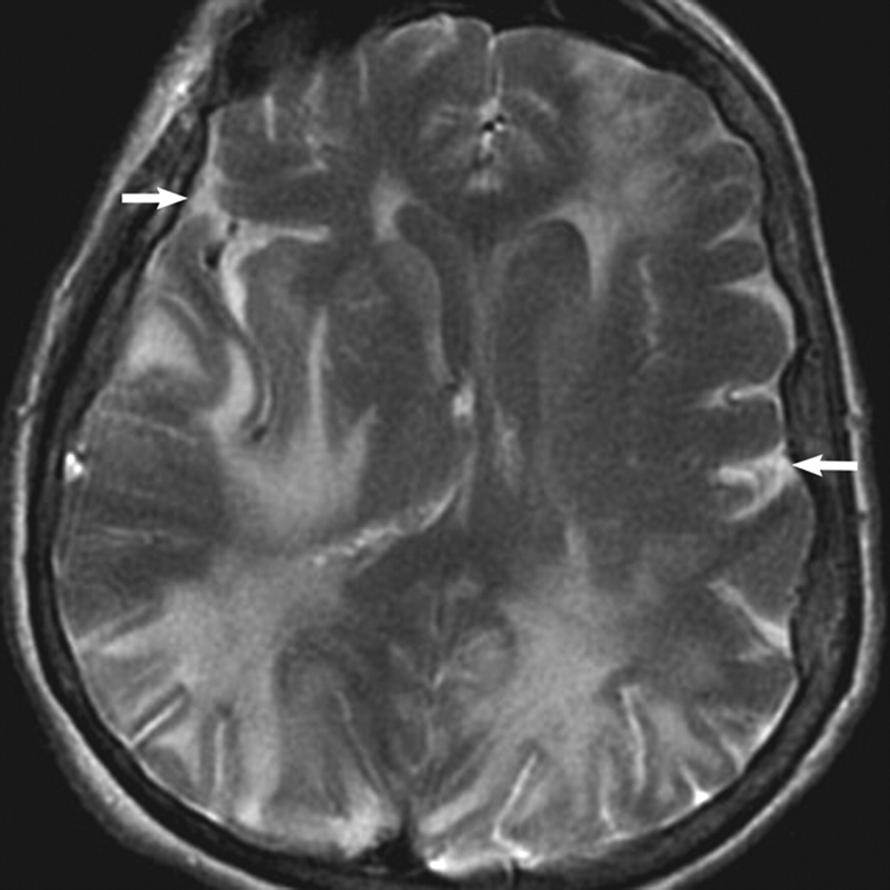
The patient underwent a second contrast-enhanced MR imaging examination, including perfusion imaging 6 hours later on the same date. Axial FLAIR images of the brain obtained immediately following the second dose of contrast revealed markedly elevated signal intensity throughout the subarachnoid spaces, which had not been present on the first study (Fig 2). CT was not performed due to no clinical concern for subarachnoid hemorrhage and given the normal-appearing FLAIR images at the time of initial presentation and imaging. A CSF assay for gadolinium was also not performed to confirm the suspicion that FLAIR hyperintensity was due to enhancement because no other indications for the procedure were sufficient to warrant consideration of this mildly invasive test.

Fig 2.
  • Download figure
  • Open in new tab
  • Download powerpoint
Fig 2.

Axial FLAIR scan obtained 6 hours later than Fig 1 demonstrates new diffuse markedly high signal intensity throughout the subarachnoid spaces (arrows). The patient received a second contrast dose (total, 0.2 mmol/kg) immediately (2 minutes) before this sequence, as part of a concurrently performed dynamic susceptibility contrast-enhanced perfusion study

The patient's admission and subsequent laboratory values revealed no evidence for renal compromise throughout her course, with serum creatinine levels ranging between 0.7 and 0.8 mg/dL.

Discussion

Many conditions have been reported to result in high signal intensity on FLAIR images in the subarachnoid spaces. Most are due to accumulation of blood, protein, cells, and/or inflammatory infiltrates within the CSF. CSF hyperintensity on FLAIR has been related to blood, proteinaceous content, inflammatory exudates or pyogenic material, anesthesia, and/or high inspired fractional oxygen concentrations (>50%).2 None of these conditions were contributory in the current patient. Single-shot, fast spin-echo FLAIR, which has been noted to result in spuriously high signal intensity in the CSF spaces, was not used in this case.3

FLAIR hyperintensity in the CSF may also result from extravascular leakage of gadolinium contrast agents, usually in the setting of an abnormal BBB. Disease states altering BBB permeability and causing CSF enhancement include acute stroke, benign and malignant brain tumors, central nervous system (CNS) infections, and multiple sclerosis.4–6 Many such types of CNS pathology have known or suspected alterations in BBB permeability. CSF enhancement is also observed in patients with compromised renal function, due to recirculation effects. CSF enhancement can occur in patients without structurally evident CNS pathology; this possibility supports the concept that the BBB is an incomplete or relative barrier. Increasing contrast dosage and delay in imaging are additional factors that contribute to extracellular contrast accumulation.2, 3, 7 Animal studies have shown that gadolinium agents cross the BBB in healthy dogs, a phenomenon that is more evident with higher contrast agent concentrations.8

Most previously reported cases of CSF gadolinium enhancement in clinical patients are seen in conjunction with higher than usual contrast doses and in the setting of poor renal clearance. Most of these cases seem to require coexisting CNS pathology from ischemic, neoplastic, or inflammatory/infectious causes.9–12 Higher circulating plasma levels of contrast alone are not expected to affect BBB permeability, but likely lower the threshold for enhancement detection on FLAIR. A delay in imaging of a few hours may be important to detection because extravascular leakage of gadolinium is not instantaneous. The relatively slow rate of fluid exchange with the interstitial compartment and/or CSF requires time to occur.

During this patient's admission, her clinical course was notable for difficult-to-control refractory hypertension requiring multiple-drug therapy to reduce blood pressure, including nitroprusside drip in the neurointensive care unit. Dilated funduscopic examination demonstrated changes of moderate hypertensive retinopathy. One day following admission, the patient's neurologic abnormalities had nearly completely resolved, and she was back to her normal baseline status.

PRES typically results in abnormal interstitial fluid accumulation in the brain, or vasogenic edema. This may be due to increasing hydrostatic pressure and/or increased microvascular permeability. There are two theories promoted to explain the pathophysiology of vasogenic edema in PRES: 1) hyperperfusion due to intravascular pressures exceeding their autoregulatory capabilities, resulting in breakthrough edema; and 2) hypoperfusion and ischemia due to excessive vasoconstriction that occurs as a response to high intravascular pressures.1 Endothelial dysfunction or injury is a common link that leads to compromise of BBB integrity and likely accounts for the extravascular leakage of contrast in the present patient.

In conclusion, this case adds PRES to the list of observed causes of CSF enhancement on FLAIR. The observation of CSF enhancement in acute PRES from hypertensive encephalopathy provides additional evidence in support of abnormally elevated microvascular permeability in this syndrome.

References

  1. ↵
    Brubaker LM, Smith JK, Lee YZ, et al. Hemodynamic and permeability changes in posterior reversible encephalopathy syndrome measured by dynamic susceptibility perfusion-weighted MR imaging. AJNR Am J Neuroradiol 2005;26:825–30
    Abstract/FREE Full Text
  2. ↵
    Frigon C, Shaw DW, Heckbert SR, et al. Supplemental oxygen causes increased signal intensity in subarachnoid cerebrospinal fluid on brain FLAIR MR images obtained in children during general anesthesia. Radiology 2004;233:51–55
    PubMed
  3. ↵
    Cianfoni A, Martin MG, Du J, et al. Artifact simulating subarachnoid and intraventricular hemorrhage on single-shot, fast spin-echo fluid-attenuated inversion recovery images caused by head movement: a trap for the unwary. AJNR Am J Neuroradiol 2006;27:843–49
    Abstract/FREE Full Text
  4. ↵
    Naul LG, Finkenstaedt M. Extensive cerebrospinal fluid enhancement with gadopentetate dimeglumine in a primitive neuroectodermal tumor. AJNR Am J Neuroradiol 1997;18:1709–11
    Abstract
  5. Hsu HL, Chen CJ. Extensive cerebrospinal fluid enhancement following gadolinium chelate administration: possible pathogenesis. Acta Radiol 2005;46:523–27
    CrossRefPubMed
  6. ↵
    Merten CL, Knitelius HO, Assheuer J, et al. MRI of acute cerebral infarcts, increased contrast enhancement with continuous infusion of gadolinium. Neuroradiology 1999;41:242–48
    CrossRefPubMed
  7. ↵
    Wiesmann M, Mayer TE, Yousry I, et al. Detection of hyperacute subarachnoid hemorrhage of the brain by using magnetic resonance imaging. J Neurosurg 2002;96:684–89
    CrossRefPubMed
  8. ↵
    Mamourian AC, Hoopes PJ, Lewis LD. Visualization of intravenously administered contrast material in the CSF on fluid-attenuated inversion-recovery MR images: an in vitro and animal-model investigation. AJNR Am J Neuroradiol 2000;21:105–11
    Abstract/FREE Full Text
  9. ↵
    Bozzao A, Floris R, Fasoli F, et al. Cerebrospinal fluid changes after intravenous injection of gadolinium chelate: assessment by FLAIR MR imaging. Eur Radiol 2003;13:592–97. Epub 2002 Jul 13
    PubMed
  10. Warach S, Latour LL. Evidence of reperfusion injury, exacerbated by thrombolytic therapy, in human focal brain ischemia using a novel imaging marker of early blood-brain barrier disruption. Stroke 2004;35 (11 suppl 1):2659–61. Epub 2004 Oct 7
    Abstract/FREE Full Text
  11. Kamran S, Bener AB, Alper D, et al. Role of fluid-attenuated inversion recovery in the diagnosis of meningitis: comparison with contrast-enhanced magnetic resonance imaging. J Comput Assist Tomogr 2004;28:68–72
    CrossRefPubMed
  12. ↵
    Taoka T, Yuh WT, White ML, et al. Sulcal hyperintensity on fluid-attenuated inversion recovery MR images in patients without apparent cerebrospinal fluid abnormality. AJR Am J Roentgenol 2001;176:519–24
    PubMed
  • Received October 22, 2007.
  • Accepted after revision November 5, 2007.
  • Copyright © American Society of Neuroradiology
View Abstract
PreviousNext
Back to top

In this issue

American Journal of Neuroradiology: 29 (3)
American Journal of Neuroradiology
Vol. 29, Issue 3
March 2008
  • Table of Contents
  • Index by author
Advertisement
Print
Download PDF
Email Article

Thank you for your interest in spreading the word on American Journal of Neuroradiology.

NOTE: We only request your email address so that the person you are recommending the page to knows that you wanted them to see it, and that it is not junk mail. We do not capture any email address.

Enter multiple addresses on separate lines or separate them with commas.
Delayed CSF Enhancement in Posterior Reversible Encephalopathy Syndrome
(Your Name) has sent you a message from American Journal of Neuroradiology
(Your Name) thought you would like to see the American Journal of Neuroradiology web site.
CAPTCHA
This question is for testing whether or not you are a human visitor and to prevent automated spam submissions.
Cite this article
B.E. Hamilton, G.M. Nesbit
Delayed CSF Enhancement in Posterior Reversible Encephalopathy Syndrome
American Journal of Neuroradiology Mar 2008, 29 (3) 456-457; DOI: 10.3174/ajnr.A0926

Citation Manager Formats

  • BibTeX
  • Bookends
  • EasyBib
  • EndNote (tagged)
  • EndNote 8 (xml)
  • Medlars
  • Mendeley
  • Papers
  • RefWorks Tagged
  • Ref Manager
  • RIS
  • Zotero
0 Responses
Respond to this article
Share
Bookmark this article
Delayed CSF Enhancement in Posterior Reversible Encephalopathy Syndrome
B.E. Hamilton, G.M. Nesbit
American Journal of Neuroradiology Mar 2008, 29 (3) 456-457; DOI: 10.3174/ajnr.A0926
del.icio.us logo Twitter logo Facebook logo Mendeley logo
  • Tweet Widget
  • Facebook Like
  • Google Plus One
Purchase

Jump to section

  • Article
    • Abstract
    • Case Report
    • Discussion
    • References
  • Figures & Data
  • Info & Metrics
  • Responses
  • References
  • PDF

Related Articles

  • No related articles found.
  • PubMed
  • Google Scholar

Cited By...

  • Diagnosis and Prediction of Relapses in Susac Syndrome: A New Use for MR Postcontrast FLAIR Leptomeningeal Enhancement
  • Investigation of leptomeningeal enhancement in MS: A postcontrast FLAIR MRI study
  • The posterior reversible encephalopathy syndrome: what's certain, what's new?
  • POSTCONTRAST FLAIR MRI DEMONSTRATES BLOOD-BRAIN BARRIER DYSFUNCTION IN PRES
  • Crossref (20)
  • Google Scholar

This article has been cited by the following articles in journals that are participating in Crossref Cited-by Linking.

  • Blood Pressure Fluctuations in Posterior Reversible Encephalopathy Syndrome
    Alejandro A. Rabinstein, Jay Mandrekar, Ryan Merrell, Osman S. Kozak, Olayemi Durosaro, Jennifer E. Fugate
    Journal of Stroke and Cerebrovascular Diseases 2012 21 4
  • Posterior reversible encephalopathy syndrome (PRES): Features on CT and MR imaging
    E. Hugonnet, D. Da Ines, H. Boby, B. Claise, V. Petitcolin, V. Lannareix, J.-M. Garcier
    Diagnostic and Interventional Imaging 2013 94 1
  • Neuroimaging features in posterior reversible encephalopathy syndrome: A pictorial review
    Morgan Ollivier, Anne Bertrand, Frédéric Clarençon, Sophie Gerber, Sandrine Deltour, Fanny Domont, Stéphanie Trunet, Didier Dormont, Delphine Leclercq
    Journal of the Neurological Sciences 2017 373
  • Investigation of leptomeningeal enhancement in MS
    Philipp Eisele, Martin Griebe, Kristina Szabo, Marc E. Wolf, Angelika Alonso, Britta Engelhardt, Michael G. Hennerici, Achim Gass
    Neurology 2015 84 8
  • Immune system activation in the pathogenesis of posterior reversible encephalopathy syndrome
    Zheng Chen, Gui-Quan Shen, Alexander Lerner, Bo Gao
    Brain Research Bulletin 2017 131
  • Spectrum of cerebral spinal fluid findings in patients with posterior reversible encephalopathy syndrome
    Lars Neeb, Jenneke Hoekstra, Matthias Endres, Bob Siegerink, Eberhard Siebert, Thomas G. Liman
    Journal of Neurology 2016 263 1
  • Diagnosis and Prediction of Relapses in Susac Syndrome: A New Use for MR Postcontrast FLAIR Leptomeningeal Enhancement
    S. Coulette, A. Lecler, E. Saragoussi, K. Zuber, J. Savatovsky, R. Deschamps, O. Gout, C. Sabben, J. Aboab, A. Affortit, F. Charbonneau, M. Obadia
    American Journal of Neuroradiology 2019 40 7
  • Childhood posterior reversible encephalopathy syndrome: Magnetic resonance imaging findings with emphasis on increased leptomeningeal FLAIR signal
    Ajay Agarwal, Gaurav Kapur, Deniz Altinok
    The Neuroradiology Journal 2015 28 6
  • Clinical characteristics of hypertensive encephalopathy in pediatric patients
    Chang Hoon Ahn, Seung-A Han, Young Hwa Kong, Sun Jun Kim
    Korean Journal of Pediatrics 2017 60 8
  • Posterior Reversible Encephalopathy Syndrome: Clinicoradiological Spectrum and Therapeutic Strategies
    Yuebing Li, Donna Jenny, John Castaldo
    Hospital Practice 2012 40 1

More in this TOC Section

  • Fast Contrast-Enhanced 4D MRA and 4D Flow MRI Using Constrained Reconstruction (HYPRFlow): Potential Applications for Brain Arteriovenous Malformations
  • Quiet PROPELLER MRI Techniques Match the Quality of Conventional PROPELLER Brain Imaging Techniques
  • Predictors of Reperfusion in Patients with Acute Ischemic Stroke
Show more Brain

Similar Articles

Advertisement

Indexed Content

  • Current Issue
  • Accepted Manuscripts
  • Article Preview
  • Past Issues
  • Editorials
  • Editors Choice
  • Fellow Journal Club
  • Letters to the Editor

Cases

  • Case Collection
  • Archive - Case of the Week
  • Archive - Case of the Month
  • Archive - Classic Case

Special Collections

  • Special Collections

Resources

  • News and Updates
  • Turn around Times
  • Submit a Manuscript
  • Author Policies
  • Manuscript Submission Guidelines
  • Evidence-Based Medicine Level Guide
  • Publishing Checklists
  • Graphical Abstract Preparation
  • Imaging Protocol Submission
  • Submit a Case
  • Become a Reviewer/Academy of Reviewers
  • Get Peer Review Credit from Publons

Multimedia

  • AJNR Podcast
  • AJNR SCANtastic
  • Video Articles

About Us

  • About AJNR
  • Editorial Board
  • Not an AJNR Subscriber? Join Now
  • Alerts
  • Feedback
  • Advertise with us
  • Librarian Resources
  • Permissions
  • Terms and Conditions

American Society of Neuroradiology

  • Not an ASNR Member? Join Now

© 2025 by the American Society of Neuroradiology All rights, including for text and data mining, AI training, and similar technologies, are reserved.
Print ISSN: 0195-6108 Online ISSN: 1936-959X

Powered by HighWire