
Article Figures & Data
Figures
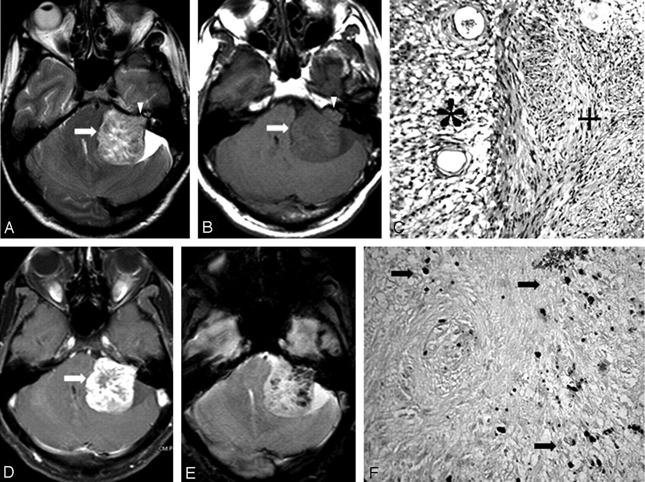
- Fig 1.
A–B, Axial T2- and T1-weighted images, respectively, of left VS show no evidence of hemorrhage (arrow). Note the extension into the IAC (arrowhead). C, Photomicrograph shows the Antoni A (+) and Antoni B (*) components in the VS (H&E ×150). D, Axial postcontrast T1-weighted image shows bright enhancement of the tumor with nonenhancing areas (arrow). E, T2*-weighted GRE shows multiple hypointense intratumoral signals suggestive of microhemorrhage. F, Photomicrograph showing discrete and confluent masses of hemosiderin pigments (arrow) in the Schwann cell tumor (Perls stain ×150).
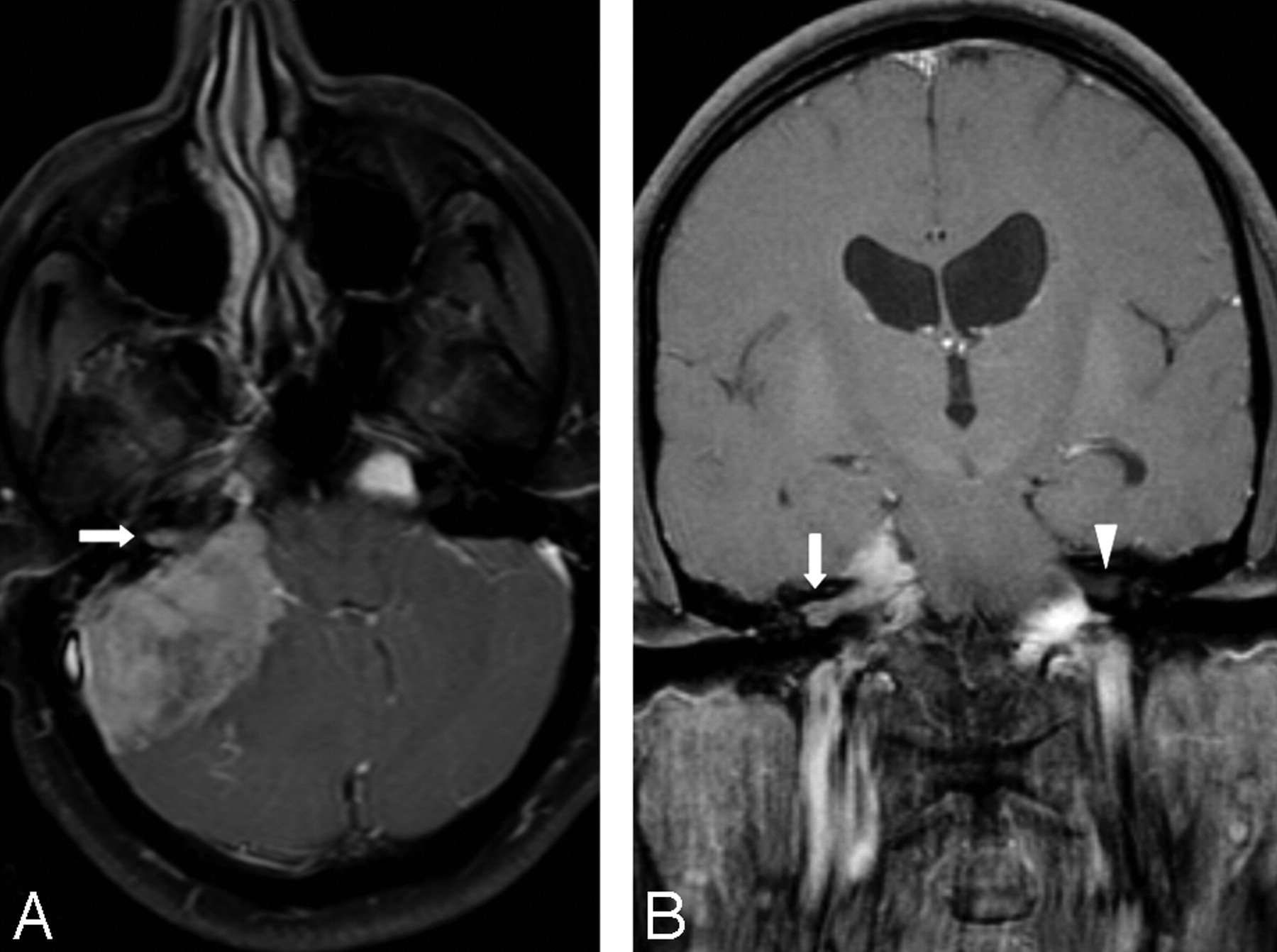
- Fig 2.
A, T1-weighted postgadolinium axial image shows right CPA meningioma with extension into the adjacent IAC (arrow). B, T1-weighted postgadolinium coronal image shows extension of the tumor into the IAC more clearly (arrow) compared with the contralateral IAC (arrowhead).
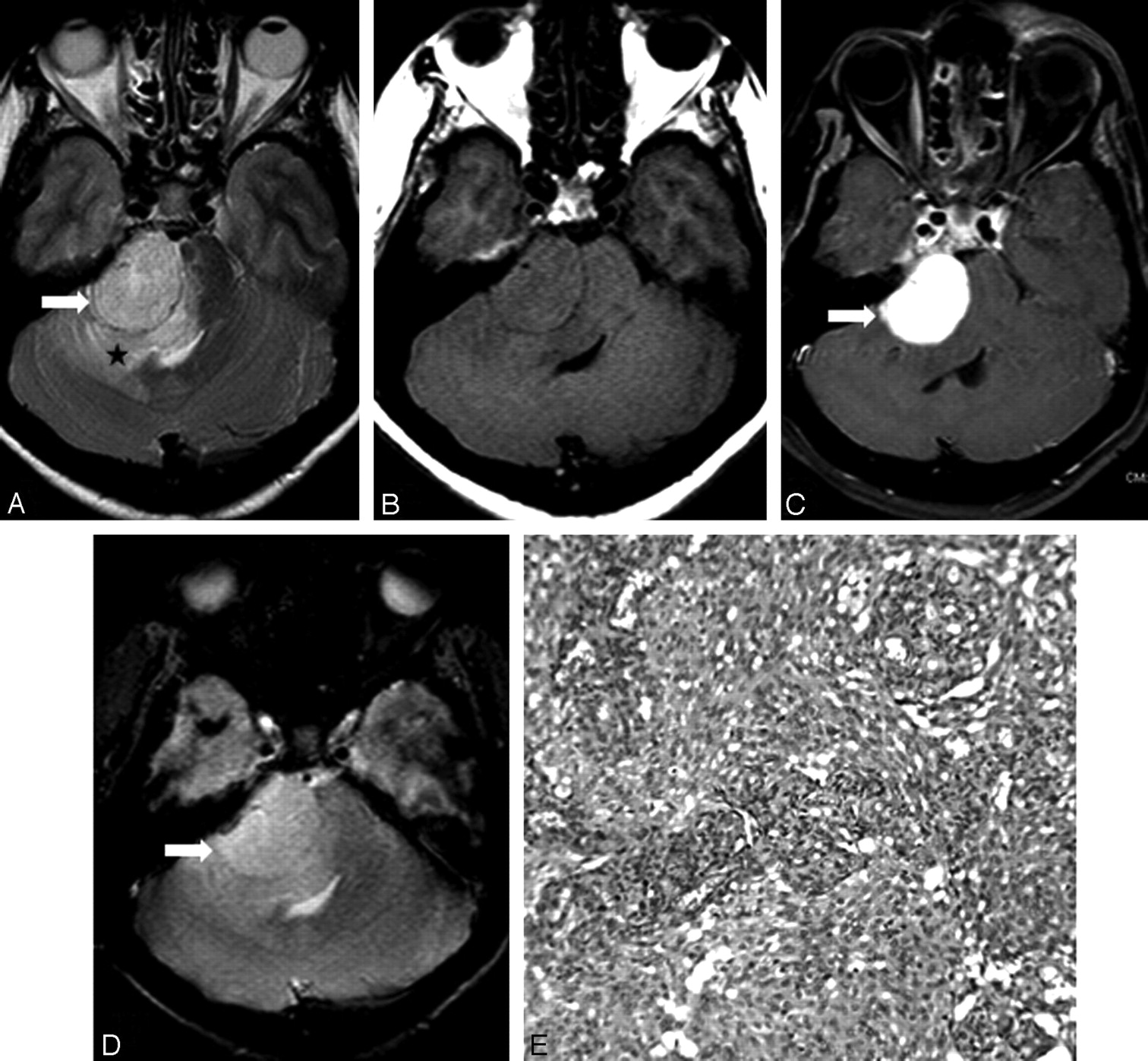
- Fig 3.
A, Axial T2-weighted TSE image shows the relatively hyperintense meningioma in the right CPA with a broad base against the petrous bone (arrow). Note the vasogenic edema of the adjacent cerebellum and middle cerebellar peduncle (star). B, On axial T1-weighted SE, the tumor is isointense to the brain. C, Postcontrast T1-weighted image at the corresponding level shows intense enhancement of the tumor (arrow). D, Axial T2*-weighted GRE does not show hypointense signals characteristic of microhemorrhage in the tumor (arrow). The tumor matrix appears homogeneous. E, Photomicrograph showing features of a meningioma in which neoplastic cells are displayed in concentric whorls and lobules of varying sizes. These neoplastic cells are supported on a vascularized connective stroma (H&E ×150).


Tables
Patient No. Age/Sex Location Size (cm) T x C x A Consistency No. of MH Size of MH (mm) Fluid Level IAC Component Sequence Showing MH Other Than T2* Surgical Treatment Pigment 1 52/M R 3.22 × 3.41 × 2.88 S 9 <3 N + N Y Y 2 42/M L 3.77 × 3.78 × 4.21 S >40 5 N + T2 & FLAIR Y Y 3 41/F R 3.03 × 3.57 × 4.14 S 17 2 N + N Y Y 4 36/F R 2.63 × 2.95 × 3.55 S 14 4 N + N Y Y 5 30/M L 3.23 × 3.48 × 3.55 S 6 5 N + N Y Y 6 60/F R 2.82 × 3.1 × 3 S 5 2 N + N Y Y 7 59/F R 3.61 × 2.43 × 2.95 S >50 4 N + N Y Y 8 38/F B/L 4.02 × 3.95 × 4.67 (R) S >50 5 N + N Y Y 2.73 × 2.52 × 3.32 (L) S 5 2 N + N N N 9 52/M R 1.45 × 1.12 × 1.12 S N N N + N N N 10 34/M R 3.68 × N × 2.88 S 28 5 N + N Y Y 11 51/M R 3.09 × 4.38 × 4.88 C 4 7 Y + T2 & FLAIR Y Y 12 23/F R 3.27 × 4.73 × 3.08 S 6 4 N + N Y Y 13 36/M R 4.40 × 4.20 × 4.03 S 10 4 N + N Y Y 14 22/M R 3.81 × 4.73 × 3.71 S 69 5 N + N Y Y 15 18/M R 5.2 × 6.09 × 6.37 S 140 8 Y + N Y Y Note:—M indicates male; F, female; R, right; L, left; B/L, bilateral; S, solid; C, cystic (in Consistency column); N, no; Y, yes; MH, microhemorrhage; IAC, internal auditory canal; T, transverse; C, craniocaudal; A, anteroposterior; FLAIR, fluid-attenuated inversion recovery; +, present.
Patient No. Age/Sex Location Size (cm) T × C × A Consistency No. of MH Size of MH (mm) Fluid Level IAC Component MH in IAC Sequence Showing MH Other Than T2* Surgical Treatment 1 59/F CPA 2.03 × 2.69 × 2.79 S N N N N N N Y 2 47/F Tentorium 4.55 × 5.64 × 4.97 S N N N N N N Y 3 44/F Falcotentorial 2.64 × 3.33 × 2.98 S N N N N N N Y 4 48/F CPA 2.22 × 3.54 × 3.32 S N N N N N N Y 5 44/M CPA 3.32 × 3.16 × 4.92 S N N N + N N Y Note:—M indicates male; F, female; S, solid; N, no; Y, yes; MH, microhemorrhage; IAC, internal auditory canal; T, transverse; C, craniocaudal; A, anteroposterior; CPA, cerebellopontine angle; +, present.


