Advertisement

Main menu

  • Home
  • Content
    • Current Issue
    • Accepted Manuscripts
    • Article Preview
    • Past Issue Archive
    • AJNR Case Collection
    • Case of the Week Archive
    • Classic Case Archive
    • Case of the Month Archive
  • Special Collections
    • Spinal CSF Leak Articles (Jan 2020-June 2024)
    • 2024 AJNR Journal Awards
    • Most Impactful AJNR Articles
  • Multimedia
    • AJNR Podcast
    • AJNR Scantastics
    • Video Articles
  • For Authors
    • Submit a Manuscript
    • Author Policies
    • Fast publishing of Accepted Manuscripts
    • Graphical Abstract Preparation
    • Manuscript Submission Guidelines
    • Imaging Protocol Submission
    • Submit a Case for the Case Collection
  • About Us
    • About AJNR
    • Editorial Board
  • More
    • Become a Reviewer/Academy of Reviewers
    • Subscribers
    • Permissions
    • Alerts
    • Feedback
    • Advertisers
    • ASNR Home
  • Other Publications
    • ajnr

User menu

  • Alerts
  • Log in

Search

  • Advanced search
American Journal of Neuroradiology

American Journal of Neuroradiology

ASHNR American Society of Functional Neuroradiology ASHNR American Society of Pediatric Neuroradiology ASSR
  • Alerts
  • Log in

Advanced Search

  • Home
  • Content
    • Current Issue
    • Accepted Manuscripts
    • Article Preview
    • Past Issue Archive
    • AJNR Case Collection
    • Case of the Week Archive
    • Classic Case Archive
    • Case of the Month Archive
  • Special Collections
    • Spinal CSF Leak Articles (Jan 2020-June 2024)
    • 2024 AJNR Journal Awards
    • Most Impactful AJNR Articles
  • Multimedia
    • AJNR Podcast
    • AJNR Scantastics
    • Video Articles
  • For Authors
    • Submit a Manuscript
    • Author Policies
    • Fast publishing of Accepted Manuscripts
    • Graphical Abstract Preparation
    • Manuscript Submission Guidelines
    • Imaging Protocol Submission
    • Submit a Case for the Case Collection
  • About Us
    • About AJNR
    • Editorial Board
  • More
    • Become a Reviewer/Academy of Reviewers
    • Subscribers
    • Permissions
    • Alerts
    • Feedback
    • Advertisers
    • ASNR Home
  • Follow AJNR on Twitter
  • Visit AJNR on Facebook
  • Follow AJNR on Instagram
  • Join AJNR on LinkedIn
  • RSS Feeds

Welcome to the new AJNR, Updated Hall of Fame, and more. Read the full announcements.


AJNR is seeking candidates for the position of Associate Section Editor, AJNR Case Collection. Read the full announcement.


OtherBRAIN

Sequential Pituitary MR Imaging in Sheehan Syndrome: Report of 2 Cases

J. Kaplun, C. Fratila, A. Ferenczi, W.C. Yang, G. Lantos, A.M. Fleckman and U.K. Schubart
American Journal of Neuroradiology May 2008, 29 (5) 941-943; DOI: https://doi.org/10.3174/ajnr.A1016
J. Kaplun
  • Find this author on Google Scholar
  • Find this author on PubMed
  • Search for this author on this site
C. Fratila
  • Find this author on Google Scholar
  • Find this author on PubMed
  • Search for this author on this site
A. Ferenczi
  • Find this author on Google Scholar
  • Find this author on PubMed
  • Search for this author on this site
W.C. Yang
  • Find this author on Google Scholar
  • Find this author on PubMed
  • Search for this author on this site
G. Lantos
  • Find this author on Google Scholar
  • Find this author on PubMed
  • Search for this author on this site
A.M. Fleckman
  • Find this author on Google Scholar
  • Find this author on PubMed
  • Search for this author on this site
U.K. Schubart
  • Find this author on Google Scholar
  • Find this author on PubMed
  • Search for this author on this site
  • Article
  • Figures & Data
  • Info & Metrics
  • Responses
  • References
  • PDF
Loading

Abstract

SUMMARY: We present the evolution of pituitary changes in the cases of 2 patients with Sheehan syndrome as assessed by MR imaging. Both patients had severe postpartum hemorrhage, symptoms of pituitary gland apoplexy, and hypopituitarism. Sequential MR imaging demonstrated evidence of ischemic infarct in the pituitary gland with enlargement followed by gradual shrinkage during several months, to pituitary atrophy.

Clinical recognition of Sheehan syndrome (hypopituitarism resulting from postpartum pituitary gland infarction after severe hypotension secondary to massive bleeding1) is often insidious, with the diagnosis being delayed for years.2 Pituitary imaging studies late in the course have revealed atrophy of the gland, resulting in a small empty sella.3,4 Rarely, patients present with pituitary apoplexy.2 MR imaging studies have been reported in only a few cases.5,6 We report the cases of 2 patients with Sheehan syndrome with acute presentations, in which the progression of pituitary gland changes was assessed by sequential MR imaging.

Case Reports

Case 1

A 29-year-old woman delivered a full-term baby. The delivery was complicated by retained placenta and profuse bleeding, requiring emergent embolization of the uterine artery. Her hemoglobin level fell to 3.8 g/dL. Several hours later, the patient developed a severe headache and nausea. A routine CT study of her head showed no abnormality. Her symptoms resolved, and she was discharged from the hospital without additional work-up. She returned on day 17 postpartum with fatigue, fever, mild headache, failure to lactate, and postural syncope. Her temperature was 101.2°F, pulse 122, and a supine blood pressure of 90/48 mm Hg. Examination showed skin pallor, a cardiac flow murmur, clear lungs, and a soft abdomen. Laboratory evaluation revealed anemia, mild hyponatremia (sodium, 134 mEq/L), and abnormal pituitary function test results (Table) consistent with panhypopituitarism.

View this table:
  • View inline
  • View popup

Results of endocrine laboratory evaluation in Cases 1 and 2

MR imaging on postpartum day 26 (Fig 1) revealed a nonenhancing, minimally hypointense lesion in the pituitary gland. MR imaging 6 months postpartum (Fig 2) showed shrinkage of the pituitary gland with CSF signal intensity in the sella (“empty sella”). The pituitary infundibulum and hypothalamus appeared normal.

Fig 1.
  • Download figure
  • Open in new tab
  • Download powerpoint
Fig 1.

Case 1. Coronal (left) and sagittal (right) gadolinium enhanced T1-weighted MR images on postpartum day 26 show central low signal intensity and peripheral enhancement in the pituitary gland.

Fig 2.
  • Download figure
  • Open in new tab
  • Download powerpoint
Fig 2.

Case 1. Coronal (left) and sagittal (right) noncontrast T1-weighted MR images 6 months later show marked diminution in the size of the pituitary gland. There is CSF signal intensity in the sella (“empty sella”).

Case 2

A 21-year-old woman delivered a full-term healthy baby after labor was induced following premature rupture of membranes. During delivery, she sustained a fourth-degree perineal laceration with severe bleeding and hypotension (blood pressure, 60/30 mm Hg). Her hemoglobin level fell to 5.5 g/dL. Two days postpartum, the patient developed fever and a severe headache. The results of blood and urine cultures, pelvic sonography, and abdominal CT studies were negative. Fever resolved within 24 hours after the patient was given broad-spectrum antibiotics. The results of pituitary function tests obtained for hyponatremia (sodium, 116 mEq/L) demonstrated hypopituitarism (Table). Replacement therapy with prednisone and levothyroxine was begun. Her headaches resolved gradually, and she was discharged from the hospital on postpartum day 9.

MR imaging obtained on postpartum day 6 (Fig 3) showed an enlarged pituitary gland with suprasellar extension to the optic chiasm. There was no evidence of hemorrhage. Postgadolinium T1-weighted images showed peripheral enhancement of the swollen pituitary gland and an irregular and poorly enhanced central portion. MR imaging 5 weeks postpartum (Fig 4) revealed a smaller pituitary gland contained within the pituitary fossa. Nonenhancing areas persisted within the pituitary gland after administration of gadolinium. The pituitary stalk appeared normal. MR imaging obtained 11 months postpartum (Fig 5) showed marked atrophy of the pituitary gland resulting in an empty sella. There was now contrast enhancement of the pituitary remnant and the distal portion of the infundibulum.

Fig 3.
  • Download figure
  • Open in new tab
  • Download powerpoint
Fig 3.

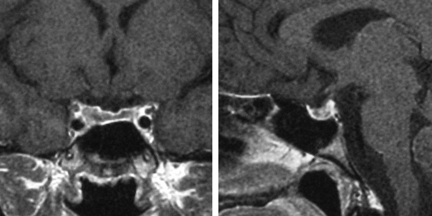
Case 2. Coronal (A) and sagittal (B) gadolinium-enhanced T1-weighted MR images on postpartum day 6 show a large low signal intensity sellar lesion with enhancement of the rim. There is extension into the suprasellar cistern, and the lesion abuts but does not compress the optic chiasm.

Fig 4.
  • Download figure
  • Open in new tab
  • Download powerpoint
Fig 4.

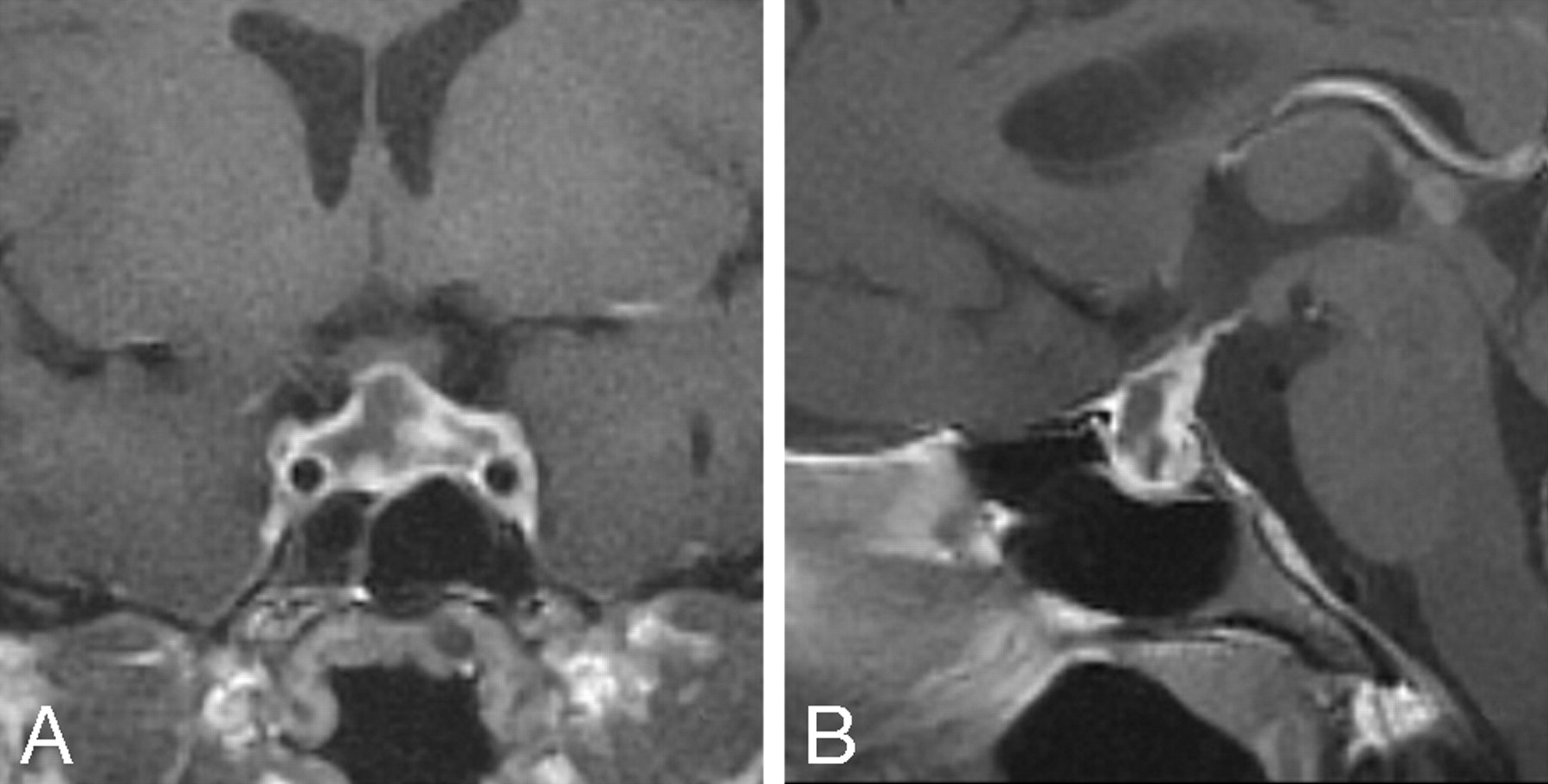
A and B, Case 2. At 5 weeks postpartum, the lesion is markedly smaller and no longer protrudes into the suprasellar cistern (image layout same as Fig 3).

Fig 5.
  • Download figure
  • Open in new tab
  • Download powerpoint
Fig 5.

A and B, Case 2. At 11 months postpartum, the sellar contents are markedly smaller, with a thin rim of enhancing tissue along the sellar floor (image layout same as Figs 3 and 4). CSF is noted in the sella (“empty sella”).

Discussion

This case report describes the clinical and MR imaging findings of 2 patients with acute hypopituitarism after severe peripartum hemorrhage. Both cases were highly suggestive of ischemic infarction of the pituitary gland (Sheehan syndrome). MR imaging during the acute phase of Sheehan syndrome has been reported in 2 isolated cases,5,6 which, to our knowledge, are the only previous reports of time-dependent findings on MR imaging.

Although the number of reported cases is limited, some characteristic findings emerge. Acutely, MR imaging reveals an enlarged pituitary gland with central hypointensity on T1-weighted images and hyperintensity on T2-weighted images. These findings are consistent with pituitary infarction. Postgadolinium, the presumed infarcted areas are interspersed with perfused tissue, which manifests irregular enhancement. Within several weeks, the swollen pituitary gland regresses and is confined to the pituitary fossa, followed by further pituitary gland atrophy for several months. The end result is an empty sella of normal size.

The findings on MR imaging observed in Sheehan syndrome are not specific. During the acute phase of lymphocytic hypophysitis, there is more uniform contrast enhancement of a sellar or suprasellar mass7; however, in approximately 5% of reported cases, a cystic appearance is observed on MR imaging.7-9 In these cases, the findings on MR imaging can resemble those seen during the acute stages of Sheehan syndrome. This is relevant because lymphocytic hypophysitis is more common in women and typically presents in the peripartum and postpartum periods.7 Although hypopituitarism in lymphocytic hypophysitis may be transient, late-stage pituitary atrophy resulting in an empty sella has been reported in a case of biopsy-proved lymphocytic hypophysitis.10 Pituitary tumors typically show uniform enhancement on MR imaging, and the clinical distinction between a pituitary tumor and Sheehan syndrome is usually not difficult. However, tumors may exhibit central necrosis, in which case findings on MR imaging may mimic those seen during the acute phase of Sheehan syndrome.

Conclusion

The cases of our 2 patients, in conjunction with the previous cases of 2 patients reported in the literature, present a picture of the evolution of the findings on MR imaging in Sheehan syndrome that is consistent with the classic pathologic description. This evolution begins acutely with nonhemorrhagic changes in signal intensity consistent with central infarction, along with peripheral and heterogeneous central enhancement in an enlarged pituitary gland. The findings are consistent with patchy central ischemic necrosis and are followed by pituitary gland atrophy and an empty sella. These findings on MR imaging characterize Sheehan syndrome and provide early confirmation of the clinical diagnosis.

Footnotes

  • Previously presented in part at: Annual Meeting of the American Association of Clinical Endocrinologists, May 1–5, 2002; Chicago, Ill.

References

  1. ↵
    Sheehan HL. Postpartum necrosis of the anterior pituitary. J Pathol Bacteriol 1937;45:189–214
    CrossRef
  2. ↵
    Keletimur F. Sheehan's syndrome. Pituitary 2003;6:181–88
    CrossRefPubMed
  3. ↵
    Fleckman AM, Schubart UK, Danziger A, et al. Empty sella of normal size in Sheehan's syndrome. Am J Med 1983;75:585–91
    CrossRefPubMed
  4. ↵
    Otsuka F, Kageyama J, Ogura T, et al. Sheehan's syndrome of more than 30 years' duration: an endocrine and MRI study of 6 cases. Endocr J 1998;45:451–58
    CrossRefPubMed
  5. ↵
    Dejager S, Gerber S, Foubert L, et al. Sheehan's syndrome: differential diagnosis in the acute phase. J Intern Med 1998;244:261–66
    CrossRefPubMed
  6. ↵
    Lavallée G, Morcos R, Palardy J, et al. MR of nonhemorrhagic postpartum pituitary apoplexy. AJNR Am J Neuroradiol 1995;16:1939–41
    Abstract
  7. ↵
    Caturegli P, Newschaffer C, Olivi A, et al. Autoimmune hypophysitis. Endocr Rev 2005;26:599–614
    CrossRefPubMed
  8. Farah JO, Rossi M, Foy PM, et al. Cystic lymphocytic hypophysitis, visual field defects and hypopituitarism. Int J Clin Pract 1999;53:643–44
    PubMed
  9. ↵
    Pérez-Núñez A, Miranda P, Arrese I, et al. Lymphocytic hypophysitis with cystic MRI appearance. Acta Neurochir (Wien) 2005;147:1297–300
    CrossRefPubMed
  10. ↵
    Ishihara T, Hino M, Kurahachi H, et al. Long-term clinical course of two cases of lymphocytic adenohypophysitis. Endocr J 1996;43:433–40
    PubMed
  • Received October 16, 2007.
  • Accepted after revision December 20, 2007.
  • Copyright © American Society of Neuroradiology
View Abstract
PreviousNext
Back to top

In this issue

American Journal of Neuroradiology: 29 (5)
American Journal of Neuroradiology
Vol. 29, Issue 5
May 2008
  • Table of Contents
  • Index by author
Advertisement
Print
Download PDF
Email Article

Thank you for your interest in spreading the word on American Journal of Neuroradiology.

NOTE: We only request your email address so that the person you are recommending the page to knows that you wanted them to see it, and that it is not junk mail. We do not capture any email address.

Enter multiple addresses on separate lines or separate them with commas.
Sequential Pituitary MR Imaging in Sheehan Syndrome: Report of 2 Cases
(Your Name) has sent you a message from American Journal of Neuroradiology
(Your Name) thought you would like to see the American Journal of Neuroradiology web site.
CAPTCHA
This question is for testing whether or not you are a human visitor and to prevent automated spam submissions.
Cite this article
J. Kaplun, C. Fratila, A. Ferenczi, W.C. Yang, G. Lantos, A.M. Fleckman, U.K. Schubart
Sequential Pituitary MR Imaging in Sheehan Syndrome: Report of 2 Cases
American Journal of Neuroradiology May 2008, 29 (5) 941-943; DOI: 10.3174/ajnr.A1016

Citation Manager Formats

  • BibTeX
  • Bookends
  • EasyBib
  • EndNote (tagged)
  • EndNote 8 (xml)
  • Medlars
  • Mendeley
  • Papers
  • RefWorks Tagged
  • Ref Manager
  • RIS
  • Zotero
0 Responses
Respond to this article
Share
Bookmark this article
Sequential Pituitary MR Imaging in Sheehan Syndrome: Report of 2 Cases
J. Kaplun, C. Fratila, A. Ferenczi, W.C. Yang, G. Lantos, A.M. Fleckman, U.K. Schubart
American Journal of Neuroradiology May 2008, 29 (5) 941-943; DOI: 10.3174/ajnr.A1016
del.icio.us logo Twitter logo Facebook logo Mendeley logo
  • Tweet Widget
  • Facebook Like
  • Google Plus One
Purchase

Jump to section

  • Article
    • Abstract
    • Case Reports
    • Discussion
    • Conclusion
    • Footnotes
    • References
  • Figures & Data
  • Info & Metrics
  • Responses
  • References
  • PDF

Related Articles

  • No related articles found.
  • PubMed
  • Google Scholar

Cited By...

  • Colonic pseudo-obstruction in a patient with Sheehans syndrome
  • Delayed diagnosis of Sheehan's syndrome in a developed country: a retrospective cohort study
  • Teaching NeuroImages: Sequential MRI of the pituitary in Sheehan syndrome
  • Crossref (33)
  • Google Scholar

This article has been cited by the following articles in journals that are participating in Crossref Cited-by Linking.

  • A case of acute Sheehan’s syndrome and literature review: a rare but life-threatening complication of postpartum hemorrhage
    Shinya Matsuzaki, Masayuki Endo, Yutaka Ueda, Kazuya Mimura, Aiko Kakigano, Tomomi Egawa-Takata, Keiichi Kumasawa, Kiyoshi Yoshino, Tadashi Kimura
    BMC Pregnancy and Childbirth 2017 17 1
  • Delayed diagnosis of Sheehan's syndrome in a developed country: a retrospective cohort study
    Cynthia Ramiandrasoa, Frédéric Castinetti, Isabelle Raingeard, Patrick Fenichel, Olivier Chabre, Thierry Brue, Blandine Courbière
    European Journal of Endocrinology 2013 169 4
  • Sheehan Syndrome Presenting as Central Diabetes Insipidus: A Rare Presentation of an Uncommon Disorder
    Saumya Kumar, Daryl Burrows, Shyam Dang, Debra Simmons
    Endocrine Practice 2011 17 1
  • Evaluation of obstetrical factors related to Sheehan syndrome
    Takahiro Matsuwaki, Khaleque Newaz Khan, Tsuneo Inoue, Atsushi Yoshida, Hideaki Masuzaki
    Journal of Obstetrics and Gynaecology Research 2014 40 1
  • Repairing A “Broken Heart” With Hormone Replacement Therapy: Case Report of Cardiogenic Shock Due to Undiagnosed Pituitary Insufficiency
    Shunzhong S. Bao, Simon J. Fisher
    Endocrine Practice 2012 18 2
  • Neuroimaging of hypophysitis: etiologies and imaging mimics
    Ryo Kurokawa, Mariko Kurokawa, Akira Baba, Moto Nakaya, Shimpei Kato, Jayapalli Bapuraj, Yasuhiro Nakata, Yoshiaki Ota, Ashok Srinivasan, Osamu Abe, Toshio Moritani
    Japanese Journal of Radiology 2023 41 9
  • A rare cause of postpartum acute hyponatremia
    Ilan Rahmani Tzvi-Ran, Judith Olchowski, Merav Fraenkel, Asher Bashiri, Leonid Barski
    Endocrinology, Diabetes & Metabolism Case Reports 2019 2019
  • Teaching Neuro Images : Sequential MRI of the pituitary in Sheehan syndrome
    Ajaykumar Morani, Hemant Parmar, Mohannad Ibrahim
    Neurology 2012 78 1
  • A novel hook-shaped enhancement on contrast-enhanced sagittal magnetic resonance image in acute Sheehan’s syndrome: a case report
    Sho Sasaki, Ichiro Fujisawa, Takashi Ishihara, Yumiko Tahara, Mariko Kazuma, Yuta Fujiwara, Toshio Iwakura, Megumu Hino, Naoki Matsuoka
    Endocrine Journal 2014 61 1
  • Imaging of Neurologic Disorders in Pregnancy
    Dara G. Jamieson, Jennifer W. McVige
    Neurologic Clinics 2020 38 1

More in this TOC Section

  • Fast Contrast-Enhanced 4D MRA and 4D Flow MRI Using Constrained Reconstruction (HYPRFlow): Potential Applications for Brain Arteriovenous Malformations
  • Quiet PROPELLER MRI Techniques Match the Quality of Conventional PROPELLER Brain Imaging Techniques
  • Predictors of Reperfusion in Patients with Acute Ischemic Stroke
Show more Brain

Similar Articles

Advertisement

Indexed Content

  • Current Issue
  • Accepted Manuscripts
  • Article Preview
  • Past Issues
  • Editorials
  • Editors Choice
  • Fellow Journal Club
  • Letters to the Editor

Cases

  • Case Collection
  • Archive - Case of the Week
  • Archive - Case of the Month
  • Archive - Classic Case

Special Collections

  • Special Collections

Resources

  • News and Updates
  • Turn around Times
  • Submit a Manuscript
  • Author Policies
  • Manuscript Submission Guidelines
  • Evidence-Based Medicine Level Guide
  • Publishing Checklists
  • Graphical Abstract Preparation
  • Imaging Protocol Submission
  • Submit a Case
  • Become a Reviewer/Academy of Reviewers
  • Get Peer Review Credit from Publons

Multimedia

  • AJNR Podcast
  • AJNR SCANtastic
  • Video Articles

About Us

  • About AJNR
  • Editorial Board
  • Not an AJNR Subscriber? Join Now
  • Alerts
  • Feedback
  • Advertise with us
  • Librarian Resources
  • Permissions
  • Terms and Conditions

American Society of Neuroradiology

  • Not an ASNR Member? Join Now

© 2025 by the American Society of Neuroradiology All rights, including for text and data mining, AI training, and similar technologies, are reserved.
Print ISSN: 0195-6108 Online ISSN: 1936-959X

Powered by HighWire