Advertisement

Main menu

  • Home
  • Content
    • Current Issue
    • Accepted Manuscripts
    • Article Preview
    • Past Issue Archive
    • AJNR Case Collection
    • Case of the Week Archive
    • Classic Case Archive
    • Case of the Month Archive
  • Special Collections
    • Spinal CSF Leak Articles (Jan 2020-June 2024)
    • 2024 AJNR Journal Awards
    • Most Impactful AJNR Articles
  • Multimedia
    • AJNR Podcast
    • AJNR Scantastics
    • Video Articles
  • For Authors
    • Submit a Manuscript
    • Author Policies
    • Fast publishing of Accepted Manuscripts
    • Graphical Abstract Preparation
    • Manuscript Submission Guidelines
    • Imaging Protocol Submission
    • Submit a Case for the Case Collection
  • About Us
    • About AJNR
    • Editorial Board
  • More
    • Become a Reviewer/Academy of Reviewers
    • Subscribers
    • Permissions
    • Alerts
    • Feedback
    • Advertisers
    • ASNR Home
  • Other Publications
    • ajnr

User menu

  • Alerts
  • Log in

Search

  • Advanced search
American Journal of Neuroradiology

American Journal of Neuroradiology

ASHNR American Society of Functional Neuroradiology ASHNR American Society of Pediatric Neuroradiology ASSR
  • Alerts
  • Log in

Advanced Search

  • Home
  • Content
    • Current Issue
    • Accepted Manuscripts
    • Article Preview
    • Past Issue Archive
    • AJNR Case Collection
    • Case of the Week Archive
    • Classic Case Archive
    • Case of the Month Archive
  • Special Collections
    • Spinal CSF Leak Articles (Jan 2020-June 2024)
    • 2024 AJNR Journal Awards
    • Most Impactful AJNR Articles
  • Multimedia
    • AJNR Podcast
    • AJNR Scantastics
    • Video Articles
  • For Authors
    • Submit a Manuscript
    • Author Policies
    • Fast publishing of Accepted Manuscripts
    • Graphical Abstract Preparation
    • Manuscript Submission Guidelines
    • Imaging Protocol Submission
    • Submit a Case for the Case Collection
  • About Us
    • About AJNR
    • Editorial Board
  • More
    • Become a Reviewer/Academy of Reviewers
    • Subscribers
    • Permissions
    • Alerts
    • Feedback
    • Advertisers
    • ASNR Home
  • Follow AJNR on Twitter
  • Visit AJNR on Facebook
  • Follow AJNR on Instagram
  • Join AJNR on LinkedIn
  • RSS Feeds

Welcome to the new AJNR, Updated Hall of Fame, and more. Read the full announcements.


AJNR is seeking candidates for the position of Associate Section Editor, AJNR Case Collection. Read the full announcement.


OtherPEDIATRICS

Prenatal and Neonatal MR Imaging Findings in Oral-Facial-Digital Syndrome Type VI

A. Poretti, U. Brehmer, I. Scheer, V. Bernet and E. Boltshauser
American Journal of Neuroradiology June 2008, 29 (6) 1090-1091; DOI: https://doi.org/10.3174/ajnr.A1038
A. Poretti
  • Find this author on Google Scholar
  • Find this author on PubMed
  • Search for this author on this site
U. Brehmer
  • Find this author on Google Scholar
  • Find this author on PubMed
  • Search for this author on this site
I. Scheer
  • Find this author on Google Scholar
  • Find this author on PubMed
  • Search for this author on this site
V. Bernet
  • Find this author on Google Scholar
  • Find this author on PubMed
  • Search for this author on this site
E. Boltshauser
  • Find this author on Google Scholar
  • Find this author on PubMed
  • Search for this author on this site
  • Article
  • Figures & Data
  • Info & Metrics
  • Responses
  • References
  • PDF
Loading

Abstract

SUMMARY: We report prenatal and neonatal neuroimaging findings in a case of oral-facial-digital syndrome type VI (OFDS VI). Prenatal MR imaging at 29 weeks' gestation showed hypoplastic cerebellar vermis and hemispheres, the molar tooth sign, and a hypothalamic hamartoma. Neonatal MR imaging confirmed these findings. The neonate developed breathing abnormalities and exhibited frontal bossing, multiple bucco-alveolar frenula, and postaxial hexadactyly of both hands. If the molar tooth sign and a hypothalamic hamartoma are present, prenatal diagnosis of OFDS VI is possible.

Oral-facial-digital syndrome type VI (OFDS VI) or Váradi-Papp syndrome (OMIM 277170) is a rare autosomal recessive syndrome1,2 belonging to Joubert syndrome and related disorders (JSRD),3 whereof less than 40 cases have been reported. The genetic basis remains unknown. JSRD and OFDS VI can present with similar neurologic symptoms such as hypotonia, delayed milestones, ataxia, cognitive impairment, irregular breathing in the neonatal period, and ocular motor apraxia4 as well as facial dysmorphic signs.5 However, postaxial polydactyly is a typical finding in OFDS VI but is only rarely observed in JSRD.3 Furthermore, tongue hamartoma, bucco-alveolar frenula, or both, are exclusive for OFDS VI.3 Characteristic neuroradiologic hallmarks of JSRD include cerebellar vermis hypoplasia in combination with the “molar tooth sign” (MTS), a complex malformation of the midbrain characterized by thickened and elongated superior cerebellar peduncles and an abnormally deep interpeduncular fossa.6 These findings are also found in OFD VI.6 Hypothalamic hamartoma (HH) is a specific, but not consistently present, feature of OFDS VI.7

With the advent of MR imaging, the diagnosis of JSRD can be made prenatally before 24 weeks' gestation.8 In this report, we describe prenatal and neonatal MR findings in OFDS VI.

Case Report

A boy was born at 38 weeks' gestation to a 34-year-old mother. He was the second child of nonconsanguineous and healthy Swiss parents. A routine fetal sonography in week 28 revealed macrocephaly and hypoplasia of the cerebellar vermis. Therefore, a fetal MR imaging examination was performed at 29 weeks' gestation. This showed hypoplastic cerebellar vermis and hemispheres (Fig 1A and B), enlarged posterior fossa, thickened and elongated superior cerebellar peduncles, corresponding to a MTS (Fig 1A), and a hypothalamic mass representing HH (Fig 1A–C). On the basis of these findings, the working diagnosis was JS or JSRD. The boy was born by cesarean delivery because of breech presentation and macrocephaly. Apgar scores were 7, 8, and 9 at 1, 5, and 10 minutes, respectively. Birth weight was 3850 g (90th–95th percentile), length 50 cm (25th–50th percentile), and head circumference 39.5 cm (3.8 cm above 97th percentile). The neonate presented with frontal bossing, low-set ears, flat nasal bridge, multiple bucco-alveolar frenula, and postaxial hexadactyly of both hands. Neurologic examination showed limb muscular hypertonia. In the first hours, he developed recurrent respiratory patterns of alternating tachypnea with respiratory rates up to 200 and apnea followed by bradycardia. MR imaging on day 2 revealed cerebellar vermis agenesis, cerebellar hemispheric hypoplasia, an enlarged fourth ventricle and fossa posterior, elongated and thickened superior cerebellar peduncles, deep interpeduncular fossa corresponding to an MTS (Fig 2A and B), and an HH (Fig 2A–C). The diagnosis of OFDS VI was made. The boy needed a few mechanical resuscitations for severe apneas and bradycardia. Because of the poor prognosis and the repeated severe apneas, the decision was made, together with the parents, that the boy not be resuscitated anymore. He died at the age of 10 weeks at home.

Fig 1.
  • Download figure
  • Open in new tab
  • Download powerpoint
Fig 1.

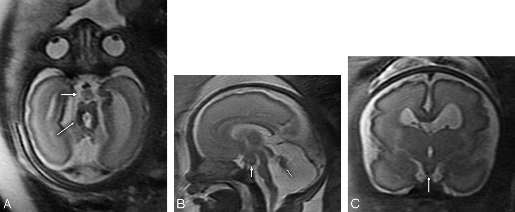
Fetal T2-weighted MR imaging at 29 gestational weeks. A, Axial MR image showing hypoplasia of both cerebellar hemispheres, the characteristic MTS (thin arrow) with thickened and elongated superior cerebellar peduncles and an abnormally deep interpeduncular fossa, and an HH (thick arrow). B, Sagittal MR image revealing significant hypoplasia of the cerebellar vermis (arrow), an enlarged fourth ventricle and posterior fossa, an HH (white arrow), and a thin brain stem. C, Coronal MR image demonstrating an HH (white arrow).

Fig 2.
  • Download figure
  • Open in new tab
  • Download powerpoint
Fig 2.

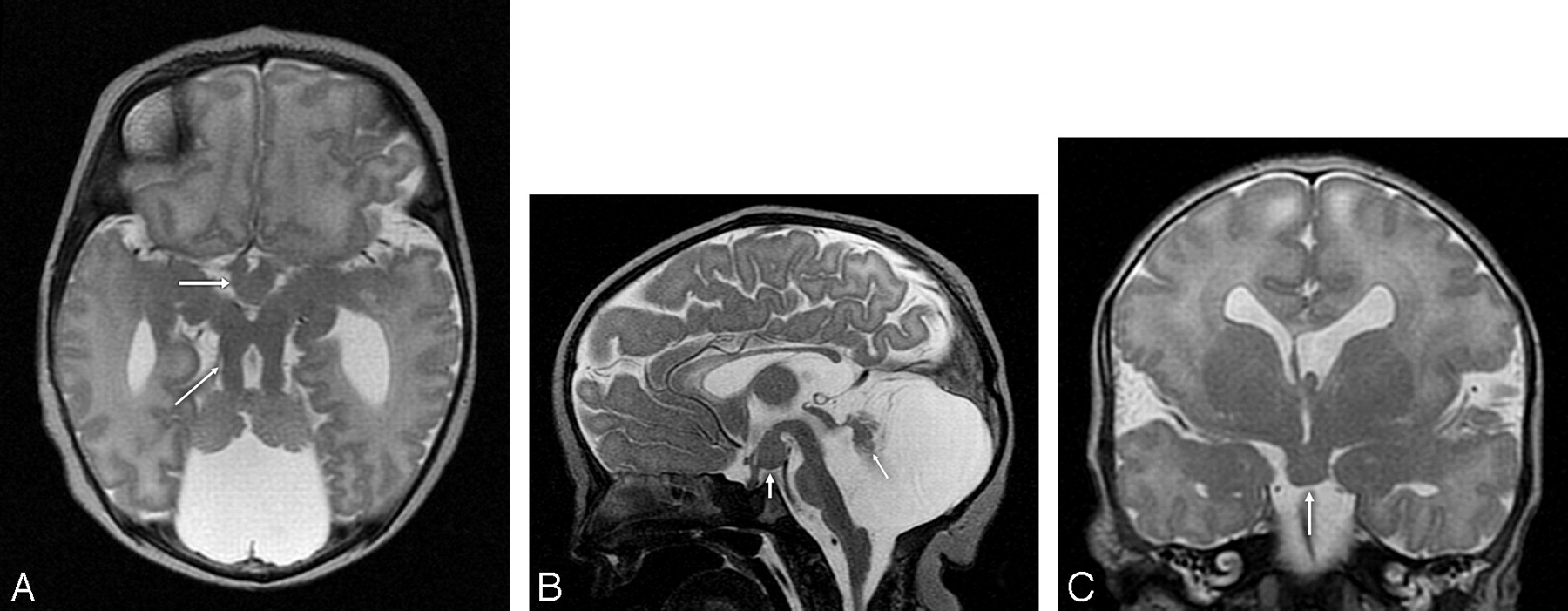
T2-weighted MR imaging at 2 days of age. A, Axial MR image showing hypoplasia of the vermis and both cerebellar hemispheres, the characteristic MTS (thin arrow) with thickened and elongated superior cerebellar peduncles, an abnormally deep interpeduncular fossa, and an HH (thick arrow). B, Sagittal MR image revealing significant hypoplasia of the cerebellar vermis and dysplasia of the remnants of the cerebellar vermis (arrow), an enlarged fourth ventricle and posterior fossa, and an HH (white arrow). In addition, the corpus callosum and the brain stem are hypoplastic, the pituitary stalk is thick, and the interthalamic adhesion is large. C, Coronal MR image demonstrating an HH (arrow), a missing left leaf of the septum pellucidum, a bulky left fornix, and a probably dysplastic cerebral cortex in the left Sylvian fissure.

Discussion

Fetal MR imaging can be very helpful in the diagnosis of posterior fossa abnormalities9 and permits superior discrimination and resolution of the different anatomic structures compared with sonography. Doherty et al8 reported prenatal MR findings of a fetus with a risk for JSRD at 21 weeks' gestation. MR imaging revealed a very small and rostrally located vermis and, inferiorly, a connection between the fourth ventricle and fossa posterior. However, a typical MTS was not seen. Fluss et al10 reported a prenatal diagnosis of JS at 27 weeks' gestation with identification of a typical MTS. In the case of our patient, the MTS was also identifiable at 29 weeks' gestation. Therefore, fetal MR imaging can identify the MTS from 27 weeks' gestation.

Two articles reported prenatal sonography findings of OFDS VI such as polydactyly and abnormalities in the posterior fossa, but prenatal MR imaging was not performed.11,12 Prenatal MR findings of OFDS VI have not been reported previously. Neuroimaging features of OFDS VI include cerebellar vermis hypoplasia and the MTS, whereas HH is occasionally found. However, most cases of HH are sporadic, and in approximately 5% of cases, HH is a finding of Pallister-Hall syndrome.13 On MR imaging, HH appears isointense compared with gray matter on T1-weighted sequences and isointense to slightly hyperintense on T2-weighted sequences.14 Prenatal diagnosis of nonsyndromic HH had been reported in 1 patient at 26 weeks' gestation.15 In the case of our patient, HH was identifiable at 29 weeks' gestation.

In conclusion, a prenatal diagnosis of OFDS VI is possible with fetal MR imaging from 29 weeks' gestation. Therefore, if prenatal MR imaging shows findings compatible with JSRD, an HH should be sought. However, because of the rarity of the disease, a prenatal diagnosis of OFDS VI can be easily missed. More cases have to be identified to further characterize the chronologic appearance of the neuroimaging features of OFDS VI.

Acknowledgments

We thank Beth Padden, MD, for help in editing the manuscript. Dr. Poretti was financially supported by a donation from the United Bank of Switzerland. This donation was made at the request of an anonymous client.

References

  1. ↵
    Váradi V, Szabo L, Papp Z. Syndrome of polydactyly, cleft lip/palate or lingual lump, and psychomotor retardation in endogamic gypsies. J Med Genet 1980;17:119–22
    Abstract/FREE Full Text
  2. ↵
    Munke M, McDonald DM, Cronister A, et al. Oral-facial-digital syndrome type VI (Váradi syndrome): further clinical delineation. Am J Med Genet 1990;35:360–69
    CrossRefPubMed
  3. ↵
    Parisi MA, Doherty D, Chance PF, et al. Joubert syndrome (and related disorders) (OMIM 213300). Eur J Hum Genet 2007;15:511–21
    CrossRefPubMed
  4. ↵
    Maria BL, Boltshauser E, Palmer SC, et al. Clinical features and revised diagnostic criteria in Joubert syndrome. J Child Neurol 1999;14:583–90
    Abstract/FREE Full Text
  5. ↵
    Braddock SR, Henley KM, Maria BL. The face of Joubert syndrome: a study of dysmorphology and anthropometry. Am J Med Genet A 2007;143:3235–42
  6. ↵
    Gleeson JG, Keeler LC, Parisi MA, et al. Molar tooth sign of the midbrain-hindbrain junction: occurrence in multiple distinct syndromes. Am J Med Genet A 2004;125:125–34
  7. ↵
    Stephan MJ, Brooks KL, Moore DC, et al. Hypothalamic hamartoma in oral-facial-digital syndrome type VI (Váradi syndrome). Am J Med Genet 1994;51:131–36
    CrossRefPubMed
  8. ↵
    Doherty D, Glass IA, Siebert JR, et al. Prenatal diagnosis in pregnancies at risk for Joubert syndrome by ultrasound and MRI. Prenat Diagn 2005;25:442–47
    CrossRefPubMed
  9. ↵
    Adamsbaum C, Moutard ML, Andre C, et al. MRI of the fetal posterior fossa. Pediatr Radiol 2005;35:124–40
    CrossRefPubMed
  10. ↵
    Fluss J, Blaser S, Chitayat D, et al. Molar tooth sign in fetal brain magnetic resonance imaging leading to the prenatal diagnosis of Joubert syndrome and related disorders. J Child Neurol 2006;21:320–24
    Abstract/FREE Full Text
  11. ↵
    Guven MA, Ceylaner S, Prefumo F, et al. Prenatal sonographic findings in a case of Varadi-Papp syndrome. Prenat Diagn 2004;24:989–91
    CrossRefPubMed
  12. ↵
    McPherson E, Zaleski C, Mascola M. Prenatal diagnosis of episodic tachypnea in an infant with OFD VI. Am J Med Genet A 2006;140:2146–49
    PubMed
  13. ↵
    Kang S, Graham JM, Jr., Olney AH, Biesecker LG. GLI3 frameshift mutations cause autosomal dominant Pallister-Hall syndrome. Nat Genet 1997;15:266–68
    CrossRefPubMed
  14. ↵
    Freeman JL, Coleman LT, Wellard RM, et al. MR imaging and spectroscopic study of epileptogenic hypothalamic hamartomas: analysis of 72 cases. AJNR Am J Neuroradiol 2004;25:450–62
    Abstract/FREE Full Text
  15. ↵
    Booth TN, Timmons C, Shapiro K, et al. Pre- and postnatal MR imaging of hypothalamic hamartomas associated with arachnoid cysts. AJNR Am J Neuroradiol 2004;25:1283–85
    Abstract/FREE Full Text
  • Received December 14, 2007.
  • Accepted after revision December 30, 2007.
  • Copyright © American Society of Neuroradiology
View Abstract
PreviousNext
Back to top

In this issue

American Journal of Neuroradiology: 29 (6)
American Journal of Neuroradiology
Vol. 29, Issue 6
June 2008
  • Table of Contents
  • Index by author
Advertisement
Print
Download PDF
Email Article

Thank you for your interest in spreading the word on American Journal of Neuroradiology.

NOTE: We only request your email address so that the person you are recommending the page to knows that you wanted them to see it, and that it is not junk mail. We do not capture any email address.

Enter multiple addresses on separate lines or separate them with commas.
Prenatal and Neonatal MR Imaging Findings in Oral-Facial-Digital Syndrome Type VI
(Your Name) has sent you a message from American Journal of Neuroradiology
(Your Name) thought you would like to see the American Journal of Neuroradiology web site.
CAPTCHA
This question is for testing whether or not you are a human visitor and to prevent automated spam submissions.
Cite this article
A. Poretti, U. Brehmer, I. Scheer, V. Bernet, E. Boltshauser
Prenatal and Neonatal MR Imaging Findings in Oral-Facial-Digital Syndrome Type VI
American Journal of Neuroradiology Jun 2008, 29 (6) 1090-1091; DOI: 10.3174/ajnr.A1038

Citation Manager Formats

  • BibTeX
  • Bookends
  • EasyBib
  • EndNote (tagged)
  • EndNote 8 (xml)
  • Medlars
  • Mendeley
  • Papers
  • RefWorks Tagged
  • Ref Manager
  • RIS
  • Zotero
0 Responses
Respond to this article
Share
Bookmark this article
Prenatal and Neonatal MR Imaging Findings in Oral-Facial-Digital Syndrome Type VI
A. Poretti, U. Brehmer, I. Scheer, V. Bernet, E. Boltshauser
American Journal of Neuroradiology Jun 2008, 29 (6) 1090-1091; DOI: 10.3174/ajnr.A1038
del.icio.us logo Twitter logo Facebook logo Mendeley logo
  • Tweet Widget
  • Facebook Like
  • Google Plus One
Purchase

Jump to section

  • Article
    • Abstract
    • Case Report
    • Discussion
    • Acknowledgments
    • References
  • Figures & Data
  • Info & Metrics
  • Responses
  • References
  • PDF

Related Articles

  • No related articles found.
  • PubMed
  • Google Scholar

Cited By...

  • Joubert syndrome: neuroimaging findings in 110 patients in correlation with cognitive function and genetic cause
  • Fifteen years of research on oral-facial-digital syndromes: from 1 to 16 causal genes
  • Using the avian mutant talpid2 as a disease model for understanding the oral-facial phenotypes of oral-facial-digital syndrome
  • Joubert Syndrome and Related Disorders: Spectrum of Neuroimaging Findings in 75 Patients
  • Interpeduncular Heterotopia in Joubert Syndrome: A Previously Undescribed MR Finding
  • Malformations of Cerebral Cortical Development in Oral-Facial-Digital Syndrome Type VI
  • Crossref (33)
  • Google Scholar

This article has been cited by the following articles in journals that are participating in Crossref Cited-by Linking.

  • Primary cilia in neurodevelopmental disorders
    Enza Maria Valente, Rasim O. Rosti, Elizabeth Gibbs, Joseph G. Gleeson
    Nature Reviews Neurology 2014 10 1
  • Joubert Syndrome and Related Disorders: Spectrum of Neuroimaging Findings in 75 Patients
    A. Poretti, T.A.G.M. Huisman, I. Scheer, E. Boltshauser
    American Journal of Neuroradiology 2011 32 8
  • Fifteen years of research on oral–facial–digital syndromes: from 1 to 16 causal genes
    Ange-Line Bruel, Brunella Franco, Yannis Duffourd, Julien Thevenon, Laurence Jego, Estelle Lopez, Jean-François Deleuze, Diane Doummar, Rachel H Giles, Colin A Johnson, Martijn A Huynen, Véronique Chevrier, Lydie Burglen, Manuela Morleo, Isabelle Desguerres, Geneviève Pierquin, Bérénice Doray, Brigitte Gilbert-Dussardier, Bruno Reversade, Elisabeth Steichen-Gersdorf, Clarisse Baumann, Inusha Panigrahi, Anne Fargeot-Espaliat, Anne Dieux, Albert David, Alice Goldenberg, Ernie Bongers, Dominique Gaillard, Jesús Argente, Bernard Aral, Nadège Gigot, Judith St-Onge, Daniel Birnbaum, Shubha R Phadke, Valérie Cormier-Daire, Thibaut Eguether, Gregory J Pazour, Vicente Herranz-Pérez, Jaclyn S Goldstein, Laurent Pasquier, Philippe Loget, Sophie Saunier, André Mégarbané, Olivier Rosnet, Michel R Leroux, John B Wallingford, Oliver E Blacque, Maxence V Nachury, Tania Attie-Bitach, Jean-Baptiste Rivière, Laurence Faivre, Christel Thauvin-Robinet
    Journal of Medical Genetics 2017 54 6
  • Joubert syndrome: brain and spinal cord malformations in genotyped cases and implications for neurodevelopmental functions of primary cilia
    Gordana Juric-Sekhar, Jonathan Adkins, Dan Doherty, Robert F. Hevner
    Acta Neuropathologica 2012 123 5
  • C5orf42 is the major gene responsible for OFD syndrome type VI
    Estelle Lopez, Christel Thauvin-Robinet, Bruno Reversade, Nadia El Khartoufi, Louise Devisme, Muriel Holder, Hélène Ansart-Franquet, Magali Avila, Didier Lacombe, Pascale Kleinfinger, Irahara Kaori, Jun-Ichi Takanashi, Martine Le Merrer, Jelena Martinovic, Catherine Noël, Mohammad Shboul, Lena Ho, Yeliz Güven, Ferechté Razavi, Lydie Burglen, Nadège Gigot, Véronique Darmency-Stamboul, Julien Thevenon, Bernard Aral, Hülya Kayserili, Frédéric Huet, Stanislas Lyonnet, Cédric Le Caignec, Brunella Franco, Jean-Baptiste Rivière, Laurence Faivre, Tania Attié-Bitach
    Human Genetics 2014 133 3
  • Mutations in ARMC9, which Encodes a Basal Body Protein, Cause Joubert Syndrome in Humans and Ciliopathy Phenotypes in Zebrafish
    Julie C. Van De Weghe, Tamara D.S. Rusterholz, Brooke Latour, Megan E. Grout, Kimberly A. Aldinger, Ranad Shaheen, Jennifer C. Dempsey, Sateesh Maddirevula, Yong-Han H. Cheng, Ian G. Phelps, Matthias Gesemann, Himanshu Goel, Ohad S. Birk, Talal Alanzi, Rifaat Rawashdeh, Arif O. Khan, Michael J. Bamshad, Deborah A. Nickerson, Stephan C.F. Neuhauss, William B. Dobyns, Fowzan S. Alkuraya, Ronald Roepman, Ruxandra Bachmann-Gagescu, Dan Doherty
    The American Journal of Human Genetics 2017 101 1
  • Diffusion tensor imaging and fiber tractography in brain malformations
    Andrea Poretti, Avner Meoded, Andrea Rossi, Charles Raybaud, Thierry A. G. M. Huisman
    Pediatric Radiology 2013 43 1
  • Delineation and Diagnostic Criteria of Oral-Facial-Digital Syndrome Type VI
    Andrea Poretti, Giuseppina Vitiello, Raoul CM Hennekam, Filippo Arrigoni, Enrico Bertini, Renato Borgatti, Francesco Brancati, Stefano D'Arrigo, Francesca Faravelli, Lucio Giordano, Thierry AGM Huisman, Miriam Iannicelli, Gerhard Kluger, Marten Kyllerman, Magnus Landgren, Melissa M Lees, Lorenzo Pinelli, Romina Romaniello, Ianina Scheer, Christoph E Schwarz, Ronen Spiegel, Daniel Tibussek, Enza Maria Valente, Eugen Boltshauser
    Orphanet Journal of Rare Diseases 2012 7 1
  • Hypothalamic hamartomas. Part 1. Clinical, neuroimaging, and neurophysiological characteristics
    Sandeep Mittal, Monika Mittal, José Luis Montes, Jean-Pierre Farmer, Frederick Andermann
    Neurosurgical Focus 2013 34 6
  • Joubert syndrome: neuroimaging findings in 110 patients in correlation with cognitive function and genetic cause
    Andrea Poretti, Joseph Snow, Angela C Summers, Aylin Tekes, Thierry A G M Huisman, Nafi Aygun, Kathryn A Carson, Dan Doherty, Melissa A Parisi, Camilo Toro, Deniz Yildirimli, Meghana Vemulapalli, Jim C Mullikin, Andrew R Cullinane, Thierry Vilboux, William A Gahl, Meral Gunay-Aygun
    Journal of Medical Genetics 2017 54 8

More in this TOC Section

  • Comparison of Image Quality and Radiation Dose in Pediatric Temporal Bone CT Using Photon-Counting Detector CT and Energy-Integrating Detector CT
  • SyMRI & MR Fingerprinting in Brainstem Myelination
  • Dual-Layer Detector CT for PEDS Image Quality
Show more Pediatrics

Similar Articles

Advertisement

Indexed Content

  • Current Issue
  • Accepted Manuscripts
  • Article Preview
  • Past Issues
  • Editorials
  • Editors Choice
  • Fellow Journal Club
  • Letters to the Editor

Cases

  • Case Collection
  • Archive - Case of the Week
  • Archive - Case of the Month
  • Archive - Classic Case

Special Collections

  • Special Collections

Resources

  • News and Updates
  • Turn around Times
  • Submit a Manuscript
  • Author Policies
  • Manuscript Submission Guidelines
  • Evidence-Based Medicine Level Guide
  • Publishing Checklists
  • Graphical Abstract Preparation
  • Imaging Protocol Submission
  • Submit a Case
  • Become a Reviewer/Academy of Reviewers
  • Get Peer Review Credit from Publons

Multimedia

  • AJNR Podcast
  • AJNR SCANtastic
  • Video Articles

About Us

  • About AJNR
  • Editorial Board
  • Not an AJNR Subscriber? Join Now
  • Alerts
  • Feedback
  • Advertise with us
  • Librarian Resources
  • Permissions
  • Terms and Conditions

American Society of Neuroradiology

  • Not an ASNR Member? Join Now

© 2025 by the American Society of Neuroradiology All rights, including for text and data mining, AI training, and similar technologies, are reserved.
Print ISSN: 0195-6108 Online ISSN: 1936-959X

Powered by HighWire