
Article Figures & Data
Figures
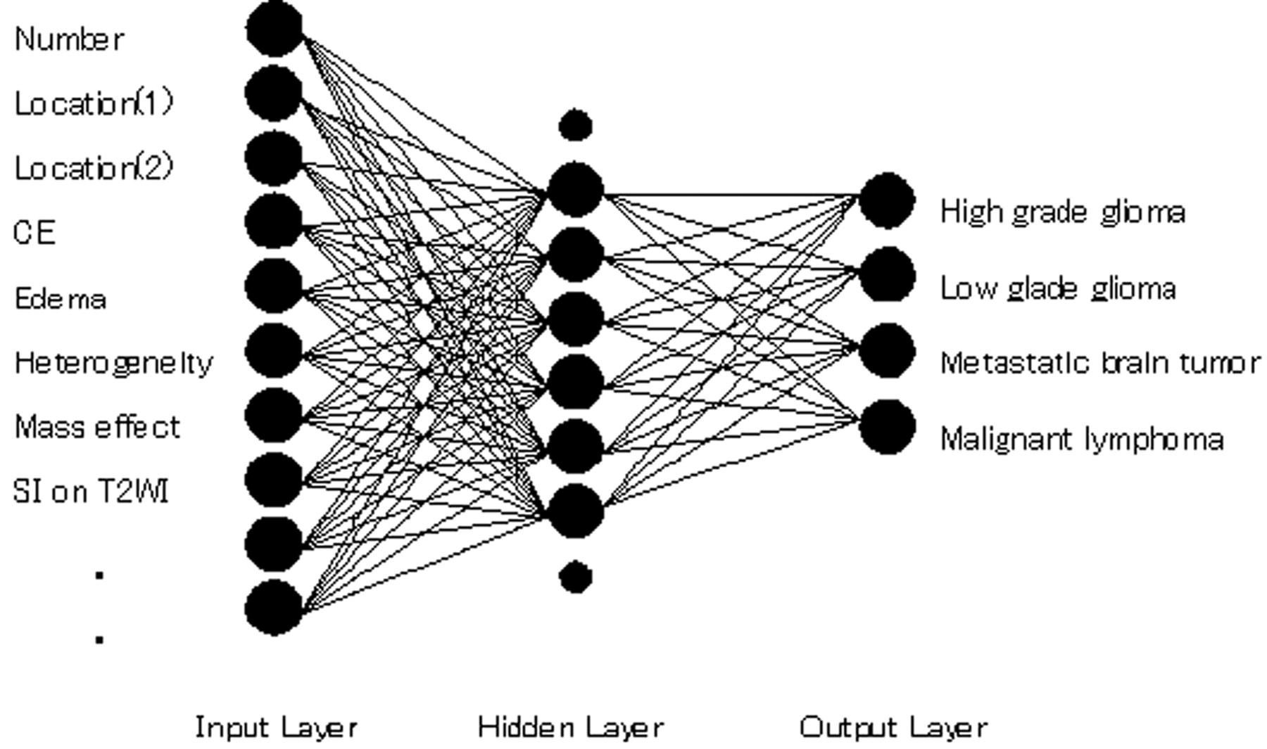
- Fig 1.
Diagram of the basic structure of the ANN. Although only 10 input units and 8 hidden units are shown for illustration, the ANN consists of 15 input units and 9 hidden units.
- Fig 2.
MR images of 2 actual cases. A, Case 1: MR images of a 44-year-old woman with a glioblastoma confirmed on pathologic examination (WHO grade IV). Left image: T2WI shows a heterogeneously hyperintense mass with central necrosis and surrounding signal intensity abnormality likely related to tumor extension and edema. Middle and right images: Precontrast and postcontrast T1WIs show hemorrhagic mass and peripheral enhancement with central necrosis, characteristic of glioblastoma. B, Case 2: MR images of a 62-year-old woman with proved metastatic brain tumor from lung cancer. Left image: T2WI shows a cystic frontoparietal mass with mixed-aged hemorrhage. Middle and right images: Precontrast and postcontrast T1WIs show a thin layer of peripheral enhancement. Surgery disclosed adenocarcinoma.
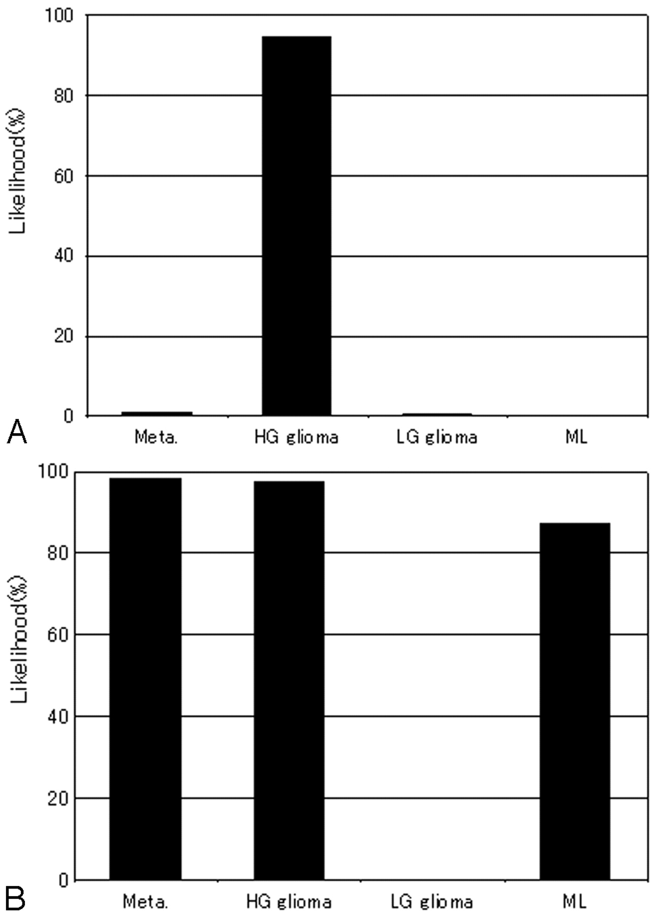
- Fig 3.
ANN output obtained on the basis of 2 radiologists' ratings of MR features and clinical information for the 2 cases shown in Fig 2. Each graph shows the largest output values among the 4 categories corresponding to the correct diagnoses. A, Case 1: The likelihood of high-grade glioma is very high. ANN led to the correct diagnosis. B, Case 2: The likelihood of metastasis is approximately equivalent to high-grade glioma and malignant lymphoma. ANN might fail to lead to the correct diagnosis.
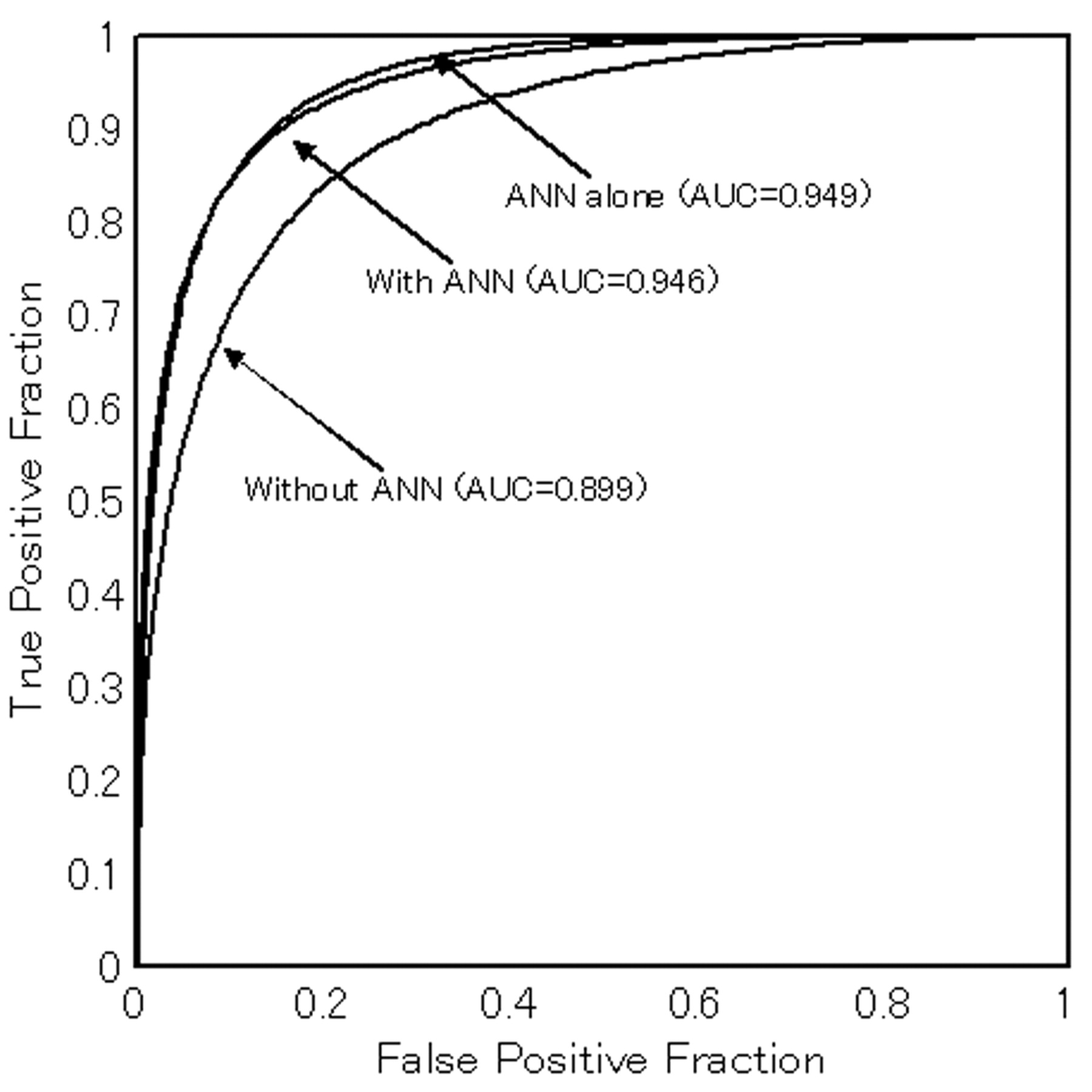
- Fig 4.
Average AUC values and binormal ROC curves for observers with and without ANN output (averaged plot values for all readers). Those for ANN alone are also indicated. Note that observer performance improves significantly with ANN output.
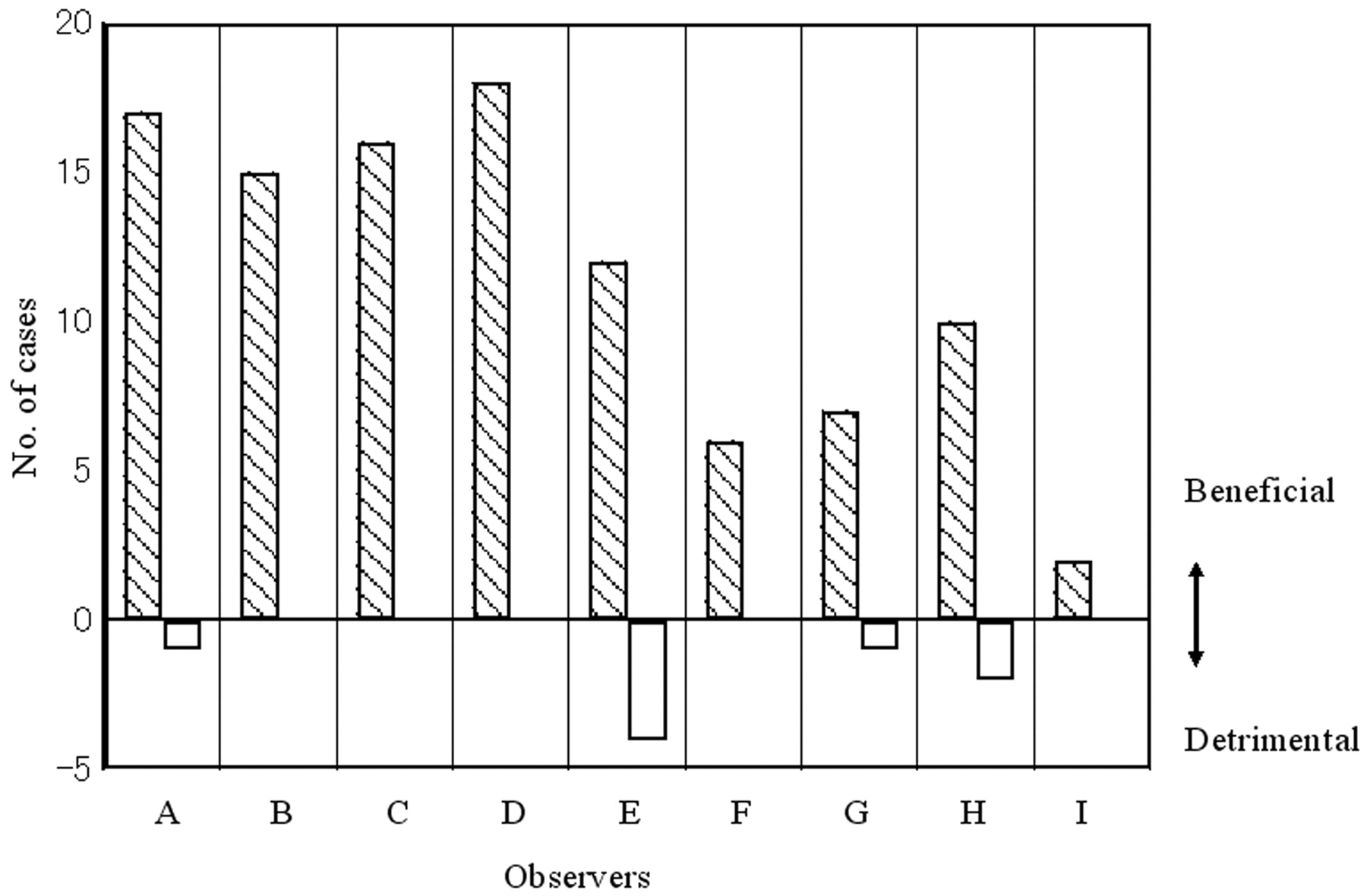
- Fig 5.
The number of correctly diagnosed cases for which observers' rankings changed because of ANN output. Positive values indicate that ANN was beneficial, whereas negative values indicate that ANN was detrimental. ANN output clearly improved the performance.


Tables
Rating score −0.9 −0.45 0 0.45 0.9 Age (y) 0–40 41–60 61– Location (1) Frontal, parietal, or temporal lobe Other areas Location (2) Cortical layer Subcortical white matter Other areas History of malignancy (−) (+) Number 1 2 3 < SI on T2WI CSF CSF >, gray matter < Gray matter White matter White matter > Edema Mild Moderate Marked Heterogeneity Mild Moderate Marked Hemorrhage (−) Equivocal (+) Border definition Infiltrative Poorly circumscribed Well circumscribed Mass effect Mild Moderate Marked CE (−) Mild Marked Ring enhancement (−) Equivocal (+) Tumor extent Localized region Intermediate Extensive Cyst formation (−) Equivocal (+) Note:—SI indicates signal intensity; T2WI, T2-weighted image; CE, contrast enhancement.
- Table 2:
AUC values for diagnostic accuracy of 9 radiologists without and with output of ANN
Observer Without ANN With ANN Difference P* Precertification radiologists A 0.891 0.945 0.054 < .001 B 0.840 0.938 0.098 < .001 C 0.850 0.950 0.099 < .001 D 0.897 0.947 0.057 < .001 Average 0.870 0.947 0.077 Board certified radiologists E 0.935 0.972 0.037 < .001 F 0.887 0.915 0.028 < .001 G 0.965 0.979 0.015 < .001 H 0.917 0.940 0.023 < .001 I 0.911 0.922 0.010 < .001 Average 0.923 0.946 0.023 Overall average 0.899 0.946 0.047 < .001 Note:—AUC indicates area under the curve; ANN, artificial neural network;.
* Statistically significant with jackknife method by use of DBM MRMC (multiple readers and multiple cases algorithm developed by Metz et al12–16).
- Table 3:
Sensitivity, specificity, and accuracy of 9 radiologists without and with output of ANN
Observer Sensitivity (%) Specificity (%) Accuracy (%) Without ANN With ANN P* Without ANN With ANN P* Without ANN With ANN P* Precertification radiologists A 79.4 87.3 89.4 94.2 86.9 92.4 B 73.0 85.7 87.6 91.8 83.9 90.3 C 74.6 88.1 87.8 93.7 84.5 92.3 D 75.4 88.9 91.0 95.0 87.1 93.5 Average 75.6 87.5 <.005 89.0 93.7 <.005 85.6 92.1 <.005 Board certified radiologists E 81.7 92.1 92.6 95.5 89.9 94.6 F 71.4 80.2 87.0 90.7 83.1 88.1 G 88.9 93.7 93.4 94.2 92.3 94.0 H 84.1 88.9 88.6 94.2 87.5 92.9 I 77.8 79.4 89.9 90.7 86.9 87.9 Average 80.8 86.8 0.19 90.3 93.1 0.11 87.9 91.5 0.13 Overall average 78.5 87.1 <.005 89.7 93.3 <.005 86.9 91.8 <.005 Note:—ANN indicates artificial neural network.
* Student t test for paired data.


