
Article Figures & Data
Figures
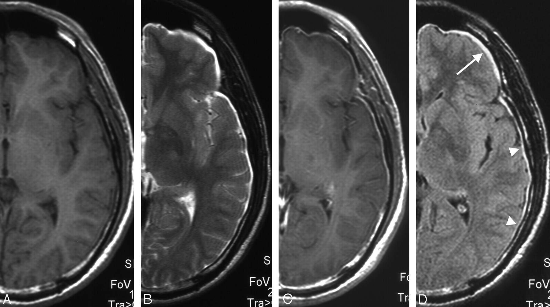
- Fig 1.
Patient 5. Initial axial, half cut, and magnified MR images of a 39-year-old man with spontaneous intracranial hypotension who presented with a 1-month history of orthostatic headache, nausea, vomiting, and diplopia. Supportive treatment resolved the symptoms. A, T1-weighted image showing diffusely thickened dura mater as isointense. Subdural lesions are unclear. B, T2-weighted image showing that bilateral subdural effusion/hematomas could not be discriminated from CSF. C, T1-weighted image with gadolinium clearly showing diffuse pachymeningeal enhancement. D, FLAIR image showing diffuse pachymeningeal hyperintensity (arrowheads) and very thin bilateral subdural effusion/hematomas in the frontal region (arrow).
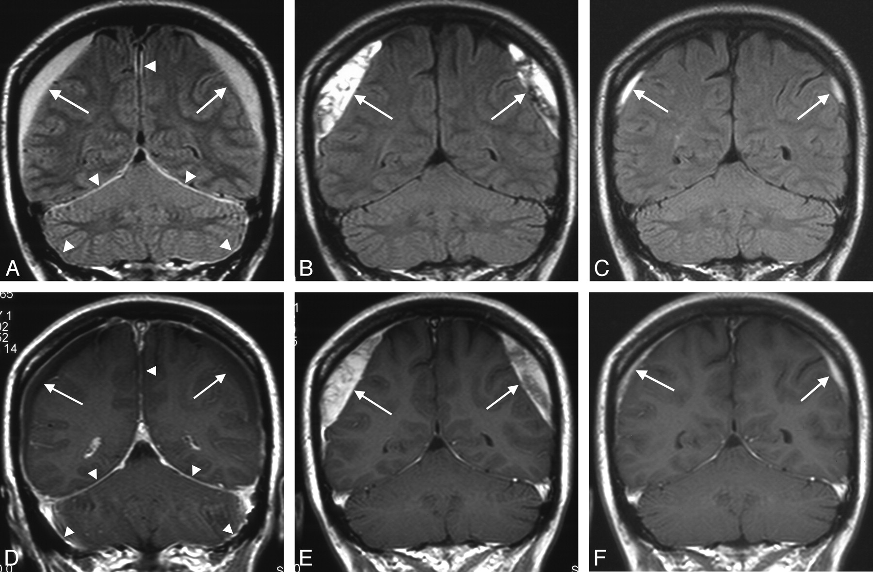
- Fig 2.
Patient 4. Chronologic FLAIR and T1-weighted MR images of a 37-year-old woman with spontaneous intracranial hypotension. She had a 1-month history of orthostatic headache. Cervical epidural blood patch resolved the symptoms. A, Initial FLAIR image showing diffuse pachymeningeal hyperintensity at the falx, tentorium, and dura of the posterior fossa (arrowheads) and bilateral thick subdural effusion/hematomas (arrows). B, Second FLAIR image performed 1 month after effective blood patch showing disappearance of the diffuse pachymeningeal hyperintensity but thickening of the subdural effusion/hematomas (arrows). C, Follow-up FLAIR image showing remnant subdural effusion/hematomas (arrows). D, Initial T1-weighted image with gadolinium showing DPME at the falx, tentorium, and dura of the posterior fossa (arrowheads) and bilateral thick subdural effusion/hematomas (arrows). E, Second T1-weighted image taken 1 month after completely effective blood patch showing slight remnant of DPME and thickening of the subdural effusion/hematomas (arrows). F, Follow-up T1-weighted image showing remnant subdural effusion/hematomas (arrows).
- Fig 3.
Patient 8. Chronologic FLAIR and T1-weighted MR images of a 47-year-old woman with spontaneous intracranial hypotension. She had a 1-week history of orthostatic headache. Lumbar epidural blood patch resolved the symptoms. A, Initial FLAIR image showing diffuse pachymeningeal hyperintensity (arrowheads). B, Second FLAIR image taken 1 week after completely effective blood patch showing thickened diffuse pachymeningeal hyperintensity (arrowheads) and partial subdural effusion/hematomas (arrows). C, Follow-up FLAIR image showing disappearance of the diffuse pachymeningeal hyperintensity and subdural effusion/hematomas. D, Initial T1-weighted image with gadolinium showing DPME (arrowheads). E, Second T1-weighted image taken 1 week after effective blood patch showing disappearance of DPME (arrowheads) and partial subdural effusion/hematomas (arrows). F, Follow-up T1-weighted image showing disappearance of the DPME and subdural effusion/hematomas.


Tables
- Table 1:
Distribution of diffuse pachymeningeal hyperintensity on FLAIR imaging and/or bilateral subdural effusion/hematomas in patients with spontaneous intracranial hypotension
No./Age (y)/Sex Diffuse Pachymeningeal Hyperintensity on FLAIR Imaging Subdural Effusion/Hematomas 1/36/F bil. OC, falx bil. OC 2/59/M bil. OC, falx, tentorium, PFC bil. OC 3/37/F bil. OC, falx, tentorium, PFC bil. frontal convexity 4/37/F bil. OC, falx, tentorium, PFC bil. frontoparietal convexity 5/39/M bil. OC, falx, tentorium, PFC bil. frontal convexity 6/45/F bil. OC ND 7/48/F bil. OC, falx, tentorium, PFC bil. OC 8/47/F bil. OC, falx bil. OC* Note:—OC indicates over the supratentorial convexity; PFC, posterior fossa convexity; ND, not detectable; bil., bilateral.
* Subdural effusion/hematomas were not found in initial study but were observed in the second study.
- Table 2:
Summary of changes in MR imaging appearance in patients with spontaneous intracranial hypotension
Pt. No. DPME DPMHF Maximum Thickness of Subdural Effusion/Hematomas Initial Second Follow-Up Initial Second Follow-Up Initial Second Follow-Up 1 Yes Yes Slightly Yes Yes NP bil. thick bil. very thick ND 2 Yes NP† NP Yes NP† ND thick/thin* NP† ND 3 Yes NP ND Yes Slightly ND bil. thin bil. thin ND 4 Yes Slightly Slightly Yes ND ND bil. thick bil. very thick bil. thin 5 Yes Slightly ND Yes ND ND bil. very thin bil. very thin ND 6 Yes Slightly ND Yes ND ND ND (DPMHF) ND ND 7 Yes Remain Slightly Yes Remain ND thick/thin* thick/ND (DPMHF)* thin/ND* 8 Yes Yes Slightly Yes Yes ND ND (DPMHF) bil. very thin ND Note:—DPME indicates diffuse pachymeningeal enhancement; DPMHF, diffuse pachymeningeal hyperintensity on FLAIR imaging; NP, not performed; ND, not detectable; Slightly, slightly remain; bil., bilateral; very thin, ≤3 mm; thin, >3 mm–≤6 mm; thick, >6–≤10 mm; very thick, >10 mm.
* Left side/right side.
† Only in patient 2, second study was performed after strict bed rest but before successful epidural blood patch.
In this issue


Jump to section
Related Articles
Cited By...
- MRI Findings after Recent Image-Guided Lumbar Puncture: The Rate of Dural Enhancement and Subdural Collections
- Delayed diagnosis of bilateral subdural effusions complicating intracranial hypotension in a patient presenting with post lumbar puncture headache
- Postoperative Intraspinal Subdural Collections after Pediatric Posterior Fossa Tumor Resection: Incidence, Imaging, and Clinical Features
- Reply:
- Dural Hyperintensity on Fluid-Attenuated Inversion Recovery in Spontaneous Intracranial Hypotension