Advertisement

Main menu

  • Home
  • Content
    • Current Issue
    • Accepted Manuscripts
    • Article Preview
    • Past Issue Archive
    • AJNR Case Collection
    • Case of the Week Archive
    • Classic Case Archive
    • Case of the Month Archive
  • Special Collections
    • Spinal CSF Leak Articles (Jan 2020-June 2024)
    • 2024 AJNR Journal Awards
    • Most Impactful AJNR Articles
  • Multimedia
    • AJNR Podcast
    • AJNR Scantastics
    • Video Articles
  • For Authors
    • Submit a Manuscript
    • Author Policies
    • Fast publishing of Accepted Manuscripts
    • Graphical Abstract Preparation
    • Manuscript Submission Guidelines
    • Imaging Protocol Submission
    • Submit a Case for the Case Collection
  • About Us
    • About AJNR
    • Editorial Board
  • More
    • Become a Reviewer/Academy of Reviewers
    • Subscribers
    • Permissions
    • Alerts
    • Feedback
    • Advertisers
    • ASNR Home
  • Other Publications
    • ajnr

User menu

  • Alerts
  • Log in

Search

  • Advanced search
American Journal of Neuroradiology

American Journal of Neuroradiology

ASHNR American Society of Functional Neuroradiology ASHNR American Society of Pediatric Neuroradiology ASSR
  • Alerts
  • Log in

Advanced Search

  • Home
  • Content
    • Current Issue
    • Accepted Manuscripts
    • Article Preview
    • Past Issue Archive
    • AJNR Case Collection
    • Case of the Week Archive
    • Classic Case Archive
    • Case of the Month Archive
  • Special Collections
    • Spinal CSF Leak Articles (Jan 2020-June 2024)
    • 2024 AJNR Journal Awards
    • Most Impactful AJNR Articles
  • Multimedia
    • AJNR Podcast
    • AJNR Scantastics
    • Video Articles
  • For Authors
    • Submit a Manuscript
    • Author Policies
    • Fast publishing of Accepted Manuscripts
    • Graphical Abstract Preparation
    • Manuscript Submission Guidelines
    • Imaging Protocol Submission
    • Submit a Case for the Case Collection
  • About Us
    • About AJNR
    • Editorial Board
  • More
    • Become a Reviewer/Academy of Reviewers
    • Subscribers
    • Permissions
    • Alerts
    • Feedback
    • Advertisers
    • ASNR Home
  • Follow AJNR on Twitter
  • Visit AJNR on Facebook
  • Follow AJNR on Instagram
  • Join AJNR on LinkedIn
  • RSS Feeds

Welcome to the new AJNR, Updated Hall of Fame, and more. Read the full announcements.


AJNR is seeking candidates for the position of Associate Section Editor, AJNR Case Collection. Read the full announcement.


Research ArticleHEAD & NECK

MR Imaging of Nonmalignant Polyps and Masses of the Nasopharynx and Sphenoid Sinus after Radiotherapy for Nasopharyngeal Carcinoma

A.D. King, A.T. Ahuja, S.-F. Leung, J. Abrigo, J.K.T. Wong, W.S. Poon, K.S. Woo, H.S. Chan and G.M.K. Tse
American Journal of Neuroradiology June 2008, 29 (6) 1209-1214; DOI: https://doi.org/10.3174/ajnr.A1043
A.D. King
  • Find this author on Google Scholar
  • Find this author on PubMed
  • Search for this author on this site
A.T. Ahuja
  • Find this author on Google Scholar
  • Find this author on PubMed
  • Search for this author on this site
S.-F. Leung
  • Find this author on Google Scholar
  • Find this author on PubMed
  • Search for this author on this site
J. Abrigo
  • Find this author on Google Scholar
  • Find this author on PubMed
  • Search for this author on this site
J.K.T. Wong
  • Find this author on Google Scholar
  • Find this author on PubMed
  • Search for this author on this site
W.S. Poon
  • Find this author on Google Scholar
  • Find this author on PubMed
  • Search for this author on this site
K.S. Woo
  • Find this author on Google Scholar
  • Find this author on PubMed
  • Search for this author on this site
H.S. Chan
  • Find this author on Google Scholar
  • Find this author on PubMed
  • Search for this author on this site
G.M.K. Tse
  • Find this author on Google Scholar
  • Find this author on PubMed
  • Search for this author on this site
  • Article
  • Figures & Data
  • Supplemental
  • Info & Metrics
  • Responses
  • References
  • PDF
Loading

Article Figures & Data

Figures

  • Tables
  • Fig 1.
    • Download figure
    • Open in new tab
    • Download powerpoint
    Fig 1.

    Axial T1-weighted postcontrast MR image in a 44-year-old man with a small contrast-enhancing polyp (arrow) arising from the posterior wall of the lower nasopharynx 11 years after chemoradiotherapy. Posttreatment scarring is present in the left lateral nasopharyngeal wall, partially effacing the parapharyngeal fat, and around both carotid sheaths.

  • Fig 2.
    • Download figure
    • Open in new tab
    • Download powerpoint
    Fig 2.

    Coronal T1-weighted postcontrast MR image in a 71-year-old man, 11 years after radiation therapy, with a contrast enhancing polyp (arrow) with less enhancing stellate areas centrally, arising at the junction of the sphenoid sinus and roof of the nasopharynx where there is an osteoradionecrotic bony defect.

  • Fig 3.
    • Download figure
    • Open in new tab
    • Download powerpoint
    Fig 3.

    A, Coronal and (B) axial T1-weighted postcontrast MR image in a 40-year-old man with a 5-cm rapidly growing contrast-enhancing polyp with a more central area of reduced contrast enhancement radiating to the periphery, 2 years after treatment with conventional radiation therapy plus a stereotactic radiation therapy boost. Inflammatory changes and retained secretions are present in the sphenoid sinus.
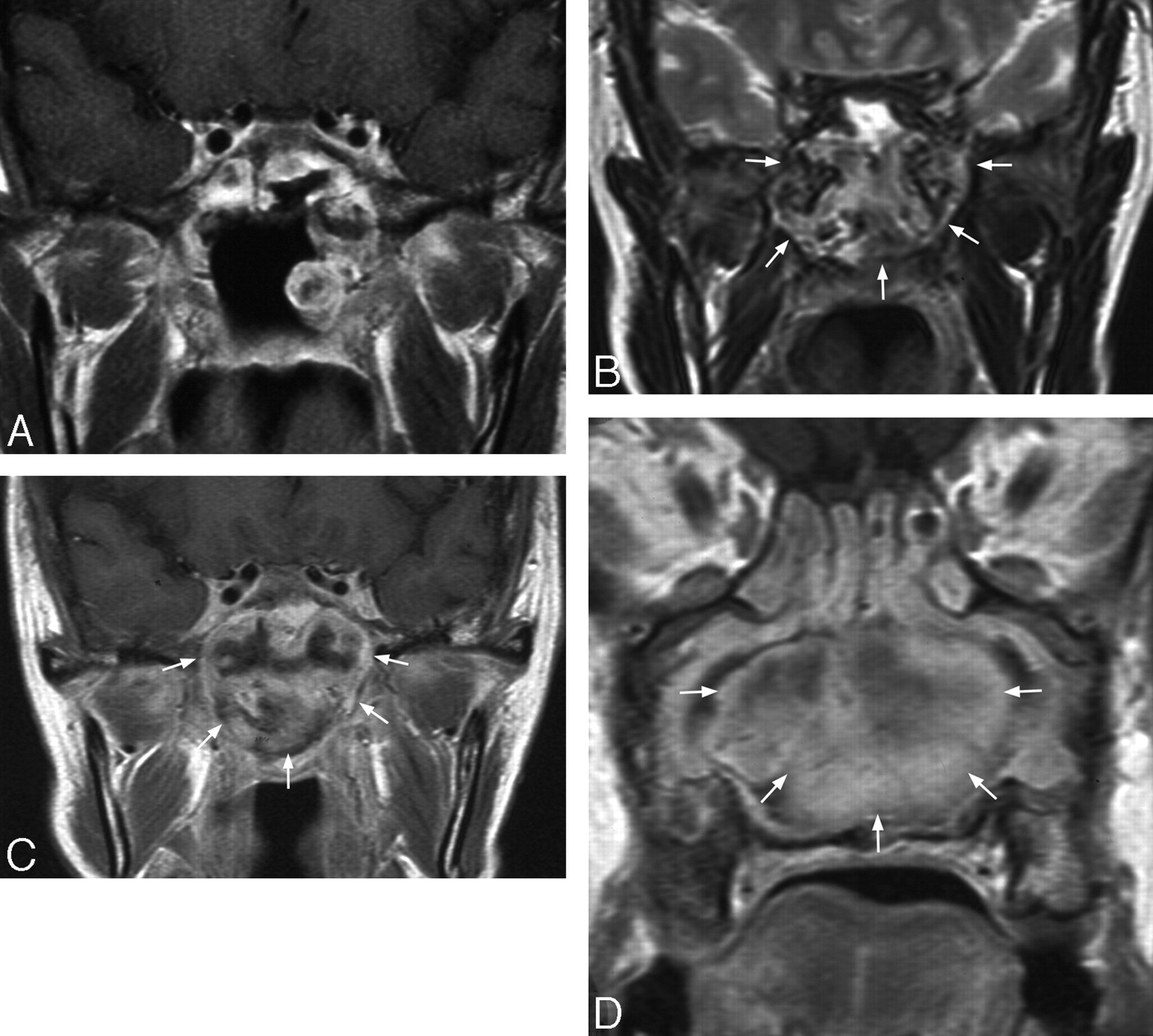
  • Fig 4.
    • Download figure
    • Open in new tab
    • Download powerpoint
    Fig 4.

    Coronal T1-weighted postcontrast MR image in a 42-year-old woman with enhancing polyps in the left lateral wall of the nasopharynx and sphenoid sinus and a large defect in the sphenoid sinus floor, 11 years after conventional radiation therapy and 3 years after a nasopharyngectomy for local tumor recurrence (A). These polyps remained static before rapidly increasing in size on MR imaging 32 months later to form a large heterogeneous mass in the nasopharynx expanding into the sphenoid sinus and nasal cavity. On the T2-weighted image, the mass shows heterogeneous mixed signal intensity (B), and on the T1-weighted image postcontrast there is heterogeneous enhancement (C) with a less enhancing stellate area centrally in the nasal cavity component (D).
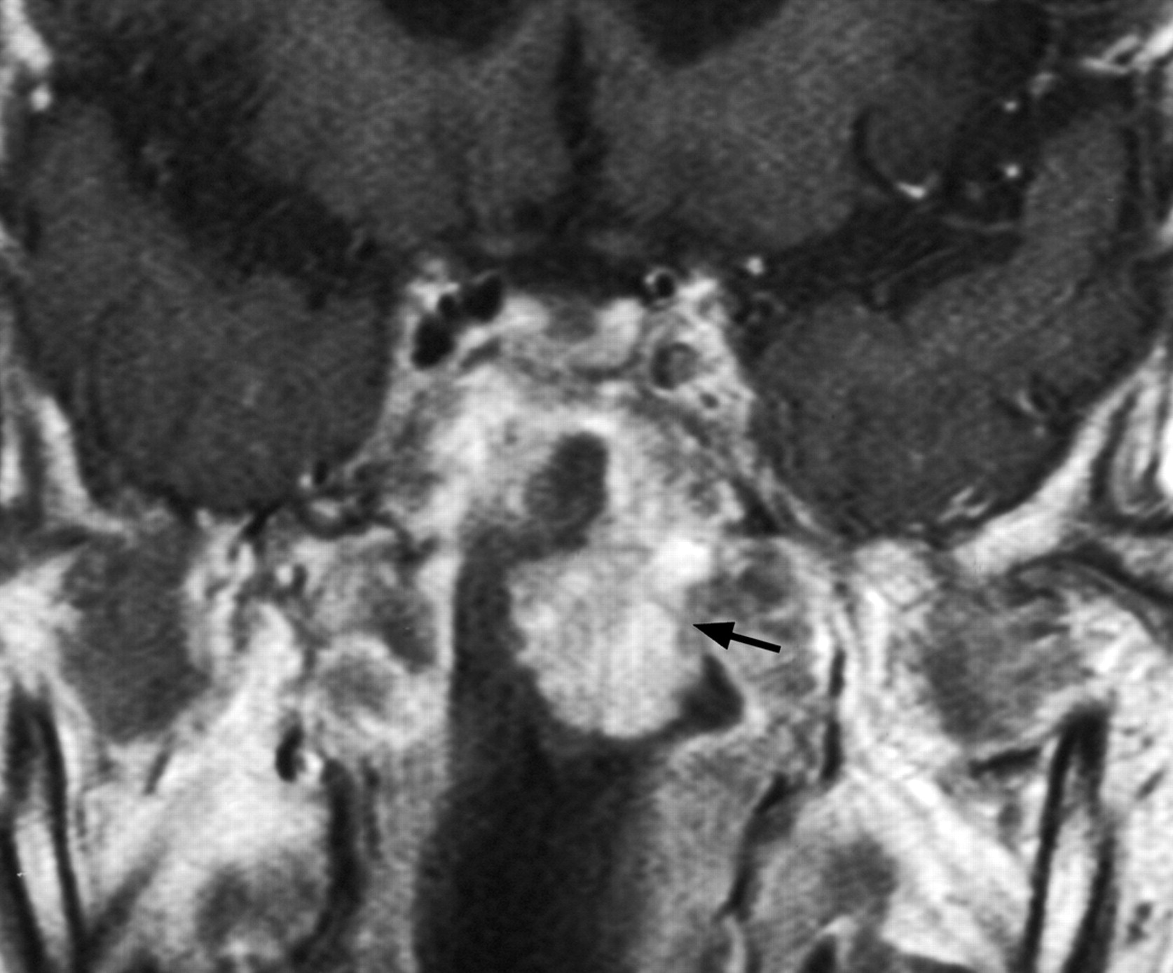
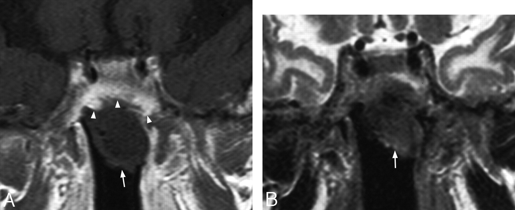
  • Fig 5.
    • Download figure
    • Open in new tab
    • Download powerpoint
    Fig 5.

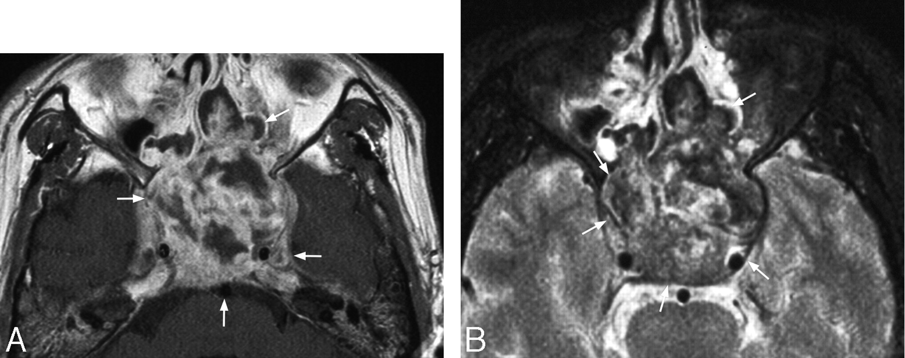
    A, Coronal T1-weighted postcontrast MR image in a 69-year-old man with osteoradionecrosis causing a large defect in the sphenoid sinus floor and a nonenhancing mass (arrow) within the sphenoid sinus that also shows thickened enhancing mucosa in the roof of the sinus (arrowheads), 5 years after radiation therapy (“rhinolith” at surgery). B, Coronal T2-weighted MR image in the same patient showing the mass is of low-intermediate signal intensity (arrow) and radiation-induced injury in the white matter of the inferior aspects of both temporal lobes.

  • Fig 6.
    • Download figure
    • Open in new tab
    • Download powerpoint
    Fig 6.

    A, Axial T1-weighted postcontrast MR image in a 39-year-old man with a heterogeneous enhancing mass causing expansion of the sphenoid sinus (arrows) 4 years after radiation therapy. B, Axial T2-weighted MR image in the same patient showing a heterogeneous mass (arrows) with foci of very low T2 signal intensity presumed to be old hemorrhage.

  • Fig 7.
    • Download figure
    • Open in new tab
    • Download powerpoint
    Fig 7.

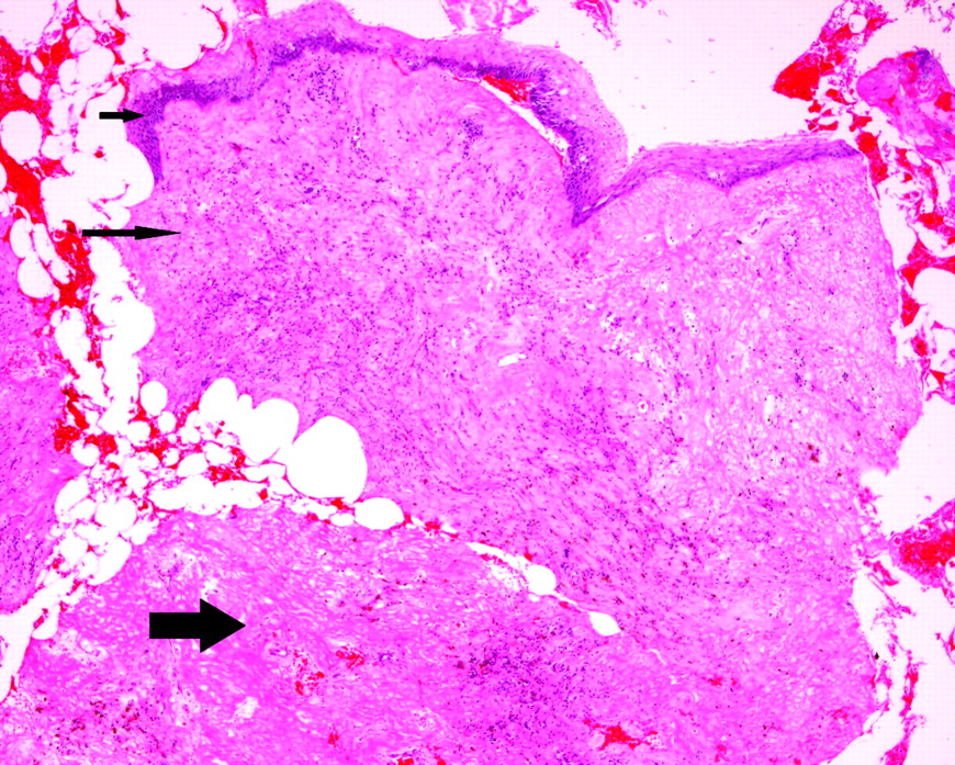
    Photomicrograph showing a polypoidal mass that is lined by benign squamous epithelium (short arrow) with proliferation of granulation tissue in the underlying stroma (long arrow) together with a fibrin deposit (thick arrow).

Tables

  • Figures
  • Radiologic findings, histology, and follow-up in 6 patients with a nonmalignant mass in the sphenoid sinus

    CaseRadiologic FeaturesHistologyFollow-Up
    Size, cm% Granulation Tissue:FibrinCells (0–3) MC, IC, and EC
    6Mucosal thickening and enhancement along the walls of the sphenoid sinus and a large bony defect in the floor forming a direct communication with the nasopharynx; mass inside the sinus cavity/nasopharyngeal roof of low nonenhancing signal intensity on T1 weighted images and heterogeneous, mainly intermediate, signal intensity on T2 weighted images “rhinolith” at surgery (Fig 5)2.050:50MC (0) IC (1) EC (2)No local tumor recurrence on endoscopy or MR imaging at 10 mo
    7See radiologic features in 6 above0.580:20MC (0) IC (3) EC (2)Multiple biopsies over the next 24 mo showed no local tumor recurrence, re-epithelialization of the nasopharyngeal roof with no tumor recurrence after 104 mo
    8See radiologic features in 6 above1.230:70MC (0) IC (3) EC (3)No local tumor recurrence on endoscopy; died from meningitis 3 y later
    9See radiologic features in 6 above plus polypoidal enhancing mucosal thickening in the wall of the sphenoid sinus extending into the nasopharynx2.040:60MC (0) IC (3) EC (1)No local tumor recurrence on endoscopy at 27 mo
    10Sphenoid sinus is expanded and filled by a heterogeneous mass that on T2 is of mixed signal intensity (high, intermediate, and low) on T1 is mainly of homogeneous intermediate signal intensity (with a few small areas of high T1 retained secretions) and mixed contrast enhancement ranging from marked enhancement to no enhancement (Fig 6)2.090:10MC (0) IC (2) EC (2)No local tumor recurrence on endoscopy but developed blindness and died 48 mo later after repeated CSF leaks and intracranial infections
    11Mass in the sphenoid sinus progressively increasing in size to 5 cm over 3 years; signal intensity of the mass is heterogeneous with mixed signal intensity on T2 (very low signal areas, especially centrally, and very high signal areas, especially peripherally), mainly homogeneous intermediate signal intensity on T1 (with a few small areas of high T1 retained secretions), and mixed contrast enhancement ranging from marked enhancement to no enhancement3.010:90MC (0) IC (1) EC (1) Organized hematomaStatic appearances on MR imaging at 39 months and no local tumor recurrence on endoscopy at 44 mo
    • Note:—No cells (0), some cells (1), many cells (2), abundant cells (3). MC indicates malignant cells; IC, inflammatory cells; EC, epithelial cells.

PreviousNext
Back to top

In this issue

American Journal of Neuroradiology: 29 (6)
American Journal of Neuroradiology
Vol. 29, Issue 6
June 2008
  • Table of Contents
  • Index by author
Advertisement
Print
Download PDF
Email Article

Thank you for your interest in spreading the word on American Journal of Neuroradiology.

NOTE: We only request your email address so that the person you are recommending the page to knows that you wanted them to see it, and that it is not junk mail. We do not capture any email address.

Enter multiple addresses on separate lines or separate them with commas.
MR Imaging of Nonmalignant Polyps and Masses of the Nasopharynx and Sphenoid Sinus after Radiotherapy for Nasopharyngeal Carcinoma
(Your Name) has sent you a message from American Journal of Neuroradiology
(Your Name) thought you would like to see the American Journal of Neuroradiology web site.
CAPTCHA
This question is for testing whether or not you are a human visitor and to prevent automated spam submissions.
Cite this article
A.D. King, A.T. Ahuja, S.-F. Leung, J. Abrigo, J.K.T. Wong, W.S. Poon, K.S. Woo, H.S. Chan, G.M.K. Tse
MR Imaging of Nonmalignant Polyps and Masses of the Nasopharynx and Sphenoid Sinus after Radiotherapy for Nasopharyngeal Carcinoma
American Journal of Neuroradiology Jun 2008, 29 (6) 1209-1214; DOI: 10.3174/ajnr.A1043

Citation Manager Formats

  • BibTeX
  • Bookends
  • EasyBib
  • EndNote (tagged)
  • EndNote 8 (xml)
  • Medlars
  • Mendeley
  • Papers
  • RefWorks Tagged
  • Ref Manager
  • RIS
  • Zotero
0 Responses
Respond to this article
Share
Bookmark this article
MR Imaging of Nonmalignant Polyps and Masses of the Nasopharynx and Sphenoid Sinus after Radiotherapy for Nasopharyngeal Carcinoma
A.D. King, A.T. Ahuja, S.-F. Leung, J. Abrigo, J.K.T. Wong, W.S. Poon, K.S. Woo, H.S. Chan, G.M.K. Tse
American Journal of Neuroradiology Jun 2008, 29 (6) 1209-1214; DOI: 10.3174/ajnr.A1043
del.icio.us logo Twitter logo Facebook logo Mendeley logo
  • Tweet Widget
  • Facebook Like
  • Google Plus One
Purchase

Jump to section

  • Article
    • Abstract
    • Methods
    • Results
    • Discussion
    • Footnotes
    • References
  • Figures & Data
  • Supplemental
  • Info & Metrics
  • Responses
  • References
  • PDF

Related Articles

  • PubMed
  • Google Scholar

Cited By...

  • No citing articles found.
  • Crossref (12)
  • Google Scholar

This article has been cited by the following articles in journals that are participating in Crossref Cited-by Linking.

  • MRI and CT of Nasopharyngeal Carcinoma
    Ahmed Abdel Khalek Abdel Razek, Ann King
    American Journal of Roentgenology 2012 198 1
  • Outcome of Patients Undergoing Salvage Surgery for Recurrent Nasopharyngeal Carcinoma: A Meta-analysis
    Shorook Na’ara, Moran Amit, Salem Billan, Jacob T. Cohen, Ziv Gil
    Annals of Surgical Oncology 2014 21 9
  • CT and MRI of radiation-induced sarcomas of the head and neck following radiotherapy for nasopharyngeal carcinoma
    Pei-qiang Cai, Yao-pan Wu, Li Li, Rong Zhang, Chuan-miao Xie, Pei-hong Wu, Jie-hua Xu
    Clinical Radiology 2013 68 7
  • MRI of radiation-induced tumors of the head and neck in post-radiation nasopharyngeal carcinoma
    Jill M. Abrigo, Ann D. King, Sing Fai Leung, Alexander C. Vlantis, Jeffrey K. T. Wong, Michael C. F. Tong, Gary M. K. Tse, Anil T. Ahuja
    European Radiology 2009 19 5
  • Recurrent Nasopharyngeal Carcinoma
    Robert I. Smee, Nicola S. Meagher, Kathryn Broadley, Timothy Ho, Janet R. Williams, G. Patrick Bridger
    American Journal of Clinical Oncology 2010 33 5
  • Magnetic resonance imaging following endoscopic nasopharyngectomy with a potassium-titanyl-phosphate (KTP) laser for early locally recurrent nasopharyngeal carcinoma
    Ya-Fang Chen, Yu-Fen Wang, Cheng-Ping Wang, Jenq-Yuh Ko, Chun-Wei Wang, Hon-Man Liu
    Neuroradiology 2013 55 11
  • Nasopharyngeal granulomatous mass after radiotherapy for nasopharyngeal carcinoma
    Lu Yang, Houyong Li, Huan Wang, Huankang Zhang, Shuyi Wang, Allison N. Fry, Wade W. Han, Dehui Wang
    Auris Nasus Larynx 2016 43 3
  • Is there a link between nasal polyposis and increased risk for sinonasal malignancy?
    Deena Pourang, Michael Batech, Kian Karimi, Javed Sheikh, Shefali Samant
    Annals of Allergy, Asthma & Immunology 2018 120 4
  • Chronic Invasive Fungal Sinusitis Mimicking Malignancy Post-Radiotherapy: A Case Report
    Aloysius W. Y. Lim, Tee Sin Lee, Jia Jia Wee, C. Y. Maria Pang
    Indian Journal of Otolaryngology and Head & Neck Surgery 2024 76 5
  • Masses of the Paranasal Sinuses After Treatment of Nasopharyngeal Carcinoma: A Case Report
    Maria Silvia Lazio, Angelo Cannavicci, Massimo Squadrelli Saraceno, Giandomenico Maggiore
    Indian Journal of Otolaryngology and Head & Neck Surgery 2019 71 S1

More in this TOC Section

  • WHO Classification Update: Nasal&Skull Base Tumors
  • Peritumoral Signal in Vestibular Schwannomas
  • Chondrosarcoma vs Synovial Chondromatosis: Imaging
Show more HEAD & NECK

Similar Articles

Advertisement

Indexed Content

  • Current Issue
  • Accepted Manuscripts
  • Article Preview
  • Past Issues
  • Editorials
  • Editors Choice
  • Fellow Journal Club
  • Letters to the Editor

Cases

  • Case Collection
  • Archive - Case of the Week
  • Archive - Case of the Month
  • Archive - Classic Case

Special Collections

  • Special Collections

Resources

  • News and Updates
  • Turn around Times
  • Submit a Manuscript
  • Author Policies
  • Manuscript Submission Guidelines
  • Evidence-Based Medicine Level Guide
  • Publishing Checklists
  • Graphical Abstract Preparation
  • Imaging Protocol Submission
  • Submit a Case
  • Become a Reviewer/Academy of Reviewers
  • Get Peer Review Credit from Publons

Multimedia

  • AJNR Podcast
  • AJNR SCANtastic
  • Video Articles

About Us

  • About AJNR
  • Editorial Board
  • Not an AJNR Subscriber? Join Now
  • Alerts
  • Feedback
  • Advertise with us
  • Librarian Resources
  • Permissions
  • Terms and Conditions

American Society of Neuroradiology

  • Not an ASNR Member? Join Now

© 2025 by the American Society of Neuroradiology All rights, including for text and data mining, AI training, and similar technologies, are reserved.
Print ISSN: 0195-6108 Online ISSN: 1936-959X

Powered by HighWire