
Article Figures & Data
Figures
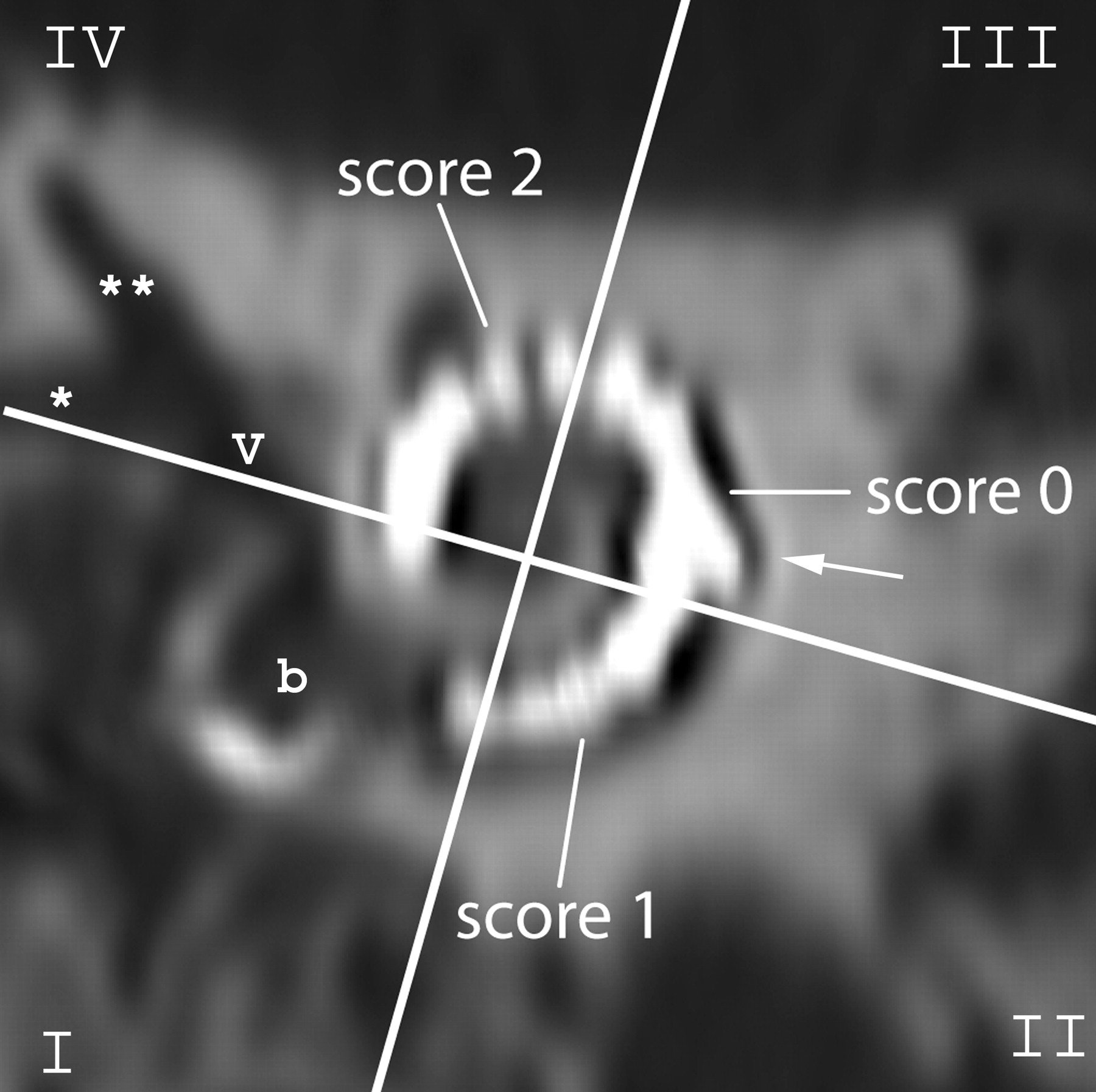
- Fig 1.
Scoring of the visibility of electrode contacts and anatomic structures: on a MPR perpendicular to the modiolus of an MSCT image of the implanted human cadaver temporal bone, the cochlea is divided in 4 quadrants (white crosslines). The quadrants are numbered counterclockwise, and the round window niche is located in the first quadrant (I-IV). A quantitative score from 0 to 2 was given to each electrode contact according to its visibility. Cochlear structures, such as the inner and outer wall, were scored per quadrant. The kinking of the electrode is localized in the third quadrant (arrow). b indicates basal turn of the cochlea at the level of the round window; v, vestibule, *horizontal semicircular canal (SCC); **superior SCC.
- Fig 2.
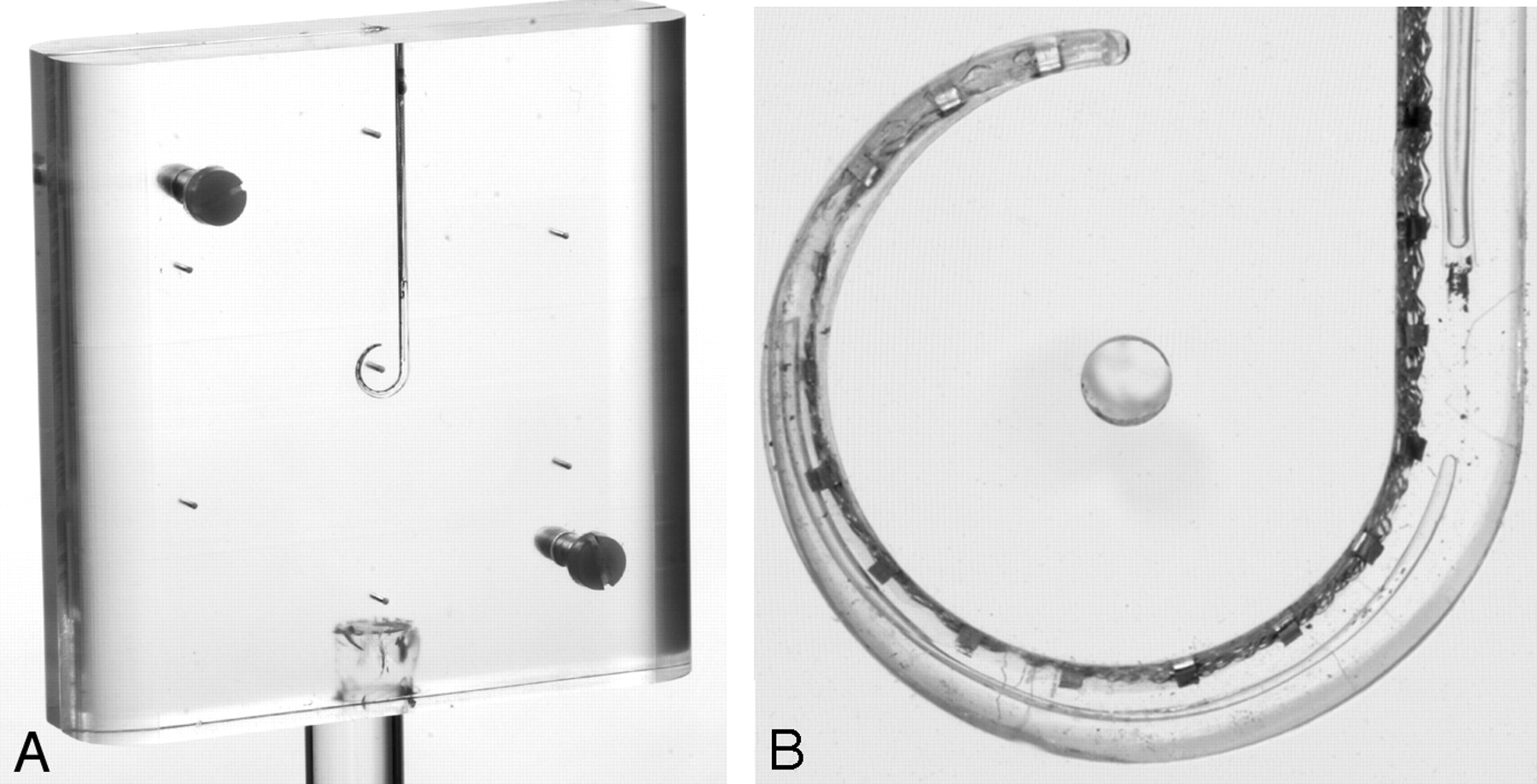
Photograph (A) and 1 high-resolution optical image (B) of the PMMA phantom containing a cochlear implant.
- Fig 3.
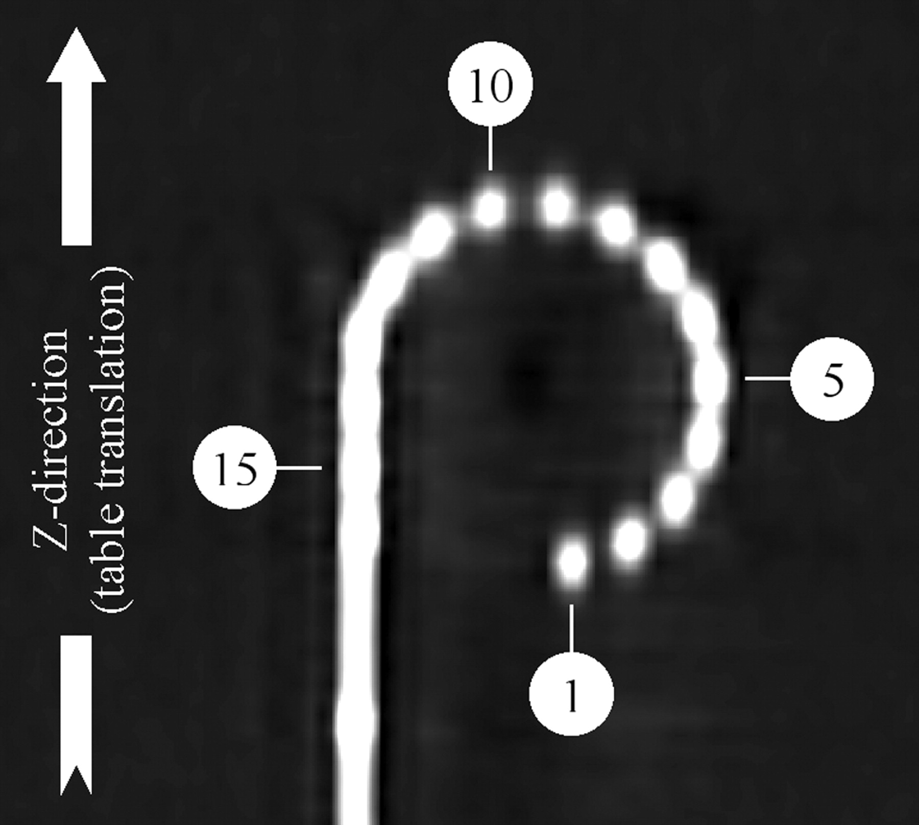
MSCT of the PMMA phantom: 16 electrode contacts are numbered from the tip to the base: electrode numbers 1, 5, 10, and 15 are indicated.
- Fig 4.
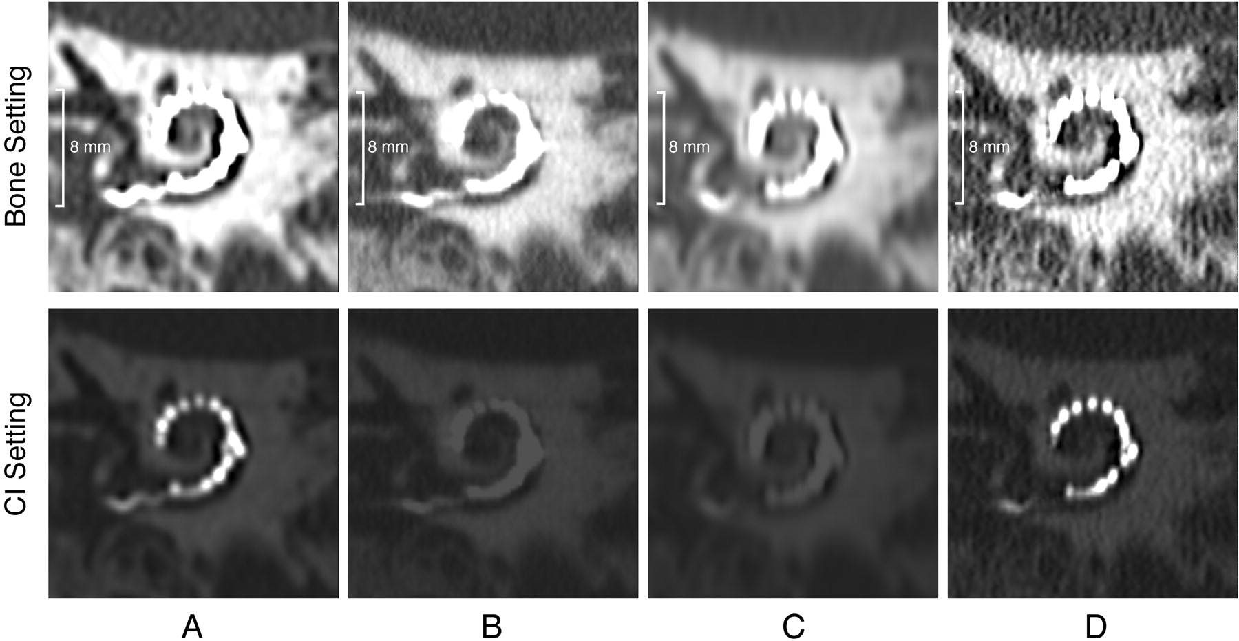
MPR CT images of the human cadaver temporal bone along the plane of the electrode array on Aquilion-64 (A), Brilliance 64 (B), LightSpeed-64 (C) (also seen in Fig 1), and Sensation-64 (D). The upper set of images is displayed with W/L 3000/800 and shows cochlear anatomic structures, such as the semicircular canals, well in all of the scanners. The lower set of images shows the chosen manually adjusted window/level setting for visualization of the electrode contacts. This illustrates that a wide range of HUs is essential for visualization of the CI.
- Fig 5.
Graphic presentation of the mean differences over 4 observers (y-axis) between electrode position as manually indicated and the position in the computer model (in millimeters). Values are shown per electrode contact (x-axis) for each scanner (Aquilion-64, yellow; Brilliance-64, blue; LightSpeed-64, red; Sensation-64, green). See also Table 3.
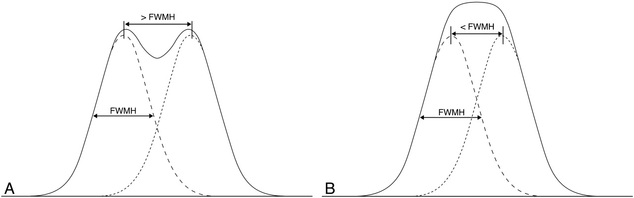
- Fig 6.
Clinical implications of the PSF: the dotted curves depict the pixel value of a single electrode contact through the center. Summation of these curves renders a curve (black line). When the distance between 2 electrodes is larger than FWHM, the resulting curve still shows 2 maximum values, and the electrode contacts can be separated in the image (A). If the distance between 2 electrode contacts is smaller than FWHM, the resulting curve shows a single peak; the electrode contact cannot be visualized separately (B).


Tables
- Table 1:
Acquisition and reconstruction protocols and effective radiation dose for cochlear implant imaging on 64-section scanners
Variable Aquilion 64 Brilliance 64 LightSpeed 64 Sensation 64 Acquisition protocol Tube voltage, kV 120 140 140 120 Tube current, mA 200 200 335 135 Beam collimation, mm 4 × 0.5 2 × 0.55 32 × 0.625 12 × 0.6 Pitch 0.75 0.5 0.531 0.45 Rotation time, s 0.5 0.5 0.6 1.0 Scan FOV 240 500 320 500 Dose of acquisition protocol Effective dose, mSv 1.4 2.0 1.8 1.3 Reconstruction protocol Section thickness, mm 0.5 0.55 0.6 0.6 Section interval, mm 0.3 0.3 0.3 0.3 Kernel FC84 Filter D BonePlus U90u Reconstruction matrix 5122 7682 5122 5122 Recon FOV PMMA phantom, mm 100 Recon FOV PSF phantom, mm 50 Recon FOV cadaver head, mm 80 Note:—FOV indicates field of view; PMMA, polymethylmethacrylate; PSF, point spread function.
Scoring Parameters Scoring Method Aquilion 64 Brilliance 64 LightSpeed 64 Sensation 64 Overall impression VAS 7.8 (1.0) 3.2 (1.0) 3.0 (1.1) 7.9 (0.7) Axial oblique MPR, distinction of electrode contacts Quantitative score 23.2 (2.7) 12.6 (4.4) 8.0 (3.9) 24.2 (4.8) Coronal oblique MPR, distinction of electrode contacts Quantitative score 23.7 (3.6) 14.2 (3.7) 10.2 (3.3) 25.6 (3.3) Kinking VAS 6.1 (2.1) 1.7 (1.5) 1.3 (1.2) 6.8 (2.0) Inner cochlear wall 4 quadrants VAS 6.9 (1.3) 4.9 (1.5) 4.1 (2.0) 6.2 (1.1) 6.6 (1.1) 4.7 (1.3) 4.3 (1.7) 5.7 (1.4) 6.2 (1.2) 3.9 (1.5) 3.9 (1.1) 5.3 (1.5) 6.4 (1.4) 3.6 (1.7) 3.9 (1.4) 5.5 (1.9) Outer cochlear walls 4 quadrants VAS 7.7 (1.1) 6.8 (1.4) 5.5 (1.7) 7.4 (0.9) 7.6 (1.2) 6.8 (1.1) 5.5 (1.5) 7.4 (1.0) 7.1 (1.4) 5.2 (1.7) 4.6 (1.6) 6.8 (1.8) 6.6 (1.9) 4.8 (1.8) 4.1 (1.8) 6.4 (1.9) Note:—VAS indicates visual analogue scale; MPR, multiplanar reconstruction.
Electrode Contact Aquilion 64 Brilliance 64 LightSpeed 64 Sensation 64 1 0.07 (0.02) 0.11 (0.02) 0.07 (0.02) 0.03 (0.01) 2 0.05 (0.02) 0.12 (0.01) 0.09 (0.03) 0.05 (0.02) 3 0.09 (0.01) 0.13 (0.00) 0.09 (0.03) 0.06 (0.01) 4 0.10 (0.02) 0.09 (0.02) 0.14 (0.02) 0.08 (0.02) 5 0.12 (0.02) 0.07 (0.01) 0.10 (0.04) 0.09 (0.02) 6 0.11 (0.02) 0.09 (0.03) 0.12 (0.01) 0.09 (0.01) 7 0.11 (0.03) 0.09 (0.05) 0.13 (0.02) 0.10 (0.01) 8 0.14 (0.03) 0.07 (0.02) 0.20 (0.04) 0.13 (0.02) 9 0.13 (0.03) 0.08 (0.01) 0.18 (0.03) 0.13 (0.02) 10 0.14 (0.02) 0.10 (0.01) 0.16 (0.03) 0.12 (0.01) 11 0.08 (0.01) 0.10 (0.03) 0.12 (0.02) 0.08 (0.01) 12 0.11 (0.01) 0.13 (0.07) 0.15 (0.02) 0.10 (0.00) 13 0.10 (0.02) 0.15 (0.05) 0.25 (0.10) 0.09 (0.01) 14 0.10 (0.01) 0.10 (0.02) 0.24 (0.11) 0.11 (0.00) 15 0.11 (0.01) 0.12 (0.05) 0.31 (0.15) 0.10 (0.01) 16 0.11 (0.00) 0.16 (0.12) 0.61 (0.35) 0.12 (0.01) Note:—PMMA indicates polymethylmethacrylate.
Variable FWHM, in mm Aquilion 64 Brilliance 64 LightSpeed 64 Sensation 64 In-plane 0.68 0.52 0.68 0.48 Longitudinal 0.81 0.84 0.98 0.70 Ratio in-plane/longitudinal 0.84 0.62 0.69 0.69 Note:—PSF indicates point spread function; FWHM, full width half maximum.


