
Article Figures & Data
Figures
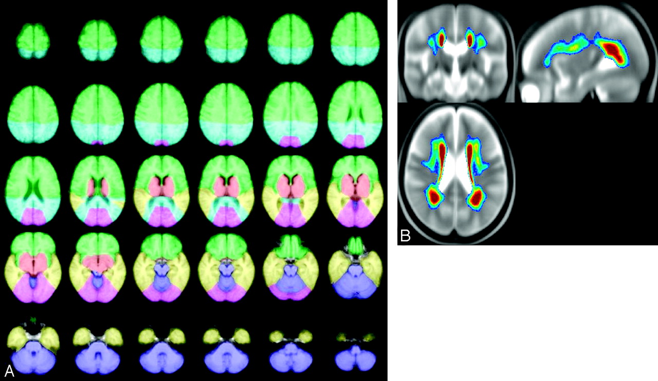
- Fig 1.
The left panel shows axial views illustrating the delineation of 6 anatomic regions: frontal (green), parietal (turquoise), temporal (yellow), basal ganglia (red), occipital lobes (pink), infratentorial regions (purple); see details in text. The right panel shows the average distribution of ARWMC projected onto orthogonal sections of the group-averaged T2-weighted image. The color-coding indicates the frequency of ARWMC occurrence, ranging from 5% (dark blue) to 30% and above (dark red).
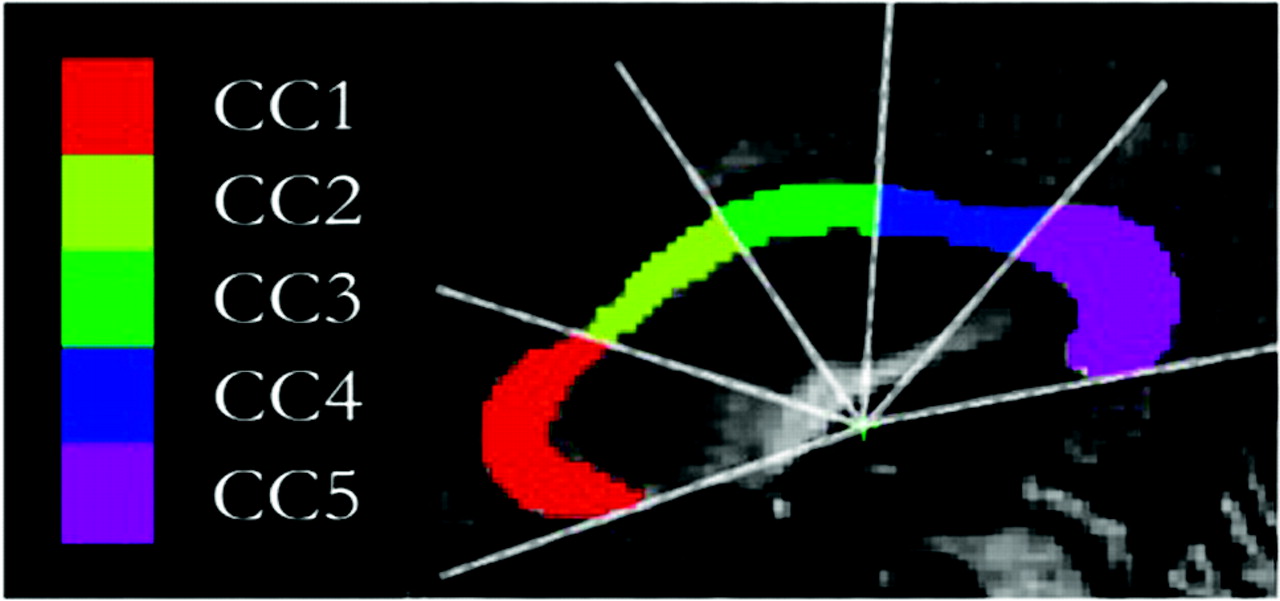
- Fig 2.
Segmentation and subdivision of the CC area into 5 distinct subregions obtained from the normalized midsagittal T1 (MPRAGE) scans. A radial partitioning scheme is used for regional analyses of the CC. CC1 indicates rostrum and genu; CC2, rostral body; CC3, midbody; CC4, isthmus; CC5, splenium.
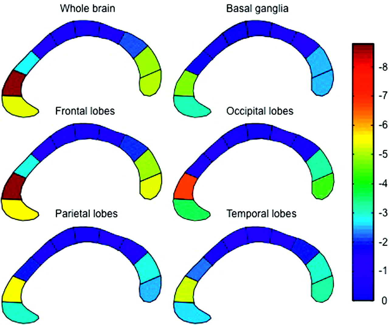
- Fig 3.
The correlation between the area of each of the 10 CC subregions and the volume of ARWMC in each of 5 hemispheric regions and the whole brain. To obtain a better illustration of the regional specificity between CC atrophy and ARWMC, we subdivided the CC into 10 subregions and not 5 subregions as reported in Fig 2. The color code from blue to red indicates the magnitude of regression coefficients.
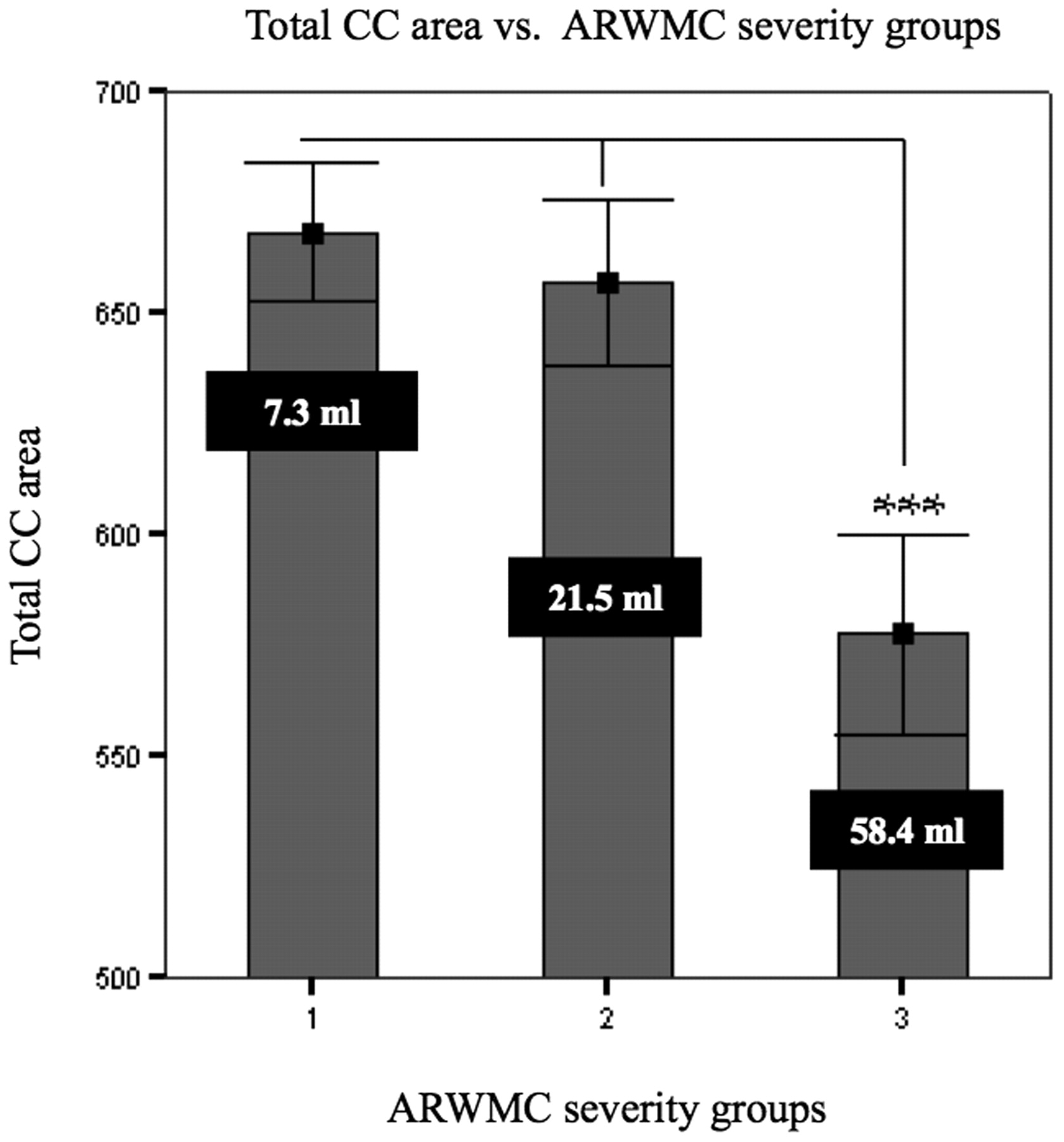
- Fig 4.
Comparison of mean total callosal area measured on normalized MR imaging in nondisabled elderly subjects, classified by severity of ARWMC (Fazekas ratings 1, 2, and 3). Vertical bars indicate SDs. The white figures indicate the mean ARWMC volumes. Triple asterisks indicate P < .001 (ANOVA test).


Tables
Characteristic No. Female 313 (54%) Age (years) 74.1 (±5) MMSE score (0–30) 27.4 (±2) Handedness (No. of subjects)† Left 27 (4.7%) Right 524 (90.8%) Ambidextrous 26 (4.5%) ARWMC ratings (No. of subjects) Mild (score 1) 260 (45%) Moderate (score 2) 178 (31%) Severe (score 3) 140 (24%) ARWMC volume (mL) Mild (score 1) 7.3 (±5.3) Moderate (score 2) 21.5 (±8.2) Severe (score 3) 58.4 (±27.2) No. of lacunes (No. of subjects) 0 308 (47%) 1–3 195 (34%) ≥4 75 (13%) No. of infarcts (No. of subjects) 0 523 (90%) 1 43 (7%) ≥2 12 (3%) Note:—MMSE indicates Mini-Mental State Examination; ARWMC, age-related white matter changes.
* Values are mean (±SD) or numbers (percentages).
† Available for n = 577.
- Table 2:
Correlation coefficients (r) between CC areas and ARWMC volume measured in standard space*
Areas Mean ARWMC (mL) Mean CC Areas vs Mean ARWMC (mL) in the Brain (r) CC1 CC2 CC3 CC4 CC5 CC Total Mean CC area (mm2) 179.3 ± 40.1 93.8 ± 25.2 90.8 ± 24.0 91.5 ± 27.3 184.0 ± 39.8 642.6 ± 135.4 ARWMC Basal ganglia region 0.7 ± 0.9 −0.111 −0.077 0.039 −0.038 −0.162‡ −0.095 Frontal lobes 14.7 ± 15.4 −0.181§ −0.078 0.027 −0.07 −0.232§ −0.144† Occipital lobes 1.2 ± 1.6 −0.181‡ −0.107 −0.004 −0.04 −0.223§ −0.146† Parietal lobes 6.0 ± 7.4 −0.137† −0.097 −0.004 −0.067 −0.201§ −0.132† Temporal lobes 1.3 ± 1.9 −0.154‡ −0.157‡ −0.067 −0.104 −0.206§ −0.168‡ Total ARWMC 24.0 ± 25.0 −0.175‡ −0.076 0.035 −0.042 −0.230§ −0.134† Note:—CC indicates corpus callosum; ARWMC, age-related white matter changes.
* All CC and ARWMC values were normalized to the same head size. Values are mean CC areas and mean ARWMC volume (± SD), relative volume of the ARWMC volume assessed relative to the entire anatomic region, correlation coefficients (r), and significance level. In addition to the log-transformed ARWMC per brain region, we entered the following covariates: number of lacunes per brain region, number of infarcts, age, handedness, recruiting center, and ratings of sulcal and ventricular atrophy.
† P < .01.
‡ P < .001.
§ P < .0001 (uncorrected).
In this issue


Jump to section
Related Articles
Cited By...
- Tractography at 3T MRI of Corpus Callosum Tracts Crossing White Matter Hyperintensities
- Leukoaraiosis Burden Significantly Modulates the Association Between Infarct Volume and National Institutes of Health Stroke Scale in Ischemic Stroke
- Leukoaraiosis Predicts Cortical Infarct Volume After Distal Middle Cerebral Artery Occlusion
- MRI characteristics and scoring in HDLS due to CSF1R gene mutations
- Brain atrophy accelerates cognitive decline in cerebral small vessel disease: The LADIS study
- Diffusion-Weighted Imaging and Cognition in the Leukoariosis and Disability in the Elderly Study
- Changes in white matter as determinant of global functional decline in older independent outpatients: three year follow-up of LADIS (leukoaraiosis and disability) study cohort