Advertisement

Main menu

  • Home
  • Content
    • Current Issue
    • Accepted Manuscripts
    • Article Preview
    • Past Issue Archive
    • AJNR Case Collection
    • Case of the Week Archive
    • Classic Case Archive
    • Case of the Month Archive
  • Special Collections
    • Spinal CSF Leak Articles (Jan 2020-June 2024)
    • 2024 AJNR Journal Awards
    • Most Impactful AJNR Articles
  • Multimedia
    • AJNR Podcast
    • AJNR Scantastics
    • Video Articles
  • For Authors
    • Submit a Manuscript
    • Author Policies
    • Fast publishing of Accepted Manuscripts
    • Graphical Abstract Preparation
    • Manuscript Submission Guidelines
    • Imaging Protocol Submission
    • Submit a Case for the Case Collection
  • About Us
    • About AJNR
    • Editorial Board
  • More
    • Become a Reviewer/Academy of Reviewers
    • Subscribers
    • Permissions
    • Alerts
    • Feedback
    • Advertisers
    • ASNR Home
  • Other Publications
    • ajnr

User menu

  • Alerts
  • Log in

Search

  • Advanced search
American Journal of Neuroradiology

American Journal of Neuroradiology

ASHNR American Society of Functional Neuroradiology ASHNR American Society of Pediatric Neuroradiology ASSR
  • Alerts
  • Log in

Advanced Search

  • Home
  • Content
    • Current Issue
    • Accepted Manuscripts
    • Article Preview
    • Past Issue Archive
    • AJNR Case Collection
    • Case of the Week Archive
    • Classic Case Archive
    • Case of the Month Archive
  • Special Collections
    • Spinal CSF Leak Articles (Jan 2020-June 2024)
    • 2024 AJNR Journal Awards
    • Most Impactful AJNR Articles
  • Multimedia
    • AJNR Podcast
    • AJNR Scantastics
    • Video Articles
  • For Authors
    • Submit a Manuscript
    • Author Policies
    • Fast publishing of Accepted Manuscripts
    • Graphical Abstract Preparation
    • Manuscript Submission Guidelines
    • Imaging Protocol Submission
    • Submit a Case for the Case Collection
  • About Us
    • About AJNR
    • Editorial Board
  • More
    • Become a Reviewer/Academy of Reviewers
    • Subscribers
    • Permissions
    • Alerts
    • Feedback
    • Advertisers
    • ASNR Home
  • Follow AJNR on Twitter
  • Visit AJNR on Facebook
  • Follow AJNR on Instagram
  • Join AJNR on LinkedIn
  • RSS Feeds

Welcome to the new AJNR, Updated Hall of Fame, and more. Read the full announcements.


AJNR is seeking candidates for the position of Associate Section Editor, AJNR Case Collection. Read the full announcement.


OtherBRAIN

Anomalous J-Modulation Effects on Amino Acids in Clinical 3T MR Spectroscopy

H.-S. Liu, H.-W. Chung, C.-J. Juan, S.-Y. Tsai, C.-Y. Wang, C.-C. Chan, G.-S. Huang, M.-C. Chou, C.-S. Lee, C.-W. Ko, N.-Y. Cho and C.-Y. Chen
American Journal of Neuroradiology October 2008, 29 (9) 1644-1648; DOI: https://doi.org/10.3174/ajnr.A1131
H.-S. Liu
  • Find this author on Google Scholar
  • Find this author on PubMed
  • Search for this author on this site
H.-W. Chung
  • Find this author on Google Scholar
  • Find this author on PubMed
  • Search for this author on this site
C.-J. Juan
  • Find this author on Google Scholar
  • Find this author on PubMed
  • Search for this author on this site
S.-Y. Tsai
  • Find this author on Google Scholar
  • Find this author on PubMed
  • Search for this author on this site
C.-Y. Wang
  • Find this author on Google Scholar
  • Find this author on PubMed
  • Search for this author on this site
C.-C. Chan
  • Find this author on Google Scholar
  • Find this author on PubMed
  • Search for this author on this site
G.-S. Huang
  • Find this author on Google Scholar
  • Find this author on PubMed
  • Search for this author on this site
M.-C. Chou
  • Find this author on Google Scholar
  • Find this author on PubMed
  • Search for this author on this site
C.-S. Lee
  • Find this author on Google Scholar
  • Find this author on PubMed
  • Search for this author on this site
C.-W. Ko
  • Find this author on Google Scholar
  • Find this author on PubMed
  • Search for this author on this site
N.-Y. Cho
  • Find this author on Google Scholar
  • Find this author on PubMed
  • Search for this author on this site
C.-Y. Chen
  • Find this author on Google Scholar
  • Find this author on PubMed
  • Search for this author on this site
  • Article
  • Figures & Data
  • Info & Metrics
  • Responses
  • References
  • PDF
Loading

Abstract

SUMMARY: The signal-intensity loss from anomalous J-modulation effects due to chemical-shift displacement was investigated on amino acid groups (alanine, valine, leucine, and isoleucine) at 3T by using point-resolved 1H spectroscopy in patients with brain abscess and phantom experiments. With a larger chemical shift between methyl and methine resonances, alanine shows a greater effect of signal-intensity cancellation compared with other amino acids around 0.9 ppm, resulting in noninverted doublets at a TE of 144 ms.

High-field MR imaging systems such as 3T strength aid in the detection of brain metabolites due to increased sensitivity. However, the clinical 3T system also increases the spatial mismatch of section-selective radio-frequency pulses due to a larger chemical shift difference at higher fields and reduced radio-frequency pulse bandwidths.1–4 The issue of limited radio-frequency bandwidths is especially prominent with the increasing popularity of parallel imaging with array coils, whose receive-only function demands radio-frequency pulse transmission from the generally inefficient body coil. One frequently encountered challenge of MR spectroscopy at high-field MR imaging systems is the anomalous J-modulation effect on the weakly coupled AX3 system, which could hamper identification of the ideally inverted lactate doublet at TE = 144 ms.1–4

Although clinical investigations of the anomalous J-modulation effects have centered on lactate1–4 with theoretic analysis described clearly in a previous article,5 the possible occurrence arising from other AX3 spin systems such as alanine or other amino acids, valine, leucine, and isoleucine, overlaid around 0.9 ppm in brain abscess,6 to our knowledge, has never been reported. With similar coupling constants exerted on the methyl protons around 7 Hz,7 these metabolites containing methyl and methine groups also form a lactate-like coupled system with a TE-dependent phase evolution of the methyl resonance. In this study, we aimed to use both in vivo and in vitro experiments to show that the anomalous J-modulation effect analogous to lactate is also present for alanine, but to a much lesser extent for other amino acids, on a clinical 3T MR imaging scanner.

Description of Technique and Results

We performed experiments on a 3T scanner (Philips Medical Systems, Best, the Netherlands) using the quadrature body (Q-body) coil for transmission and an 8-channel phased-array modified sensitivity encoding head coil for receiving, by using point-resolved 1H spectroscopy sequence (PRESS). The Q-body coil had a maximum transmission B1 field of 13.5 μT (∼590 Hz). Single-voxel MR spectral data were obtained with TR/TE = 2000/40, 144, and/or 288 ms with 128 acquisitions (total scanning time, 4 minutes 56 seconds). Two patients with abscesses underwent MR spectroscopic examinations on this system. To investigate the anomalous J-modulation effect on alanine and amino acid groups, we performed in vitro experiments on four 1.5-L bottles of aqueous solution phantom containing 10 mmol/L of alanine, valine, leucine, and isoleucine, respectively. The phantom experiments were performed by using parameters identical to those of the patients, except that the voxel size was fixed at 25 × 25 × 25 mm3.

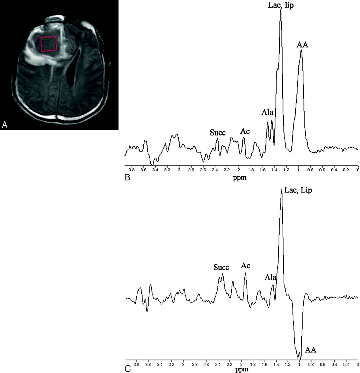
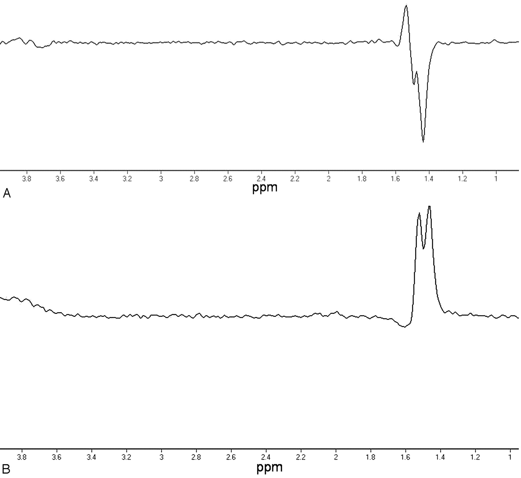
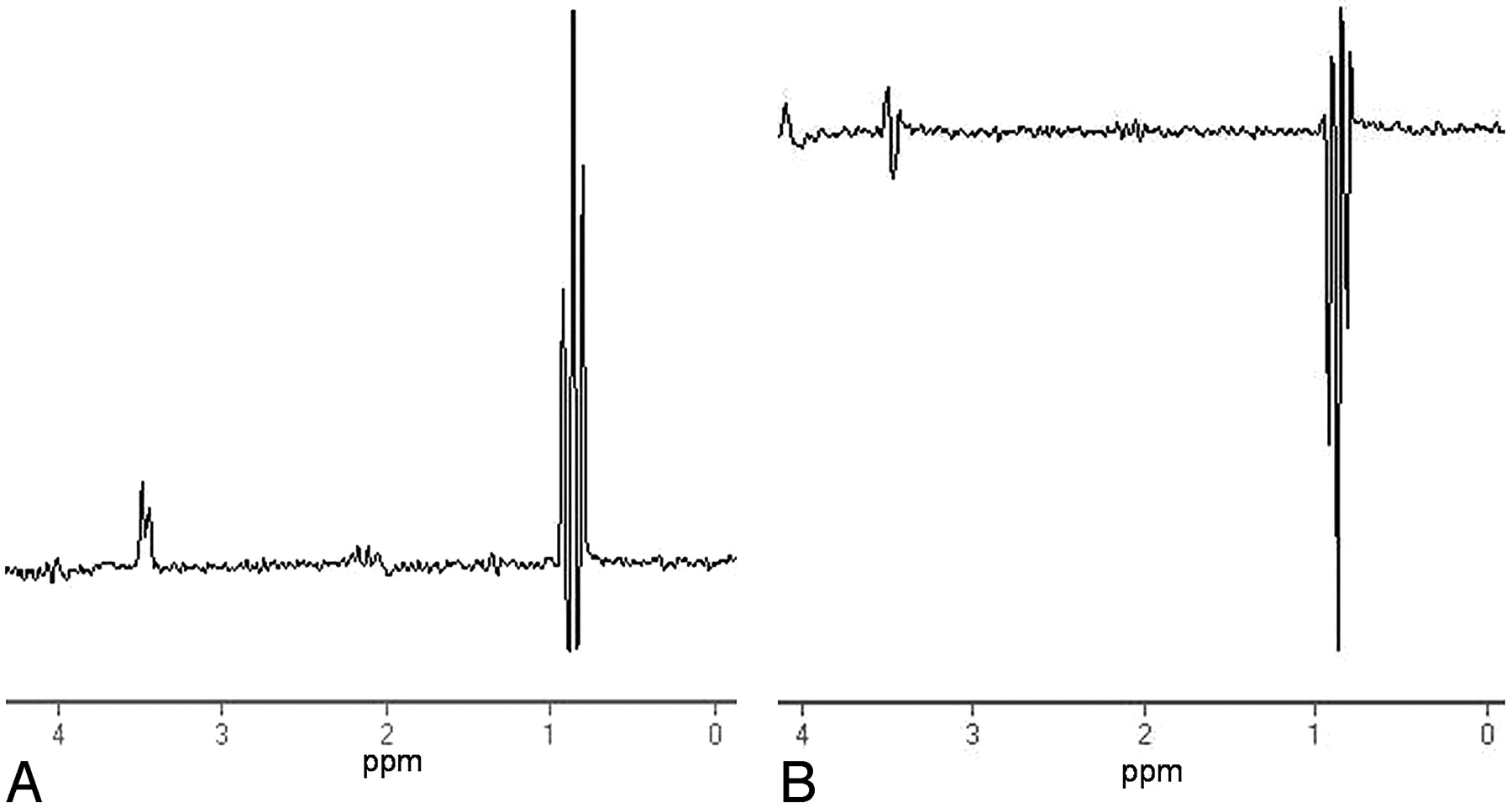
Figs 1 and 2 show the data from a 19-year-old man and a 42-year-old woman with brain abscess, respectively. The doublet at 1.47 ppm due to the presence of alanine was clearly visible in the 40-ms and 288-ms TE MR spectra (Figs 1B and 2C). However, for the first patient (Fig 1C), there was a positive peak centered on the same resonance which, if from alanine, is expected to exhibit an opposite sign of resonance (ie, inverted doublet) at TE = 144 ms. The spectrum acquired from the second patient with TE = 144 ms (Fig 2B) also exhibited signal-intensity canceling, whereas the spectrum acquired with TE = 288 ms showed substantially larger and clearer alanine resonances than that with TE = 144 ms. The expected inverted doublet of alanine in the 144-ms TE spectra was hardly detectable. Similarly, both of these in vivo spectra also show no clearly identifiable phase inversion of lactate around 1.33 ppm in the TE = 144 ms spectra. In contrast, note that the amino acid groups near 0.9 ppm for the 2 subjects showed clearly inverted spectral lines in the TE = 144 ms spectra without noticeable reduction in signal intensity. The in vivo observations are consistent with results from the phantom experiments, in which an incomplete inverted phase at TE = 144 ms was seen for the methyl protons in alanine with reduced amplitude, as opposed to the strong signals in TE = 40 ms and TE = 288 ms spectra (Fig 3), whereas valine (Fig 4), leucine, and isoleucine (not shown) showed clear inverted peaks at TE = 144 ms for the methyl protons with an amplitude comparable with that in TE = 40 ms and TE = 288 ms spectra.

Fig 1.
  • Download figure
  • Open in new tab
  • Download powerpoint
Fig 1.

Transverse T2 fluid-attenuated inversion recovery MR image (A) and single-voxel spectra obtained in a 19-year-old male patient with brain abscess by using PRESS localization at 3T with TE = 40 ms (B) and TE = 144 ms (C). Succ indicates succinate; Ac, acetate; Ala, alanine; Lac, lactate; Lip, lipids; AA, amino acids.

Fig 2.
  • Download figure
  • Open in new tab
  • Download powerpoint
Fig 2.

Transverse postcontrast T1-weighted MR image (A) and single-voxel proton MR spectra obtained in a 42-year-old female patient with brain abscess by using PRESS localization at 3T with TE = 144 ms (B) and TE = 288 ms (C). Ala, indicates alanine; Lac, lactate; AA, amino acids.

Fig 3.
  • Download figure
  • Open in new tab
  • Download powerpoint
Fig 3.

Spectra from a phantom that contained 10 mmol/L of alanine to illustrate the anomalous J-modulation effect of the PRESS sequence at TE = 144 ms obtained from a 3T scanner, with the identical setting in the receiver gain at TE = 144 (A) and TE = 288 ms (B). Note at 1.47 ppm the incompletely inverted phase and reduced amplitude of the methyl doublet in the TE = 144 ms spectrum, evidence of the presence of an anomalous J-modulation effect.

Fig 4.
  • Download figure
  • Open in new tab
  • Download powerpoint
Fig 4.

Spectra from a phantom that contained only valine at TE = 40 ms (A) and TE = 144 ms (B). The complex doublets between 0.90 and 1.06 ppm show a clearly inverted phase at TE = 144 ms.

Discussion

In this study, we demonstrated that the anomalous J-modulation effect analogous to lactate is also present for alanine on a clinical 3T scanner. Our inference suggests that the degree of reduction in sensitivity for alanine detection at TE = 144 ms is similar to that of lactate detection, due to a similar chemical shift difference between the methyl and the methine group protons (2.78 ppm or 36 Hz at 3T for lactate, and 2.31 ppm or 296 Hz at 3T for alanine, respectively).7 On our system in which the refocusing radio-frequency bandwidth is 590 Hz, >50% of the methine protons within the section profile may thus exceed the spectral width without experiencing radio-frequency refocusing for each of the two 180° pulses.3,5 Depending on whether the methine protons within the excitation volume experience the second and/or the third radio-frequency pulses in the PRESS sequence, the excitation volume thus is divided into 4 partial volumes, each with a different phase evolution that leads to destructive interference and consequent signal-intensity loss particularly prominent at TE = 144 ms (1–3, 5).

In contrast to the situation of alanine, the chemical shift difference between the methyl and methine resonances is approximately 1.23 ppm (157 Hz at 3T) for valine and 0.70 ppm (89 Hz at 3T) for leucine, respectively,7 more than a factor of 2 smaller than that found for lactate. Consequently, the signal-intensity cancellation effect is anticipated to be substantially weaker than that for lactate and alanine. In other words, spectra acquired with TE = 144 ms may not show clearly noticeable anomalous J-modulation for the amino acids near the 0.90-ppm region in the spectra, as shown in both our in vivo and in vitro data.

Alanine is potentially a pathologic marker of abscess resulting from bacterial infections of all kinds.6 It has also been observed following ischemia8 or in meningioma.9 Although alanine is not regarded as the most important biomarker for the differential diagnosis between tumor and abscess,8 a number of animal studies have addressed its potential role in monitoring metabolic alterations following brain injuries.10,11 Furthermore, the concentration of alanine in the brain, if detectable by in vivo MR spectroscopy, is usually small compared with that of lactate in the presence of prominent anaerobic glycolysis. This means that with destructive signal-intensity loss due to anomalous J-modulation effects, the identification of alanine at TE = 144 ms by using PRESS is even more prone to error than the situation expected for lactate. Special care in spectral interpretation related to alanine is therefore recommended at high-field systems such as 3T.

Footnotes

  • Supported in part by National Science Council under grants NSC-95-2314-B-002-221-MY3 and NSC-95-2314-B-016-036-MY2.

References

  1. ↵
    Lange T, Dydak U, Roberts TPL, et al. Pitfalls in lactate measurements at 3T. AJNR Am J Neuroradiol 2006;27:895–901
    Abstract/FREE Full Text
  2. Edden RA, Schar M, Hillis AE, et al. Optimized detection of lactate at high fields using inner volume saturation. Magn Reson Med 2006;56:912–17
    CrossRefPubMed
  3. ↵
    Kelley DA, Wald LL, Star-Lack JM. Lactate detection at 3T: compensating J coupling effects with BASING. J Magn Reson Imaging 1999;9:732–37
    CrossRefPubMed
  4. ↵
    Klomp DW, van der Graaf M, Willemsen MA, et al. Transmit/receive headcoil for optimal 1H MR spectroscopy of the brain in paediatric patients at 3T. MAGMA 2004;17:1–4. Epub 2004 May 24
    CrossRefPubMed
  5. ↵
    Yablonskiy DA, Neil JJ, Raichle ME, et al. Homonuclear J coupling effects in volume localized NMR spectroscopy: pitfalls and solutions. Magn Reson Med 1998;39:169–78
    CrossRefPubMed
  6. ↵
    Garg M, Gupta RK, Husain M, et al. Brain abscesses: etiologic categorization with in vivo proton MR spectroscopy. Radiology 2004;230:519–27
    PubMed
  7. ↵
    Govindaraju V, Yang K, Maudsley AA. Proton NMR chemical shifts and coupling constants for brain metabolites. NMR Biomed 2000;13:129–53
    CrossRefPubMed
  8. ↵
    Grand S, Passaro G, Ziegler A, et al. Necrotic tumor versus brain abscess: importance of amino acids detected at 1H MR spectroscopy-initial results. Radiology 1999;213:785–93
    CrossRefPubMed
  9. ↵
    Demir MK, Iplikcioglu AC, Dincer A, et al. Single voxel proton MR spectroscopy findings of typical and atypical intracranial meningiomas. Eur J Radiol 2006;60:48–55
    PubMed
  10. ↵
    Pascual JM, Solivera J, Prieto R, et al. Time course of early metabolic changes following diffuse traumatic brain injury in rats as detected by (1)H NMR spectroscopy. J Neurotrauma 2007;24:944–59
    CrossRefPubMed
  11. ↵
    Melo TM, Nehlig A, Sonnewald U. Metabolism is normal in astrocytes in chronically epileptic rats: a (13)C NMR study of neuronal-glial interactions in a model of temporal lobe epilepsy. J Cereb Blood Flow Metab 2005;25:1254–64
    CrossRefPubMed
  • Received September 30, 2007.
  • Accepted after revision December 11, 2007.
  • Copyright © American Society of Neuroradiology
View Abstract
PreviousNext
Back to top

In this issue

American Journal of Neuroradiology: 29 (9)
American Journal of Neuroradiology
Vol. 29, Issue 9
October 2008
  • Table of Contents
  • Index by author
Advertisement
Print
Download PDF
Email Article

Thank you for your interest in spreading the word on American Journal of Neuroradiology.

NOTE: We only request your email address so that the person you are recommending the page to knows that you wanted them to see it, and that it is not junk mail. We do not capture any email address.

Enter multiple addresses on separate lines or separate them with commas.
Anomalous J-Modulation Effects on Amino Acids in Clinical 3T MR Spectroscopy
(Your Name) has sent you a message from American Journal of Neuroradiology
(Your Name) thought you would like to see the American Journal of Neuroradiology web site.
CAPTCHA
This question is for testing whether or not you are a human visitor and to prevent automated spam submissions.
Cite this article
H.-S. Liu, H.-W. Chung, C.-J. Juan, S.-Y. Tsai, C.-Y. Wang, C.-C. Chan, G.-S. Huang, M.-C. Chou, C.-S. Lee, C.-W. Ko, N.-Y. Cho, C.-Y. Chen
Anomalous J-Modulation Effects on Amino Acids in Clinical 3T MR Spectroscopy
American Journal of Neuroradiology Oct 2008, 29 (9) 1644-1648; DOI: 10.3174/ajnr.A1131

Citation Manager Formats

  • BibTeX
  • Bookends
  • EasyBib
  • EndNote (tagged)
  • EndNote 8 (xml)
  • Medlars
  • Mendeley
  • Papers
  • RefWorks Tagged
  • Ref Manager
  • RIS
  • Zotero
0 Responses
Respond to this article
Share
Bookmark this article
Anomalous J-Modulation Effects on Amino Acids in Clinical 3T MR Spectroscopy
H.-S. Liu, H.-W. Chung, C.-J. Juan, S.-Y. Tsai, C.-Y. Wang, C.-C. Chan, G.-S. Huang, M.-C. Chou, C.-S. Lee, C.-W. Ko, N.-Y. Cho, C.-Y. Chen
American Journal of Neuroradiology Oct 2008, 29 (9) 1644-1648; DOI: 10.3174/ajnr.A1131
del.icio.us logo Twitter logo Facebook logo Mendeley logo
  • Tweet Widget
  • Facebook Like
  • Google Plus One
Purchase

Jump to section

  • Article
    • Abstract
    • Description of Technique and Results
    • Discussion
    • Footnotes
    • References
  • Figures & Data
  • Info & Metrics
  • Responses
  • References
  • PDF

Related Articles

  • No related articles found.
  • PubMed
  • Google Scholar

Cited By...

  • No citing articles found.
  • Crossref (5)
  • Google Scholar

This article has been cited by the following articles in journals that are participating in Crossref Cited-by Linking.

  • Hepatic Alanine Differentiates Nonalcoholic Steatohepatitis From Simple Steatosis in Humans and Mice: A Proton MR Spectroscopy Study With Long Echo Time
    Tae‐Hoon Kim, Hong Young Jun, Ki‐Jong Kim, Young Hwan Lee, Myeung Su Lee, Keum Ha Choi, Ki Jung Yun, Yong Yeon Jeong, Chung Hwan Jun, Eun Young Cho, Kwon‐Ha Yoon
    Journal of Magnetic Resonance Imaging 2017 46 5
  • Key concepts in MR spectroscopy and practical approaches to gaining biochemical information in children
    Loukas G. Astrakas, Maria I. Argyropoulou
    Pediatric Radiology 2016 46 7
  • MR Spectroscopy of Pediatric Brain Disorders
    Kim M. Cecil, Diana M. Lindquist
    2013
  • Repeatability of Lac+ measurements in healthy human brain at 3 T
    Ryan Armbruster, Neil Wilson, Mark A. Elliott, Fang Liu, Blake Benyard, Paul Jacobs, Anshuman Swain, Ravi Prakash Reddy Nanga, Ravinder Reddy
    NMR in Biomedicine 2024 37 9
  • Neuro-Imagerie Diagnostique
    R. Guillevin
    2018

More in this TOC Section

  • Fast Contrast-Enhanced 4D MRA and 4D Flow MRI Using Constrained Reconstruction (HYPRFlow): Potential Applications for Brain Arteriovenous Malformations
  • Quiet PROPELLER MRI Techniques Match the Quality of Conventional PROPELLER Brain Imaging Techniques
  • Predictors of Reperfusion in Patients with Acute Ischemic Stroke
Show more Brain

Similar Articles

Advertisement

Indexed Content

  • Current Issue
  • Accepted Manuscripts
  • Article Preview
  • Past Issues
  • Editorials
  • Editors Choice
  • Fellow Journal Club
  • Letters to the Editor

Cases

  • Case Collection
  • Archive - Case of the Week
  • Archive - Case of the Month
  • Archive - Classic Case

Special Collections

  • Special Collections

Resources

  • News and Updates
  • Turn around Times
  • Submit a Manuscript
  • Author Policies
  • Manuscript Submission Guidelines
  • Evidence-Based Medicine Level Guide
  • Publishing Checklists
  • Graphical Abstract Preparation
  • Imaging Protocol Submission
  • Submit a Case
  • Become a Reviewer/Academy of Reviewers
  • Get Peer Review Credit from Publons

Multimedia

  • AJNR Podcast
  • AJNR SCANtastic
  • Video Articles

About Us

  • About AJNR
  • Editorial Board
  • Not an AJNR Subscriber? Join Now
  • Alerts
  • Feedback
  • Advertise with us
  • Librarian Resources
  • Permissions
  • Terms and Conditions

American Society of Neuroradiology

  • Not an ASNR Member? Join Now

© 2025 by the American Society of Neuroradiology All rights, including for text and data mining, AI training, and similar technologies, are reserved.
Print ISSN: 0195-6108 Online ISSN: 1936-959X

Powered by HighWire