Advertisement

Main menu

  • Home
  • Content
    • Current Issue
    • Accepted Manuscripts
    • Article Preview
    • Past Issue Archive
    • Video Articles
    • AJNR Case Collection
    • Case of the Week Archive
    • Case of the Month Archive
    • Classic Case Archive
  • Special Collections
    • AJNR Awards
    • Low-Field MRI
    • Alzheimer Disease
    • ASNR Foundation Special Collection
    • Photon-Counting CT
    • View All
  • Multimedia
    • AJNR Podcasts
    • AJNR SCANtastic
    • Trainee Corner
    • MRI Safety Corner
    • Imaging Protocols
  • For Authors
    • Submit a Manuscript
    • Submit a Video Article
    • Submit an eLetter to the Editor/Response
    • Manuscript Submission Guidelines
    • Statistical Tips
    • Fast Publishing of Accepted Manuscripts
    • Graphical Abstract Preparation
    • Imaging Protocol Submission
    • Author Policies
  • About Us
    • About AJNR
    • Editorial Board
    • Editorial Board Alumni
  • More
    • Become a Reviewer/Academy of Reviewers
    • Subscribers
    • Permissions
    • Alerts
    • Feedback
    • Advertisers
    • ASNR Home

User menu

  • Alerts
  • Log in

Search

  • Advanced search
American Journal of Neuroradiology

American Journal of Neuroradiology

ASHNR American Society of Functional Neuroradiology ASHNR American Society of Pediatric Neuroradiology ASSR
  • Alerts
  • Log in

Advanced Search

  • Home
  • Content
    • Current Issue
    • Accepted Manuscripts
    • Article Preview
    • Past Issue Archive
    • Video Articles
    • AJNR Case Collection
    • Case of the Week Archive
    • Case of the Month Archive
    • Classic Case Archive
  • Special Collections
    • AJNR Awards
    • Low-Field MRI
    • Alzheimer Disease
    • ASNR Foundation Special Collection
    • Photon-Counting CT
    • View All
  • Multimedia
    • AJNR Podcasts
    • AJNR SCANtastic
    • Trainee Corner
    • MRI Safety Corner
    • Imaging Protocols
  • For Authors
    • Submit a Manuscript
    • Submit a Video Article
    • Submit an eLetter to the Editor/Response
    • Manuscript Submission Guidelines
    • Statistical Tips
    • Fast Publishing of Accepted Manuscripts
    • Graphical Abstract Preparation
    • Imaging Protocol Submission
    • Author Policies
  • About Us
    • About AJNR
    • Editorial Board
    • Editorial Board Alumni
  • More
    • Become a Reviewer/Academy of Reviewers
    • Subscribers
    • Permissions
    • Alerts
    • Feedback
    • Advertisers
    • ASNR Home
  • Follow AJNR on Twitter
  • Visit AJNR on Facebook
  • Follow AJNR on Instagram
  • Join AJNR on LinkedIn
  • RSS Feeds

AJNR Awards, New Junior Editors, and more. Read the latest AJNR updates

Research ArticleBrain

Contrast-Enhanced MR Imaging of Brain Lesions: A Large-Scale Intraindividual Crossover Comparison of Gadobenate Dimeglumine versus Gadodiamide

H.A. Rowley, G. Scialfa, P.-y. Gao, J.A. Maldjian, D. Hassell, M.J. Kuhn, F.J. Wippold, M. Gallucci, B.C. Bowen, I.M. Schmalfuss, J. Ruscalleda, S. Bastianello and C. Colosimo
American Journal of Neuroradiology October 2008, 29 (9) 1684-1691; DOI: https://doi.org/10.3174/ajnr.A1185
H.A. Rowley
  • Find this author on Google Scholar
  • Find this author on PubMed
  • Search for this author on this site
G. Scialfa
  • Find this author on Google Scholar
  • Find this author on PubMed
  • Search for this author on this site
P.-y. Gao
  • Find this author on Google Scholar
  • Find this author on PubMed
  • Search for this author on this site
J.A. Maldjian
  • Find this author on Google Scholar
  • Find this author on PubMed
  • Search for this author on this site
D. Hassell
  • Find this author on Google Scholar
  • Find this author on PubMed
  • Search for this author on this site
M.J. Kuhn
  • Find this author on Google Scholar
  • Find this author on PubMed
  • Search for this author on this site
F.J. Wippold II
  • Find this author on Google Scholar
  • Find this author on PubMed
  • Search for this author on this site
M. Gallucci
  • Find this author on Google Scholar
  • Find this author on PubMed
  • Search for this author on this site
B.C. Bowen
  • Find this author on Google Scholar
  • Find this author on PubMed
  • Search for this author on this site
I.M. Schmalfuss
  • Find this author on Google Scholar
  • Find this author on PubMed
  • Search for this author on this site
J. Ruscalleda
  • Find this author on Google Scholar
  • Find this author on PubMed
  • Search for this author on this site
S. Bastianello
  • Find this author on Google Scholar
  • Find this author on PubMed
  • Search for this author on this site
C. Colosimo
  • Find this author on Google Scholar
  • Find this author on PubMed
  • Search for this author on this site
  • Article
  • Figures & Data
  • Info & Metrics
  • Responses
  • References
  • PDF
Loading

Article Figures & Data

Figures

  • Tables
  • Fig 1.
    • Download figure
    • Open in new tab
    • Download powerpoint
    Fig 1.

    Reader preference for gadobenate dimeglumine or gadodiamide based on blinded qualitative evaluation. Each reader expresses a highly significant (P < .0001) preference for gadobenate dimeglumine for each parameter. Note that the number of patients for whom equality is expressed is not shown.
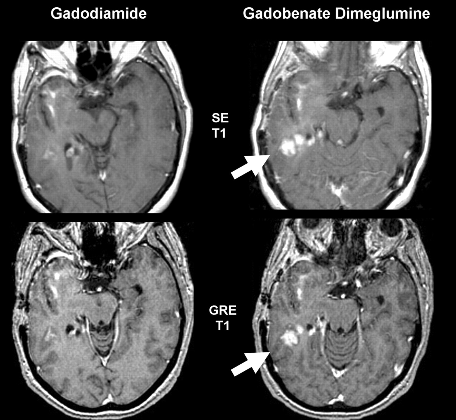
  • Fig 2.
    • Download figure
    • Open in new tab
    • Download powerpoint
    Fig 2.

    Glioblastoma with recurrent disease. This 55-year-old woman shows more conspicuous enhancement (arrows) in the right posterior temporal lobe with gadobenate dimeglumine than gadodiamide for both SE and GRE sequences. Although there are differences in angulation, changes were confirmed by review of all adjacent sections.

  • Fig 3.
    • Download figure
    • Open in new tab
    • Download powerpoint
    Fig 3.

    A 32-year-old woman with primary cerebellar glioma, which had previously been resected. A solid nodule of enhancement (arrow) is seen convincingly on the gadobenate dimeglumine–enhanced image. The clear visualization of contrast enhancement was important in postoperative decision making.

  • Fig 4.
    • Download figure
    • Open in new tab
    • Download powerpoint
    Fig 4.

    A 46-year-old man with a primary anaplastic carcinoma of the small bowel who developed sensory changes in the left upper extremity. The solitary metastasis (arrow) in the right superior frontal gyrus is only visualized on the gadobenate dimeglumine–enhanced image.

Tables

  • Figures
    • View popup
    Table 1:

    Qualitative assessments of patients with glial tumors, metastases, and extra-axial lesions

    Diagnostic Information End PointReaderGlial Tumors (n = 47)Metastases (n = 27)Extra-Axial Lesions (n = 18)
    Gadobenate Dimeglumine Preferred (%)Gadodiamide Preferred (%)PGadobenate Dimeglumine Preferred (%)Gadodiamide Preferred (%)PGadobenate Dimeglumine Preferred (%)Gadodiamide PreferredP
    Global diagnostic preference122 (46.8%)1 (2.1)<.000120 (74.1)2 (7.4)<.000114 (77.8)0.0001
    235 (74.5)1 (2.1)<.000118 (66.7)1 (3.7)<.000113 (72.2)0.0002
    325 (53.2)1 (2.1)<.000119 (70.4)2 (7.4)<.000116 (88.9)0<.0001
    Lesion border delineation115 (31.9)1 (2.1).000518 (66.7)1 (3.7)<.00011 (5.6)01.00
    218 (38.3)1 (2.1)<.000117 (63.0)0<.00013 (16.7)0.25
    316 (34.0)1 (2.1).000317 (63.0)2 (7.4).00077 (38.9)0.0156
    Definition of disease extent18 (17.0)0.007813 (48.1)1 (3.7).00181 (5.6)01.00
    27 (14.9)0.015613 (48.1)1 (3.7).00182 (11.1)0.50
    37 (14.9)0.015616 (59.3)1 (3.7).00031 (5.6)01.00
    Visualization of lesion internal morphology110 (21.3)0.00213 (48.1)1 (3.7).00182 (11.1)0.50
    214 (29.8)0.000113 (48.1)0.00023 (16.7)0.25
    38 (17.0)0.007814 (51.9)2 (7.4).00424 (22.2%)0.125
    Lesion contrast enhancement123 (48.9)1 (2.1)<.000120 (74.1)2 (7.4)<.000115 (83.3)0.0001
    235 (74.5)2 (4.3)<.000117 (63.0)1 (3.7)0.000114 (77.8)0.0001
    32 (55.3)1 (2.1)<.000120 (74.1)2 (7.4)<.000116 (88.9)0<.0001
    • View popup
    Table 2:

    Reasons for global diagnostic preference as expressed by expert blinded neuroradiologists

    Specifications of Global Diagnostic PreferenceReader 1 (n = 66)Preference Expressed Reader 2 (n = 79)Reader 3 (n= 76)
    Gadobenate Dimeglumine (n= 63) (%)Gadodiamide (n= 3) (%)Gadobenate Dimeglumine (n = 77) (%)Gadodiamide (n = 2) (%)Gadobenate Dimeglumine (n= 73) (%)Gadodiamide (n = 3) (%)
    Superior contrast enhancement63 (95.5)2 (3.0)74 (93.7)2 (2.5)73 (96.1)3 (3.9)
    Better delineation of normal structures007 (8.9)03 (3.9)0
    Better delineation of at least 1 lesion33 (50.0)1 (1.5)39 (49.4)044 (57.9)3 (3.9)
    Better visualization of lesion internal structure10 (15.2)030 (38.0)019 (25.0)0
    Detection of more lesions3 (4.8)1 (1.5)3 (3.8)1 (1.3)2 (2.6)1 (1.3)
    Greater diagnostic confidence10 (15.2)010 (12.7)1 (1.3)10 (13.2%)0
    • View popup
    Table 3:

    Comparison of CNR values on T1SE and T1GRE sequences after administration of gadobenate dimeglumine and gadodiamide for all evaluated lesions and for lesion subsets

    Lesion TypeReaderPostdose SE SequencesPostdose GRE Sequences
    Mean CNR Values% Increase in CNR with Gadobenate Dimeglumine (P)Mean CNR Values% Increase in CNR with Gadobenate Dimeglumine (P)
    Gadobenate DimeglumineGadodiamideGadobenate DimeglumineGadodiamide
    All lesions161.0746.6530.9<.000132.2622.6542.4<.0001
    246.4637.6723.3<.000123.5916.1945.7<.0001
    354.7140.6234.7<.000126.5617.8448.9<.0001
    Glial tumors147.0835.2833.5.000230.5721.6741.1.0068
    239.9532.9721.2.012522.8015.8144.2.0002
    343.8436.8219.1.099325.0820.0125.3.0466
    Metastases160.9746.5231.1.016227.4120.6332.9.0261
    246.2337.3223.9.011924.1416.0250.7<.0001
    351.2737.6336.2.009824.8115.4660.5.0004
    Extra-axial lesions180.5767.1719.9.004641.4428.8343.7.0021
    262.6149.2127.2.038827.3020.1935.2.0031
    377.1254.1842.3<.000133.9721.1061.0<.0001
    • Note:—SE indicates spin-echo; GRE, gradient recalled-echo; CNR, contrast-to-noise ratio.

PreviousNext
Back to top

In this issue

American Journal of Neuroradiology: 29 (9)
American Journal of Neuroradiology
Vol. 29, Issue 9
October 2008
  • Table of Contents
  • Index by author
Advertisement
Print
Download PDF
Email Article

Thank you for your interest in spreading the word on American Journal of Neuroradiology.

NOTE: We only request your email address so that the person you are recommending the page to knows that you wanted them to see it, and that it is not junk mail. We do not capture any email address.

Enter multiple addresses on separate lines or separate them with commas.
Contrast-Enhanced MR Imaging of Brain Lesions: A Large-Scale Intraindividual Crossover Comparison of Gadobenate Dimeglumine versus Gadodiamide
(Your Name) has sent you a message from American Journal of Neuroradiology
(Your Name) thought you would like to see the American Journal of Neuroradiology web site.
CAPTCHA
This question is for testing whether or not you are a human visitor and to prevent automated spam submissions.
Cite this article
H.A. Rowley, G. Scialfa, P.-y. Gao, J.A. Maldjian, D. Hassell, M.J. Kuhn, F.J. Wippold, M. Gallucci, B.C. Bowen, I.M. Schmalfuss, J. Ruscalleda, S. Bastianello, C. Colosimo
Contrast-Enhanced MR Imaging of Brain Lesions: A Large-Scale Intraindividual Crossover Comparison of Gadobenate Dimeglumine versus Gadodiamide
American Journal of Neuroradiology Oct 2008, 29 (9) 1684-1691; DOI: 10.3174/ajnr.A1185

Citation Manager Formats

  • BibTeX
  • Bookends
  • EasyBib
  • EndNote (tagged)
  • EndNote 8 (xml)
  • Medlars
  • Mendeley
  • Papers
  • RefWorks Tagged
  • Ref Manager
  • RIS
  • Zotero
0 Responses
Respond to this article
Share
Bookmark this article
Contrast-Enhanced MR Imaging of Brain Lesions: A Large-Scale Intraindividual Crossover Comparison of Gadobenate Dimeglumine versus Gadodiamide
H.A. Rowley, G. Scialfa, P.-y. Gao, J.A. Maldjian, D. Hassell, M.J. Kuhn, F.J. Wippold, M. Gallucci, B.C. Bowen, I.M. Schmalfuss, J. Ruscalleda, S. Bastianello, C. Colosimo
American Journal of Neuroradiology Oct 2008, 29 (9) 1684-1691; DOI: 10.3174/ajnr.A1185
del.icio.us logo Twitter logo Facebook logo Mendeley logo
  • Tweet Widget
  • Facebook Like
  • Google Plus One
Purchase

Jump to section

  • Article
    • Abstract
    • Materials and Methods
    • Results
    • Discussion
    • Conclusion
    • Footnotes
    • References
  • Figures & Data
  • Info & Metrics
  • Responses
  • References
  • PDF

Related Articles

  • No related articles found.
  • PubMed
  • Google Scholar

Cited By...

  • Reply:
  • The Benefits of High Relaxivity for Brain Tumor Imaging: Results of a Multicenter Intraindividual Crossover Comparison of Gadobenate Dimeglumine with Gadoterate Meglumine (The BENEFIT Study)
  • Gadolinium Contrast Agents for CNS Imaging: Current Concepts and Clinical Evidence
  • Multicenter, Intraindividual Comparison of Single-Dose Gadobenate Dimeglumine and Double-Dose Gadopentetate Dimeglumine for MR Angiography of the Supra-Aortic Arteries (the Supra-Aortic VALUE Study)
  • Does Higher Gadolinium Concentration Play a Role in the Morphologic Assessment of Brain Tumors? Results of a Multicenter Intraindividual Crossover Comparison of Gadobutrol versus Gadobenate Dimeglumine (the MERIT Study)
  • MR Imaging of Neoplastic Central Nervous System Lesions: Review and Recommendations for Current Practice
  • Correlations between Perfusion MR Imaging Cerebral Blood Volume, Microvessel Quantification, and Clinical Outcome Using Stereotactic Analysis in Recurrent High-Grade Glioma
  • Comparative Studies of Different Gadolinium Agents in Brain Tumors: Differences between Gadolinium Chelates and Their Possible Influence on Imaging Features
  • Diagnostic Yield of Double-Dose Gadobutrol in the Detection of Brain Metastasis: Intraindividual Comparison with Double-Dose Gadopentetate Dimeglumine
  • MR Imaging in Multiple Sclerosis: Review and Recommendations for Current Practice
  • Comparison of Gadobenate Dimeglumine and Gadodiamide in the Evaluation of Spinal Vascular Anatomy with MR Angiography
  • Crossref
  • Google Scholar

This article has not yet been cited by articles in journals that are participating in Crossref Cited-by Linking.

More in this TOC Section

  • Fast Contrast-Enhanced 4D MRA and 4D Flow MRI Using Constrained Reconstruction (HYPRFlow): Potential Applications for Brain Arteriovenous Malformations
  • Multimodal CT Provides Improved Performance for Lacunar Infarct Detection
  • Optimal MRI Sequence for Identifying Occlusion Location in Acute Stroke: Which Value of Time-Resolved Contrast-Enhanced MRA?
Show more Brain

Similar Articles

Advertisement

Indexed Content

  • Current Issue
  • Accepted Manuscripts
  • Article Preview
  • Past Issues
  • Editorials
  • Editor's Choice
  • Fellows' Journal Club
  • Letters to the Editor
  • Video Articles

Cases

  • Case Collection
  • Archive - Case of the Week
  • Archive - Case of the Month
  • Archive - Classic Case

More from AJNR

  • Trainee Corner
  • Imaging Protocols
  • MRI Safety Corner
  • Book Reviews

Multimedia

  • AJNR Podcasts
  • AJNR Scantastics

Resources

  • Turnaround Time
  • Submit a Manuscript
  • Submit a Video Article
  • Submit an eLetter to the Editor/Response
  • Manuscript Submission Guidelines
  • Statistical Tips
  • Fast Publishing of Accepted Manuscripts
  • Graphical Abstract Preparation
  • Imaging Protocol Submission
  • Evidence-Based Medicine Level Guide
  • Publishing Checklists
  • Author Policies
  • Become a Reviewer/Academy of Reviewers
  • News and Updates

About Us

  • About AJNR
  • Editorial Board
  • Editorial Board Alumni
  • Alerts
  • Permissions
  • Not an AJNR Subscriber? Join Now
  • Advertise with Us
  • Librarian Resources
  • Feedback
  • Terms and Conditions
  • AJNR Editorial Board Alumni

American Society of Neuroradiology

  • Not an ASNR Member? Join Now

© 2025 by the American Society of Neuroradiology All rights, including for text and data mining, AI training, and similar technologies, are reserved.
Print ISSN: 0195-6108 Online ISSN: 1936-959X

Powered by HighWire