
Article Figures & Data
Figures
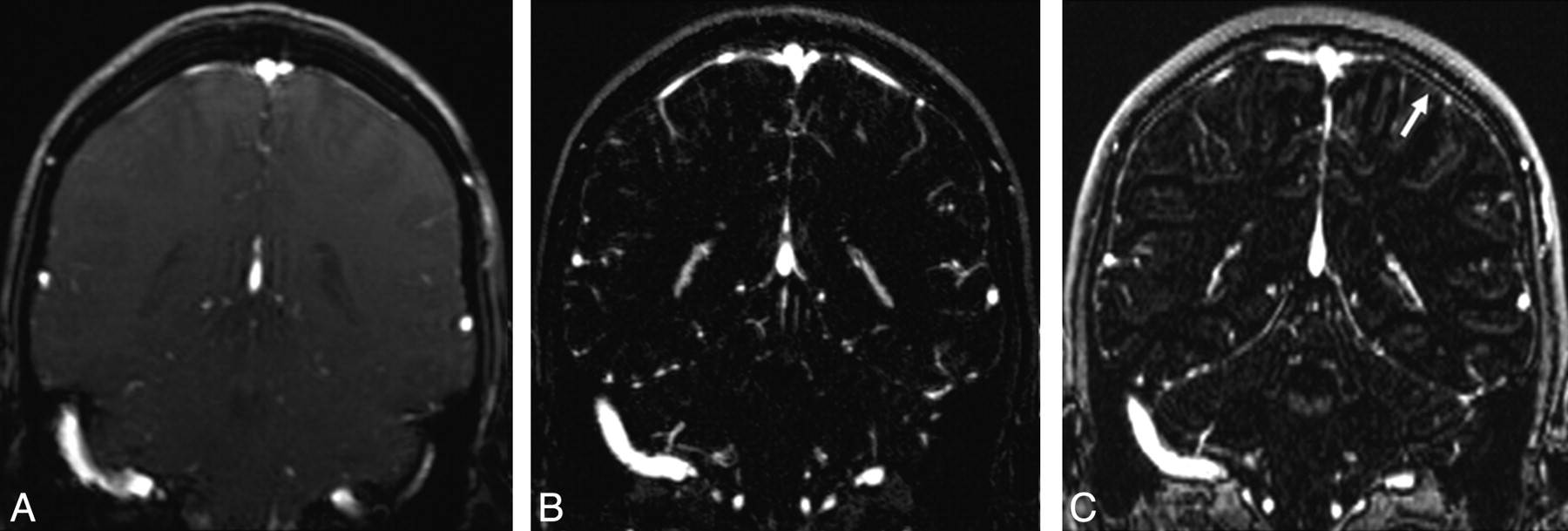
- Fig 1.
Coronal 2D MPR images obtained from 2D TOF MRV (A), CE MRV (B), and MPRAGE sequences (C). The section thickness is 2.5 mm (A), 0.7 mm (B), and 1.2 mm (C). The number of bridging veins in both hemispheres was compared. On the MPRAGE sequences (C), the bridging veins could not easily be distinguished from dural enhancement (arrow).
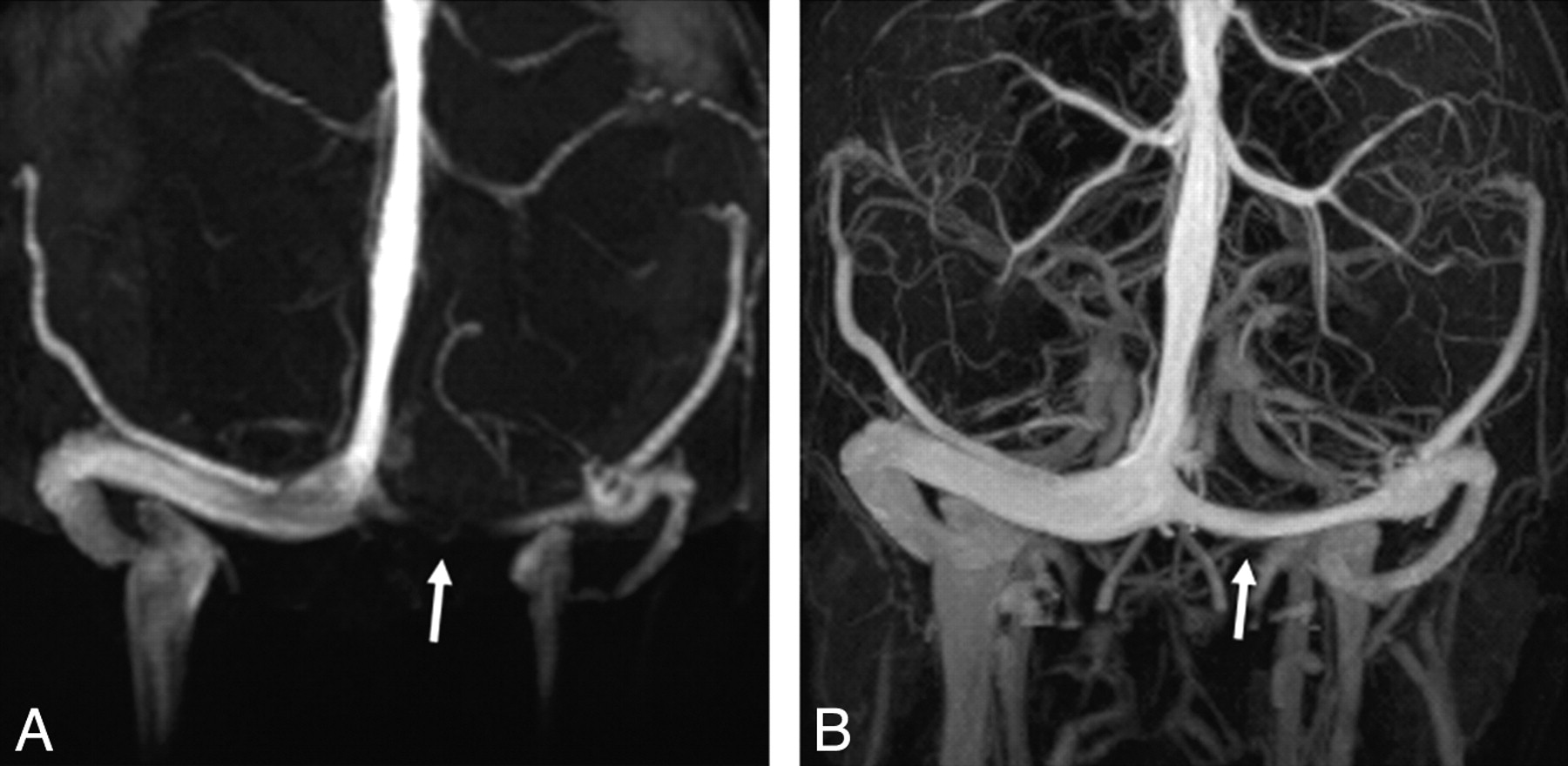
- Fig 2.
3D MIP images obtained from 2D TOF MRV (A), and CE MRV (B). Note the flow gap in the left transverse sinus on 2D TOF MRV (A) and the intense and continuous signal intensity on CE MRV (B) images (arrows).
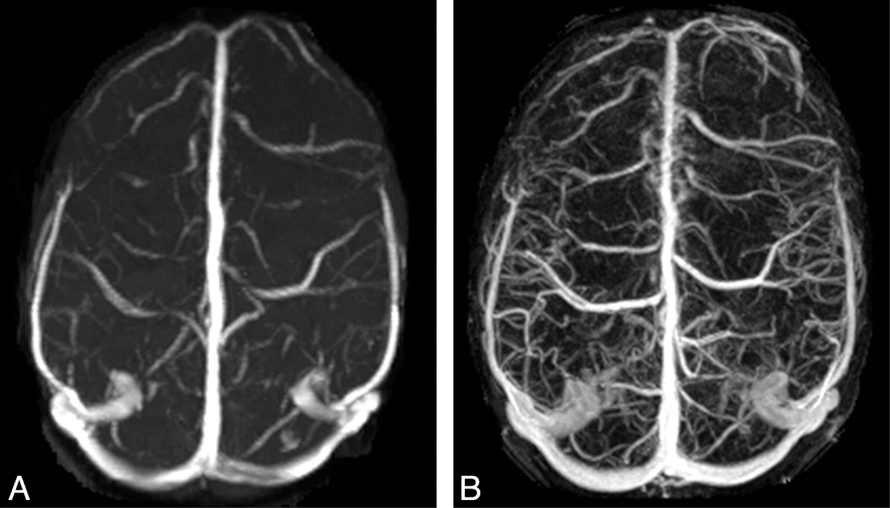
- Fig 3.
3D MIP images obtained from 2D TOF MRV (A), and CE MRV (B). The superficial veins were detected best by CE MRV (B).


Tables
Parameter CE TOF MPRAGE Sequence 3D FLASH 2D TOF 3D MPRAGE Orientation Sagittal Coronal Axial TR (ms) 3.78 28 2250 TE (ms) 1.5 6.93 3.04 Inversion time (ms) 900 Flip angle (°) 15 60 9 Bandwith (Hz/pixel) 430 65 200 Section thickness (mm) 0.4 2.5 1.2 Sections/partitions 416 128 144 Distant factor (%) −33 FOV (mm) (270–290)2 220 × 220 220 × 220 Matrix 384 × 384 256 × 256 384 × 384 GRAPPA-factor 2 2 2 Acquisition time (min. sec) 1.15 5.14 5.35 Note:—CE indicates contrast-enhanced MR venography; FLASH, fast low-angle shot; TOF, time-of-flight MR venography; MPRAGE, magnetization-prepared rapid acquisition of gradient echo; GRAPPA, generalized autocalibrating partially parallel acquisition.
- Table 2.
Comparison (P < .05) of the image quality of unpaired cerebral sinuses and veins in TOF MRV, CE MRV, and MPRAGE sequences
Venous structure Mean scores CE/TOF CE/MPRAGE MPRAGE/TOF CE/TOF/MPRAGE Superior Superior Superior Superior sagittal sinus 3.0/2.9/2.0 None CE TOF Inferior sagittal sinus 2.3/1.6/2.0 CE CE None Vein of Galen 3.0/2.6/3.0 CE None MPRAGE Straight sinus 3.0/3.0/2.7 None CE TOF Intercavernous sinus 2.6/0.4/2.1 CE CE MPRAGE Basilar plexus 0/0/0 None None None Note:—CE indicates contrast-enhanced MR venography; TOF, time-of-flight MR venography; MPRAGE, magnetization-prepared rapid acquisition of gradient echo.
- Table 3.
Comparison (P < .05) of the image quality of paired sinuses and veins and bridging veins in TOF MRV, CE MRV, and MPRAGE sequences
Venous structure Mean scores/number CE/TOF CE/MPRAGE MPRAGE/TOF CE/TOF/MPRAGE Superior Superior Superior Transverse sinuses R:3.0/2.6/3.0 L:3.0/2.2/3.0 CE None MPRAGE Sigmoid sinus R 3.0/2.4/2.0 CE CE TOF Sigmoid sinus L 3.0/2.1/2.0 CE CE None Jugular bulbs R:3.0/2.2/2.0 L:3.0/2.1/2.0 CE CE None Septal veins R 2.0/1.6/1.8 CE CE None Septal veins L 2.0/1.6/1.9 CE None MPRAGE Thalamostriate veins R:2.0/1.7/2.0 L:2.0/1.6/2.0 CE None MPRAGE Internal cerebral veins R:3.0/3.0/3.0 L:3.0/3.0/3.0 None None None Cavernous sinuses R:2.2/1.0/2.0 L:2.2/1.0/2.0 CE CE MPRAGE Sphenoparietal sinuses R:2.5/0.6/2.1 L:2.3/0.7/2.0 CE CE MPRAGE Superior petrosal sinuses R:1.5/0.6/1.5 L:1.8/0.7/1.6 CE None MPRAGE Inferior petrosal sinuses R:2.6/1.2/2.0 L:2.3/0.8/2.0 CE CE MPRAGE Basal veins of Rosenthal R:3.0/2.3/2.9 L:3.0/2.6/2.9 CE None MPRAGE Trolard veins R:2.3/1.0/1.7 L:2.5/1.1/1.8 CE CE MPRAGE Labbé vein R 3.0/2.1/2.6 CE CE MPRAGE Labbé vein L 3.0/2.4/3.0 CE None MPRAGE Sup. middle cerebral vein R:2.9/1.8/2.3 L:2.8/1.5/2.3 CE CE MPRAGE Bridging veins R:8.8/3.6/4.9 L:7.9/3.3/4.4 CE CE MPRAGE Note:—CE indicates contrast-enhanced MR venography; TOF, time-of-flight MR venography; MPRAGE, magnetization-prepared rapid acquisition of gradient echo; R, right; L, left; Sup, superior.
In this issue


Jump to section
Related Articles
Cited By...
- Depiction of the Superior Petrosal Vein Complex by 3D Contrast-Enhanced MR Angiography
- Prevalence of dural venous sinus stenosis and hypoplasia in a generalized population
- Visualization of the Internal Cerebral Veins on MR Phase-Sensitive Imaging: Comparison with 3D Gadolinium-Enhanced MR Venography and Fast-Spoiled Gradient Recalled Imaging
- Diagnosis and Management of Cerebral Venous Thrombosis: A Statement for Healthcare Professionals From the American Heart Association/American Stroke Association