
Article Figures & Data
Figures
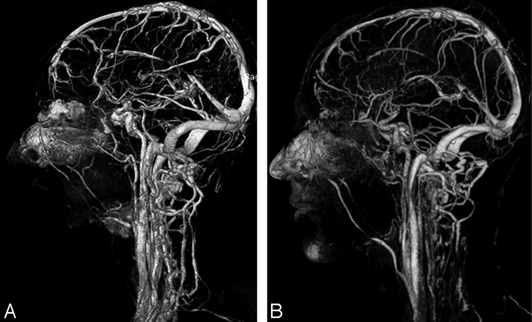
- Fig 1.
3D volume-rendered images from full-dose (15 mL) (A) and half-dose (7.5 mL) (B) contrast-enhanced MRV show diagnostic image quality of most of the cerebral venous structures.
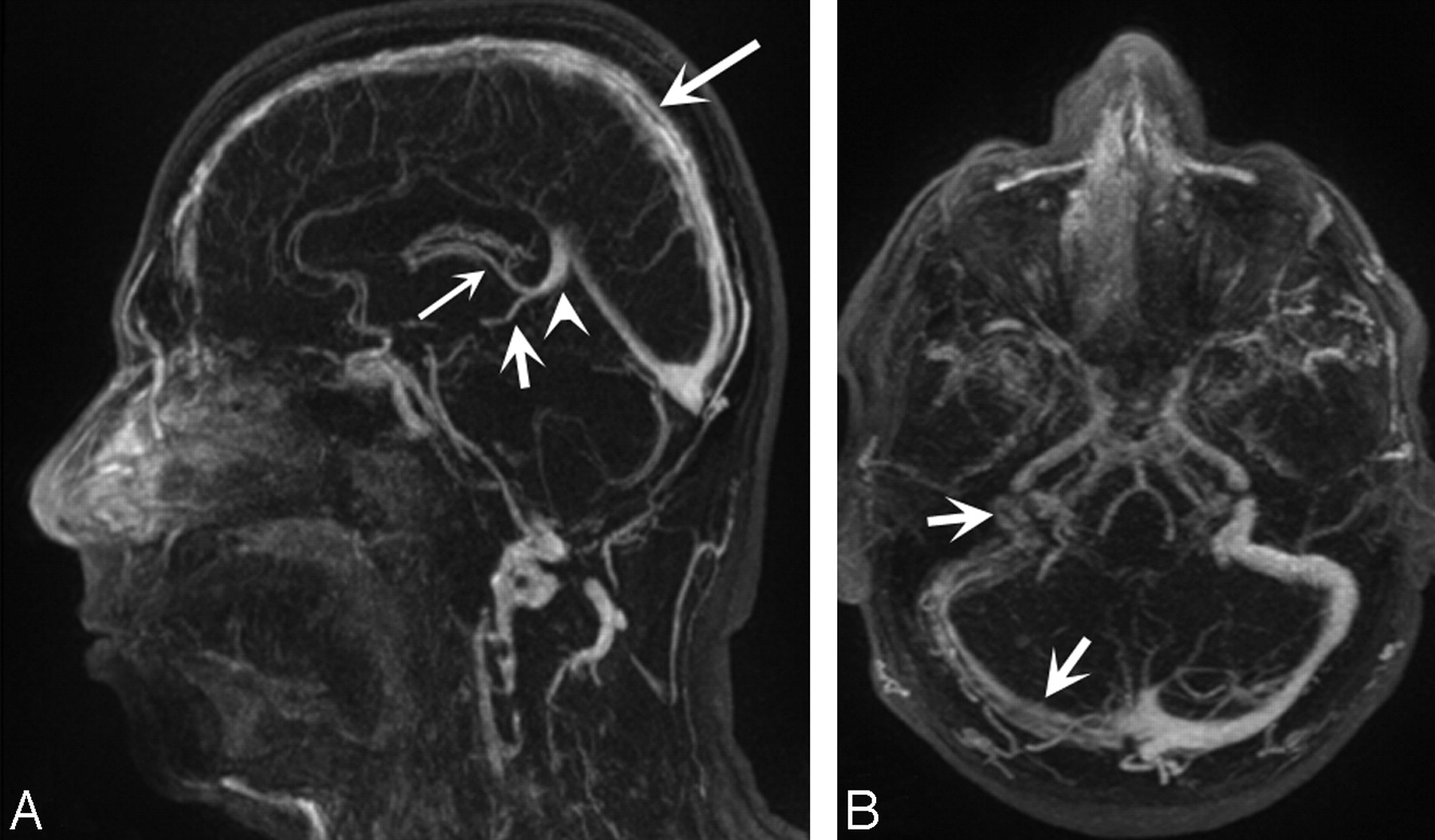
- Fig 2.
Half-dose CE-MRV. Sagittal (A) and axial (B) thin MIP images (TR/TE, 3.4/1.3 ms; flip angle, 25°) in a 28-year-old man with history of headache show filling defects within the superior sagittal sinus (A, large arrow) and transverse and sigmoid sinuses (B, small arrows) consistent with thrombosis. Note the high diagnostic quality of the vein of Galen (A, arrowhead), the basal vein of Rosenthal (A, small arrow), and the internal cerebral veins (A, thin large arrow).
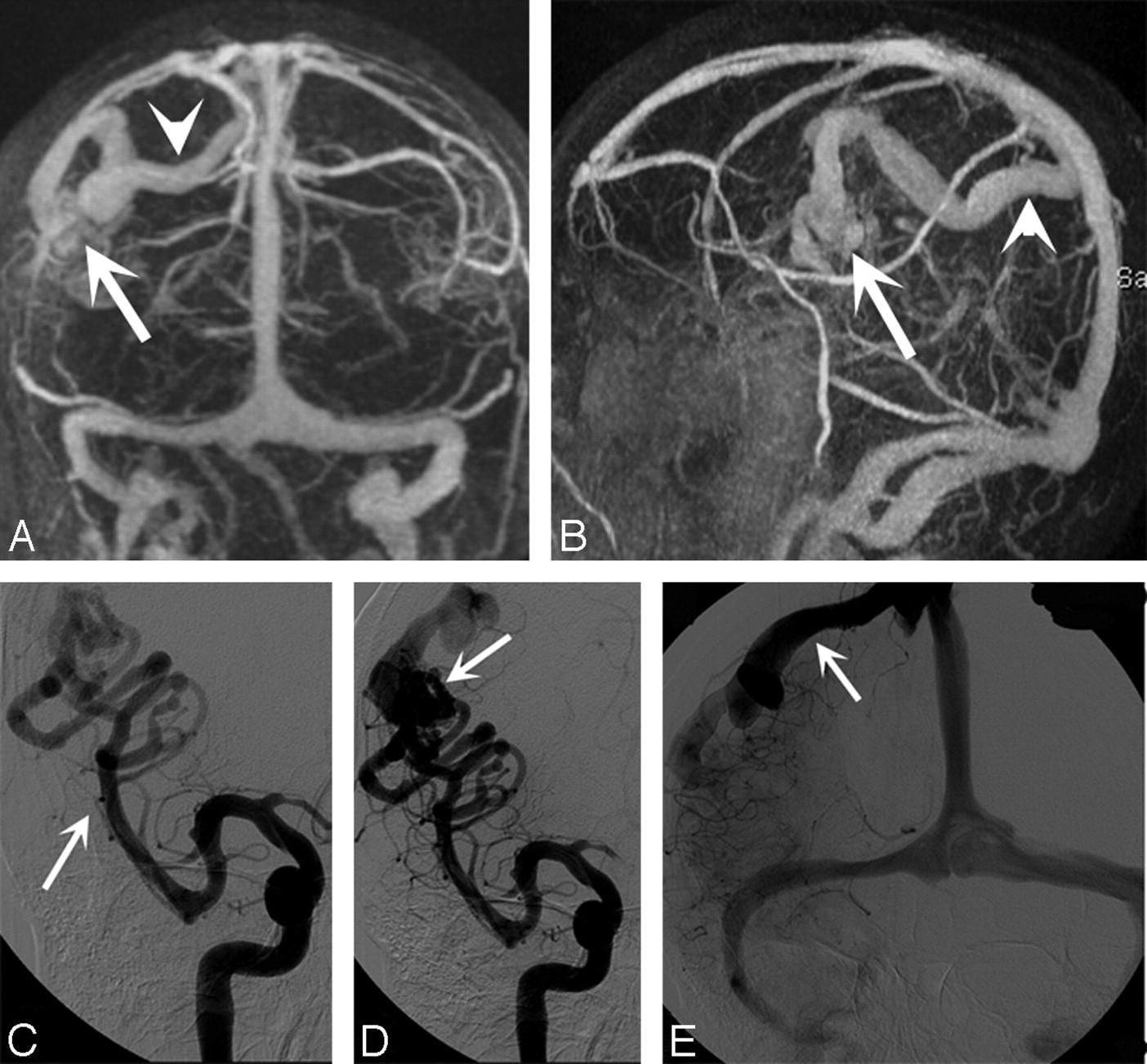
- Fig 3.
Half-dose CE-MRV. A and B, Coronal (A) and sagittal (B) full-thickness MIP images (TR/TE, 3.4/1.3 ms; flip angle, 25°) in a 27-year-old man show cerebral arteriovenous malformation nidus (A and B, arrow) and a large draining vein to the superior sagittal sinus (A and B, arrowhead). C–E, DSAs confirm the findings on CE-MRV. Feeder artery (middle cerebral artery) (C, arrow), arteriovenous malformation nidus (D, arrow), and draining vein to the superior sagittal sinus (E, arrow) are clearly visualized.


Tables
- Table 1:
Visualization of venous segments on full-dose and half-dose high-spatial-resolution CE-MRV (n = 640)*
Location Reader 1 Reader 2 Full-Dose (half-dose) CE-MRV Full-Dose (half-dose) CE-MRV Mean ± SD Median Range Mean ± SD Median Range Superior sagittal sinus 4.00 ± 0.00 (4.00 ± 0.00) 4 (4) 4–4 (4–4) 4.00 ± 0.00 (4.00 ± 0.00) 4 (4) 4–4 (4–4) Inferior sagittal sinus 3.45 ± 0.60 (3.40 ± 0.68) 3.5 (3.5) 2–4 (2–4) 3.55 ± 0.60 (3.45 ± 0.69) 4 (4) 2–4 (2–4) Transverse sinus 3.97 ± 0.16 (4.00 ± 0.00) 4 (4) 3–4 (4–4) 4.00 ± 0.00 (4.00 ± 0.00) 4 (4) 4–4 (4–4) Sigmoid sinus 3.97 ± 0.16 (3.97 ± 0.16) 4 (4) 3–4 (3–4) 4.00 ± 0.00 (4.00 ± 0.00) 4 (4) 4–4 (4–4) Straight sinus 3.95 ± 0.22 (3.90 ± 0.31) 4 (4) 3–4 (3–4) 3.90 ± 0.31 (3.95 ± 0.22) 4 (4) 3–4 (3–4) Cavernous sinus 3.72 ± 0.60 (3.70 ± 0.52) 4 (4) 2–4 (2–4) 3.70 ± 0.65 (3.62 ± 0.59) 4 (4) 2–4 (2–4) Superior petrosal sinus 3.05 ± 0.75 (2.97 ± 0.66) 3 (3) 1–4 (1–4) 2.95 ± 0.71 (2.90 ± 0.67) 3 (3) 1–4 (1–4) Torcula herophili 4.00 ± 0.00 (4.00 ± 0.00) 4 (4) 4–4 (4–4) 4.00 ± 0.00 (4.00 ± 0.00) 4 (4) 4–4 (4–4) Cortical vein 3.97 ± 0.16 (3.97 ± 0.16) 4 (4) 3–4 (3–4) 3.92 ± 0.27 (3.95 ± 0.22) 4 (4) 3–4 (3–4) Vein of Galen 3.95 ± 0.22 (3.90 ± 0.31) 4 (4) 3–4 (3–4) 3.90 ± 0.31 (3.85 ± 0.37) 4 (4) 3–4 (3–4) Internal cerebral vein 3.65 ± 0.53 (3.55 ± 0.64) 4 (4) 2–4 (2–4) 3.62 ± 0.54 (3.52 ± 0.64) 4 (4) 2–4 (2–4) Middle cerebral vein 3.25 ± 0.67 (2.37 ± 0.87) 3 (2) 2–4 (1–4) 3.25 ± 0.71 (2.37 ± 0.84) 3 (2.5) 2–4 (1–4) Basal vein of Rosenthal 3.40 ± 0.68 (3.20 ± 0.70) 3.5 (3) 2–4 (2–4) 3.35 ± 0.75 (3.35 ± 0.67) 3.5 (3) 2–4 (2–4) Septal vein 2.95 ± 0.60 (2.35 ± 0.81) 3 (2) 2–4 (1–4) 2.95 ± 0.69 (2.35 ± 0.75) 3 (2.5) 2–4 (1–3) Superior cerebellar vein 2.72 ± 0.60 (2.32 ± 0.76) 3 (2.5) 1–4 (1–3) 2.82 ± 0.68 (2.37 ± 0.77) 3 (3) 1–4 (1–4) Posterior tonsillar vein 2.87 ± 0.56 (2.37 ± 0.63) 3 (2) 2–4 (1–3) 3.00 ± 0.60 (2.45 ± 0.68) 3 (2.5) 2–4 (1–4) Inferior vermian vein 2.70 ± 0.73 (2.15 ± 0.74) 3 (2) 2–4 (1–3) 2.85 ± 0.75 (2.25 ± 0.79) 3 (2) 2–4 (1–3) Superior ophthalmic vein 3.62 ± 0.59 (3.55 ± 0.60) 4 (4) 2–4 (2–4) 3.70 ± 0.52 (3.55 ± 0.55) 4 (4) 2–4 (2–4) Thalamostriate vein 2.67 ± 0.73 (2.25 ± 0.74) 3 (2) 1–4 (1–3) 2.62 ± 0.74 (2.22 ± 0.73) 3 (2) 1–4 (1–3) Internal jugular vein 4.00 ± 0.00 (4.00 ± 0.00) 4 (4) 4–4 (4–4) 4.00 ± 0.00 (4.00 ± 0.00) 4 (4) 4–4 (4–4) Note:—CE-MRV indicates contrast-enhanced MR venography.
* The 4-point scale for evaluation of visualization of venous segments is as follows: grade 1, not visible; grade 2, partially visible, not sufficient for diagnosis; grade 3, generally homogeneous enhancement and continuity of venous structure, sufficient for diagnosis; and grade 4, excellent image quality with highly homogeneous and continuous enhancement with sharpness of vessel border allowing highly confident diagnosis. There was no significant difference in the vessel delineation scores assigned by the 2 readers (P > .05 for all segments). Analysis with the κ coefficient revealed excellent interobserver agreement for the full-dose (κ = 0.87) and half-dose (κ = 0.85) groups. Delineation scores were significantly lower for the small venous segments, including the middle cerebral, septal, superior cerebellar, inferior vermian, posterior tonsillar, and thalamostriate veins in the half-dose group compared with the single-dose group (P < .01 for all).
Full-Dose CE-MRV Half-Dose CE-MRV SNR CNR SNR CNR Superior sagittal sinus 611.5 ± 82.3 501.3 ± 77.6 322.2 ± 45.1 291.3 ± 41.6 Inferior sagittal sinus 276.8 ± 31.2 236.5 ± 29.3 158.7 ± 23.3 132.7 ± 21.5 Straight sinus 508.8 ± 77.6 461.1 ± 70.9 234.5 ± 32.6 203.8 ± 28.8 Transverse sinus 532.9 ± 81.2 479.2 ± 75.4 302.2 ± 45.6 269.3 ± 40.9 Sigmoid sinus 555.4 ± 84.6 497.6 ± 79.4 309.4 ± 48.1 261.8 ± 43.3 Vein of Galen 490.7 ± 62.3 434.2 ± 58.2 224.4 ± 29.7 194.4 ± 26.9 Internal cerebral vein 390.1 ± 58.8 342.1 ± 50.8 201.3 ± 31.3 168.6 ± 30.2 Internal jugular vein 572.6 ± 81.1 516.6 ± 72.9 334.8 ± 44.5 305.8 ± 40.8 Note:—SNR indicates signal intensity-to-noise ratio; CNR, contrast-to-noise ratio.
* All values are presented as mean ± SD. SNR and CNR values are significantly lower in the half-dose CE-MRV group compared with the single-dose CE-MRV group (P < .001 for all segments).


