
Article Figures & Data
Figures
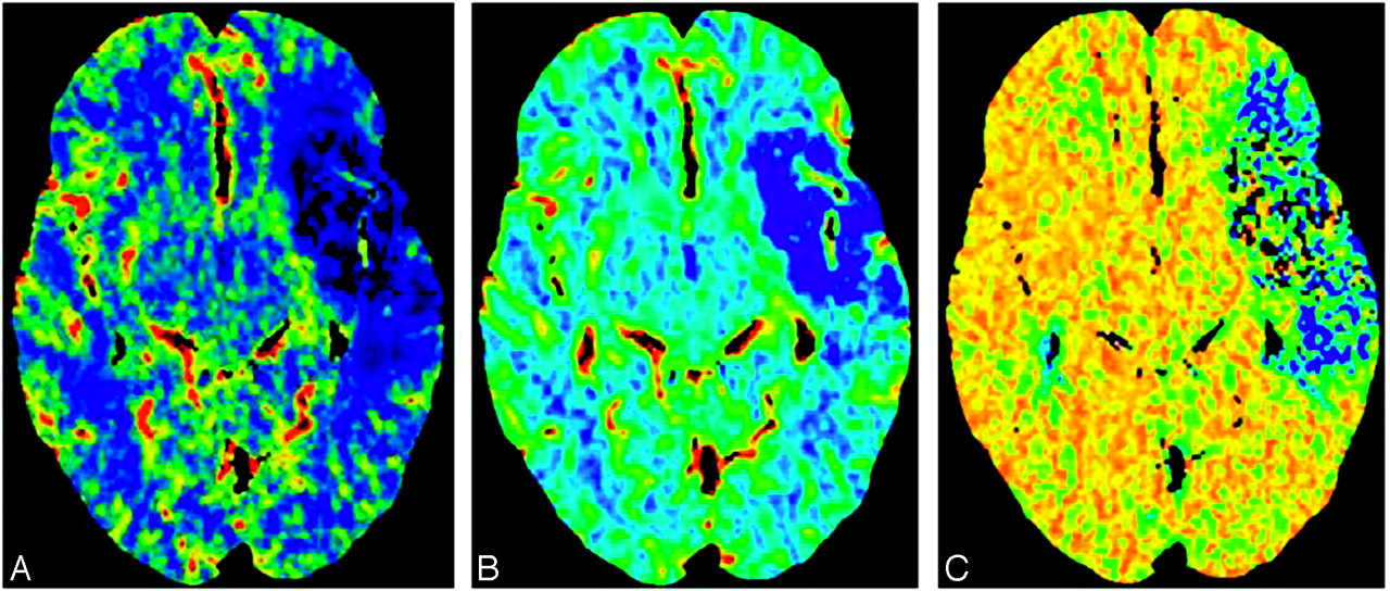
- Fig 1.
CT perfusion images obtained in a patient with acute ischemic stroke demonstrate a large perfusion defect in the left MCA distribution, with minimal CBV/MTT or CBF mismatch. A, CBF. B, CBV. C, MTT.
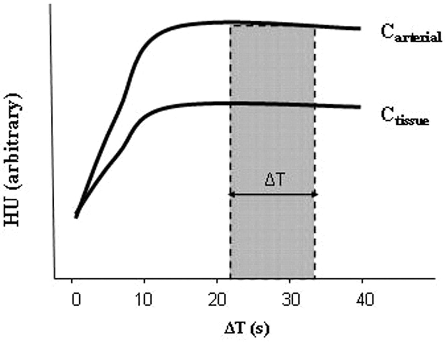
- Fig 2.
CTA source images acquired during a steady state of contrast concentration for both the arterial and tissue−time-attenuation curves (ΔT) are predominantly blood-volume− rather than blood-flow−weighted. The change in attenuation due to iodine administration is directly proportional to its concentration. CBV equals the ratio of the areas under the 2 curves, Ctissue and Carterial, respectively. This can be approximated as the ratio of the HUtissue/HUarterial when the 2 curves approach steady state.
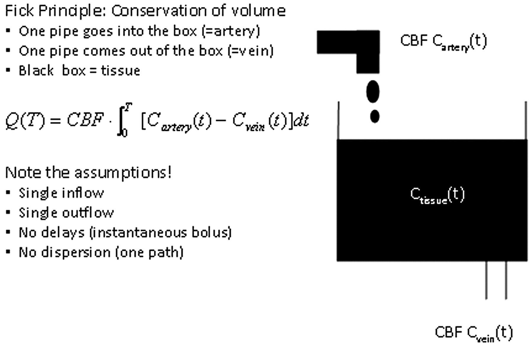
- Fig 3.
Fick Principle.


Tables
CTP MRP CTP advantages Linear relation of signal changes with contrast concentration; quantitative maps Nonlinear relation of signal changes with gadolinium concentration; nonquantitative maps Higher spatial resolution Lower spatial resolution More readily available Not as readily available MRP advantages Ionizing radiation No ionizing radiation Limited z-direction coverage Whole-brain coverage Iodinated contrast–related concerns Gadolinium contrast concerns (NSF) Complex postprocessing Less labor-intensive postprocessing Note:—NSF indicates nephrogenic systemic fibrosis; CTP, CT perfusion; MRP, MR perfusion.
Study Subjects Validation Method Results Comments Animal Studies Gobbel et al (1991)69 25 Healthy dogs Microspheres CBF: r = 0.95, P < .05 Cenic et al (1999)61 13 Rabbits Microspheres CBF: r = 0.84, P < .001, slope = 0.97 Deconvolution method Nabavi et al (1999)62 5 Healthy and 7 ischemic beagles Microspheres Healthy: CBF, r = 0.78, slope = 0.93; ischemic: r = 0.79, slope = 0.97 Deconvolution method Cenic et al (2000)64 9 Rabbits with tumors Microspheres; postmortem histology CBF: r = 0.85, P < .001, slope = 0.99 Deconvolution method Nabavi et al (2001)66 7 Ischemic rabbits CBF: r = 0.95, slope = 1.05 Deconvolution method CBV: r = 0.80, slope = 0.49 MTT: r = 0.85, slope = 0.95 Human studies Gillard et al (2000)72 2 With gliomas, 6 with AVMs PET r2 = 0.52 Maximal slope method; VPE Wintermark et al (2001)67 9 With cerebrovascular disease Xe-CT CBF: r2 = 0.79, slope = 0.87 Deconvolution method Kudo et al (2003)70 5 Healthy subjects PET CBF: r = 0.69, slope = 1.05 Deconvolution method; VPE Sase et al (2005)71 7 Healthy subjects Xe-CT CBF: r2 = 0.46–0.93 depending on the brain region, P < .05, slopes <0.81 or >1.20 Maximum slope method; VPE; different territories compared Kanazawa et al (2007)29 28 Healthy subjects Xe-CT CBF: r = 0.61–0.70, P < .01 Several territories compared Bisdas (2008)73 7 With strokes PET CBF: r = 0.77, P = .00 Deconvolution method Note:—AVM indicates arteriovenous malformation; PET, positron-emission tomography; Xe-CT, xenon-enhanced CT; VPE, vascular pixel elimination; CBF, cerebral blood flow; MTT, mean transit time.
In this issue


Jump to section
Related Articles
Cited By...
- Comparison of Automated MRI Perfusion Analysis Software: Agreement in Ischemic Penumbra Estimation and Decision-Making for Endovascular Thrombectomy
- Reducing False-Positives in CT Perfusion Infarct Core Segmentation Using Contralateral Local Normalization
- Detecting CTP Truncation Artifacts in Acute Stroke Imaging from the Arterial Input and the Vascular Output Functions
- Defining Ischemic Core in Acute Ischemic Stroke Using CT Perfusion: A Multiparametric Bayesian-Based Model
- Whole-brain Volume Perfusion Computed Tomography: Acquisition Techniques and Radiation Dose
- Admission CT perfusion may overestimate initial infarct core: the ghost infarct core concept
- Comparison of Perfusion CT Software to Predict the Final Infarct Volume After Thrombectomy
- Occult Anterograde Flow Is an Under-Recognized but Crucial Predictor of Early Recanalization With Intravenous Tissue-Type Plasminogen Activator
- Whole-Brain Adaptive 70-kVp Perfusion Imaging with Variable and Extended Sampling Improves Quality and Consistency While Reducing Dose
- A Novel Technique for the Measurement of CBF and CBV with Robot-Arm-Mounted Flat Panel CT in a Large-Animal Model
- Comparison of Computed Tomographic and Magnetic Resonance Perfusion Measurements in Acute Ischemic Stroke: Back-to-Back Quantitative Analysis
- Pretreatment Advanced Imaging in Patients with Stroke Treated with IV Thrombolysis: Evaluation of a Multihospital Data Base
- Can Iterative Reconstruction Improve Imaging Quality for Lower Radiation CT Perfusion? Initial Experience
- Differences in CT Perfusion Summary Maps for Patients with Acute Ischemic Stroke Generated by 2 Software Packages
- Application of acute stroke imaging: Selecting patients for revascularization therapy
- CT Angiographic Source Images: Flow- or Volume-Weighted?
- Evaluation of CT Perfusion in the Setting of Cerebral Ischemia: Patterns and Pitfalls
- FDA Investigates the Safety of Brain Perfusion CT
- Theoretic Basis and Technical Implementations of CT Perfusion in Acute Ischemic Stroke, Part 2: Technical Implementations