
Article Figures & Data
Figures
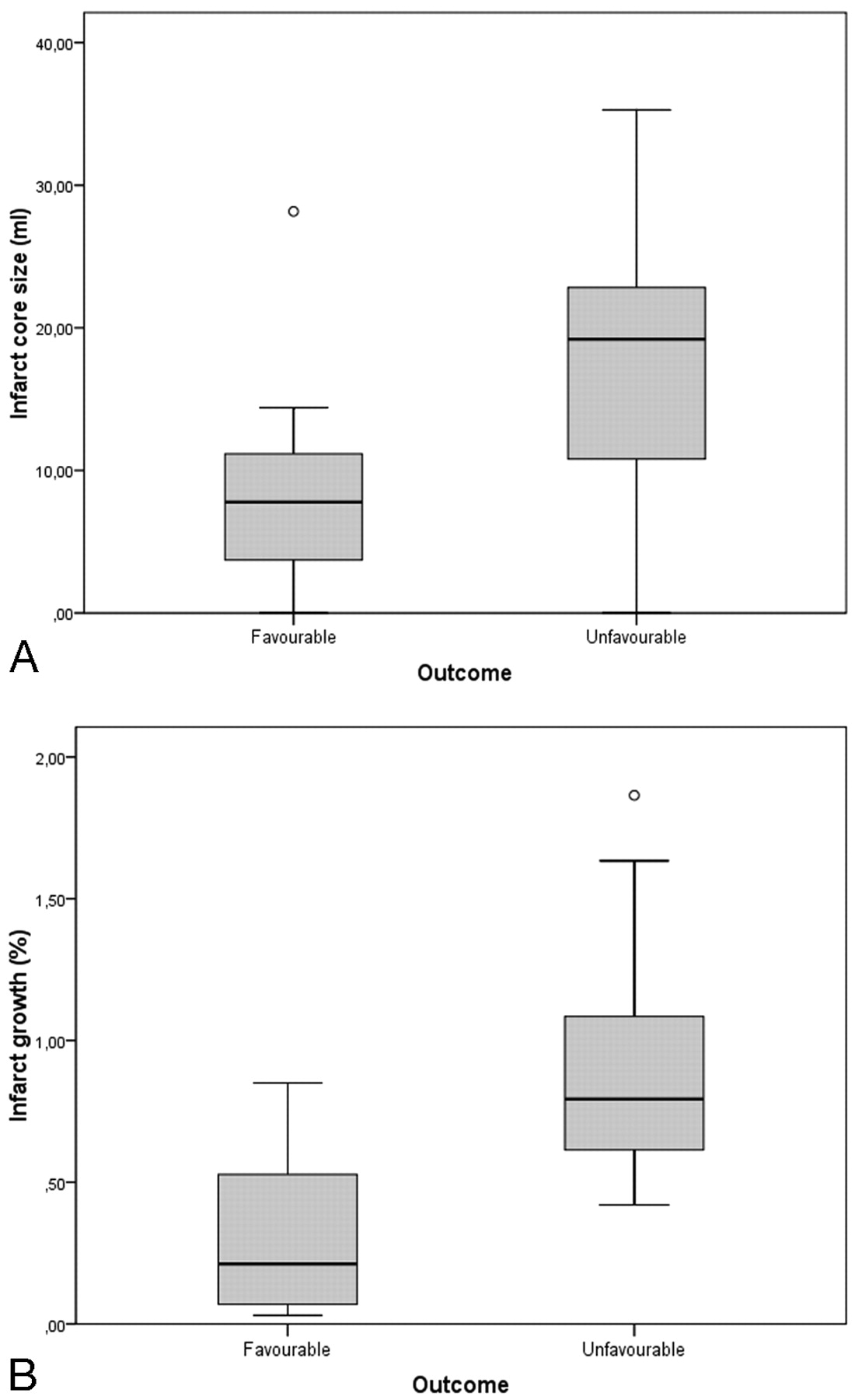
- Fig 1.
Box and whisker graphs of infarct core size according to clinical outcome (A) and infarct growth according to recanalization after IAT (B).
- Fig 2.
A 35-year-old man with left hemiplegia, imaged 3 hours after symptom onset. NCCT, CBV, CBF, and TTP maps, delayed NCCT (A−E, first level; F−L, second level); and IAT (M−O). NCCT shows mild hypoattenuation of the right lenticular nucleus. PCT color maps show small multiple areas with severely reduced CBV (CBV ratio <33%) and CBF (<11.5 mL/100 g/min) in the anterior third of the right lenticular nucleus; external capsule; and fronto-opercular, insular, posterior temporal and parietal cortices, corresponding to the infarct core (solid line). The infarct core is surrounded by a large perfusion deficit in the right MCA cortical territory, characterized by reduced CBF (color-coded blue) and increased TTP (color-coded red), indicating a TTP-CBV mismatch, which corresponds to the ischemic penumbra. IADSA, performed after PCT, shows proximal right M1 occlusion (M), with poor collateral leptomeningeals. The patient underwent intra-arterial thrombolysis with injection of rtPA and mechanical clot manipulation, followed by percutaneous transluminal angioplasty, with complete right MCA recanalization (N). A mild residual M1 stenosis is identifiable at the end of the procedure (O). Follow-up CT scans (E and L), obtained 2 days after stroke, show small multiple infarcts in the right caudate and anterior third of the lenticular nucleus; insular cortex; and temporal, frontal, and parietal cortices, which correspond to the infarct core, with consequent recovery of a large portion of the mismatch area. At 3 months, the patient was independent (mRS score, 1).


Tables
Core Penumbra P Value CBV ratio 0.25 ± 0.09 0.81 ± 0.17 <.001 CBF (mL/100 g/min) 6.9 ± 2.8 25.9 ± 2.7 <.001 CBF ratio 0.14 ± 0.05 0.49 ± 0.06 <.001 ΔTTP (s) 6.5 ± 2.63 4.32 ± 1.54 <.01 Note:—CBV indicates cerebral blood volume; CBF, cerebral blood flow; TTP, time to peak.
Core Final Infarct Infarct Growth NIHSS Score mRS Score Core 1 0.793 0.401 0.408 0.604 P < .001 P = .038 P = .035 P = .001 Final infarct 0.793 1 0.788 0.56 0.73 P < .001 P < .001 P = .002 P < .001 Infarct growth 0.401 0.788 1 0.493 0.499 P = .038 P < .001 P = .009 P = .008 NIHSS score 0.408 0.56 0.493 1 0.545 P = .035 P = .002 P = .009 P = .003 mRS score 0.604 0.73 0.499 0.545 1 P = .001 P < .001 P = .008 P = .003 Note:—NIHSS indicates National Institutes of Health Stroke Scale; mRS, modified Rankin score.
Favorable Outcome (n = 12) Unfavorable Outcome (n = 15) P Value Age (year) 46.4 ± 13.9 58.7 ± 15.2 .034 Sex (M/F) 8/4 5/10 n.s. NIHSS score 15 ± 4 21 ± 5 <.01 Serum glucose level (mg/dL) 135.6 ± 38.1 136.7 ± 29.7 n.s. Core (mL) 8.6 ± 7.8 19.8 ± 14.5 .03 TTP/CBV mismatch ratio (%) 75.4 ± 19.8 55.8 ± 23 .03 Infarct volume (mL) 11.3 ± 11.2 36.8 ± 15.7 <.01 Infarct growth (%) 31.2 ± 28 90 ± 45 <.01 Time to IAT (min) 61.2 ± 36.5 66.6 ± 35.9 n.s. Time to recanalization (min) 338 ± 50 371 ± 36 n.s Recanalization rate (TIMI, 2–3) 11/12 8/15 .03 Symptomatic hemorrhage 1 1 Thrombolytic agent n.s UK 6/17 11/17 rtPA 6/10 4/10 ASPECTS 9 8.6 n.s. Note:—n.s. indicates not significant; UK, urokinase; IAT, intra-arterial thrombolysis; rtPA, recombinant tissue plasminogen activator; ASPECTS, Alberta Stroke Program Early CT Score.
- Table 4:
Multiple regression analysis of the best model from an “all subset” model selection (R2 = 74.6%)*
Change X Change Y 95% CI P Value Core (mL) 1 SD = 13.21 +0.96 0.17–1.75 .01 NIHSS 1 SD = 5.8 +0.47 0.13–1.08 .05 Sex (male) 1 SD = 1 −1.06 −2.1 to −0.1 .035 Note:—X indicates predictors (core, NIHSS score, sex); Y, outcome measure (mRS score).
* The regression parameter is expressed as the average change of the outcome measure per 1 SD change of the predictor variable.
In this issue


Jump to section
Related Articles
Cited By...
- Assessment of computed tomography perfusion software in predicting spatial location and volume of infarct in acute ischemic stroke patients: a comparison of Sphere, Vitrea, and RAPID
- Assessment of a Bayesian Vitrea CT Perfusion Analysis to Predict Final Infarct and Penumbra Volumes in Patients with Acute Ischemic Stroke: A Comparison with RAPID
- The POST trial: initial post-market experience of the Penumbra system: revascularization of large vessel occlusion in acute ischemic stroke in the United States and Europe
- Predictive Value of RAPID Assessed Perfusion Thresholds on Final Infarct Volume in SWIFT PRIME (Solitaire With the Intention for Thrombectomy as Primary Endovascular Treatment)
- Interaction of Recanalization, Intracerebral Hemorrhage, and Cerebral Edema After Intravenous Thrombolysis
- Effect of Collaterals on Clinical Presentation, Baseline Imaging, Complications, and Outcome in Acute Stroke
- Value of Computed Tomographic Perfusion-Based Patient Selection for Intra-Arterial Acute Ischemic Stroke Treatment
- Computed Tomographic Angiography and Cerebral Blood Volume Can Predict Final Infarct Volume and Outcome After Recanalization
- Relative Cerebral Blood Volume as a Marker of Durable Tissue-at-Risk Viability in Hyperacute Ischemic Stroke
- Implementation of a patient selection protocol for intra-arterial therapy increases treatment rates in patients with acute ischemic stroke
- Prediction of Recanalization Trumps Prediction of Tissue Fate: The Penumbra: A Dual-edged Sword
- Pre-intervention triage incorporating perfusion imaging improves outcomes in patients undergoing endovascular stroke therapy: a comparison with the device trials
- Leukoaraiosis and Sex Predict the Hyperacute Ischemic Core Volume
- Location of the Clot and Outcome of Perfusion Defects in Acute Anterior Circulation Stroke Treated with Intravenous Thrombolysis
- Multiparametric MRI and CT Models of Infarct Core and Favorable Penumbral Imaging Patterns in Acute Ischemic Stroke
- Selecting stroke patients for intra-arterial therapy
- Systematic Review of Methods for Assessing Leptomeningeal Collateral Flow
- The POST trial: initial post-market experience of the Penumbra system: revascularization of large vessel occlusion in acute ischemic stroke in the United States and Europe
- Cost-effectiveness of multimodal CT for evaluating acute stroke
- Imaging in acute ischaemic stroke: essential for modern stroke care