Advertisement

Main menu

  • Home
  • Content
    • Current Issue
    • Accepted Manuscripts
    • Article Preview
    • Past Issue Archive
    • AJNR Case Collection
    • Case of the Week Archive
    • Classic Case Archive
    • Case of the Month Archive
  • Special Collections
    • Spinal CSF Leak Articles (Jan 2020-June 2024)
    • 2024 AJNR Journal Awards
    • Most Impactful AJNR Articles
  • Multimedia
    • AJNR Podcast
    • AJNR Scantastics
    • Video Articles
  • For Authors
    • Submit a Manuscript
    • Author Policies
    • Fast publishing of Accepted Manuscripts
    • Graphical Abstract Preparation
    • Manuscript Submission Guidelines
    • Imaging Protocol Submission
    • Submit a Case for the Case Collection
  • About Us
    • About AJNR
    • Editorial Board
  • More
    • Become a Reviewer/Academy of Reviewers
    • Subscribers
    • Permissions
    • Alerts
    • Feedback
    • Advertisers
    • ASNR Home
  • Other Publications
    • ajnr

User menu

  • Alerts
  • Log in
  • Log out

Search

  • Advanced search
American Journal of Neuroradiology

American Journal of Neuroradiology

ASHNR American Society of Functional Neuroradiology ASHNR American Society of Pediatric Neuroradiology ASSR
  • Alerts
  • Log in
  • Log out

Advanced Search

  • Home
  • Content
    • Current Issue
    • Accepted Manuscripts
    • Article Preview
    • Past Issue Archive
    • AJNR Case Collection
    • Case of the Week Archive
    • Classic Case Archive
    • Case of the Month Archive
  • Special Collections
    • Spinal CSF Leak Articles (Jan 2020-June 2024)
    • 2024 AJNR Journal Awards
    • Most Impactful AJNR Articles
  • Multimedia
    • AJNR Podcast
    • AJNR Scantastics
    • Video Articles
  • For Authors
    • Submit a Manuscript
    • Author Policies
    • Fast publishing of Accepted Manuscripts
    • Graphical Abstract Preparation
    • Manuscript Submission Guidelines
    • Imaging Protocol Submission
    • Submit a Case for the Case Collection
  • About Us
    • About AJNR
    • Editorial Board
  • More
    • Become a Reviewer/Academy of Reviewers
    • Subscribers
    • Permissions
    • Alerts
    • Feedback
    • Advertisers
    • ASNR Home
  • Follow AJNR on Twitter
  • Visit AJNR on Facebook
  • Follow AJNR on Instagram
  • Join AJNR on LinkedIn
  • RSS Feeds

Welcome to the new AJNR, Updated Hall of Fame, and more. Read the full announcements.


AJNR is seeking candidates for the position of Associate Section Editor, AJNR Case Collection. Read the full announcement.


OtherBRAIN

Usefulness of Susceptibility-Weighted Imaging for Voxel Placement in MR Spectroscopy

L. Josey, A. Al Sayyari, R. Buckley and A. Coulthard
American Journal of Neuroradiology April 2009, 30 (4) 752-754; DOI: https://doi.org/10.3174/ajnr.A1403
L. Josey
  • Find this author on Google Scholar
  • Find this author on PubMed
  • Search for this author on this site
A. Al Sayyari
  • Find this author on Google Scholar
  • Find this author on PubMed
  • Search for this author on this site
R. Buckley
  • Find this author on Google Scholar
  • Find this author on PubMed
  • Search for this author on this site
A. Coulthard
  • Find this author on Google Scholar
  • Find this author on PubMed
  • Search for this author on this site
  • Article
  • Figures & Data
  • Info & Metrics
  • Responses
  • References
  • PDF
Loading

Abstract

SUMMARY: MR spectroscopy is used to provide in vivo biochemical information about cerebral metabolites. Magnetic field homogeneity secondary to anatomic interfaces, hemorrhage, or necrosis may lead to suboptimal MR spectroscopy. Susceptibility-weighted imaging (SWI) can identify field inhomogeneity and could be used to guide MR spectroscopy voxel placement, leading to higher-quality MR spectroscopy examinations.

Proton MR spectroscopy is used in clinical and research MR imaging studies to help differentiate between regions of normal and abnormal brain tissue and to monitor disease progression. Usually, key cerebral metabolites such as N-acetylaspartate (NAA), choline-containing compounds (Cho), and creatine/phosphocreatine (Cr) are identified, and quantitative data or metabolite ratios are measured.1 A homogeneous magnetic field and good signal-to-noise ratio are required for high-quality spectra. Spectral quality may be compromised at both 1.5 and 3T by signal-to-noise ratio issues and by magnetic field inhomogeneity, leading to poor water suppression, increased spectral line widths, and excessive baseline roll.2,3 Interfaces between bone, air, and soft tissue cause localized perturbations in the magnetic field, and this is typically noted at the skull base adjacent to the sphenoid, frontal, and petrous air cells.

If regions of magnetic field heterogeneity are unwittingly included within the sampling voxel by the operator, MR spectroscopy may be suboptimal. Susceptibility-weighted imaging (SWI) is a relatively new imaging sequence that is increasingly routinely used.4 This sequence can detect subtle signal intensity phase differences within tissues, visualized as a signal intensity image or a phase map.4 Anatomic regions with increased susceptibility differences can contribute to magnetic field inhomogeneity. Therefore, SWI or phase maps could be used to guide placement of the MR spectroscopy sampling voxel or aid interpretation of poor spectral results.

Technique

MR imaging studies were performed at 1.5T with use of a 12-channel receiver coil (Siemens, Erlangen, Germany). Data from a healthy subject and from a patient with recurrent glioblastoma multiforme were analyzed. We acquired SWI data by using a velocity-compensated radio-frequency-spoiled high-resolution 3D gradient-echo sequence (TR, 49 ms; TE, 40 ms; 72 sections; matrix, 256.177; voxel size, 1.1 × 0.9 × 2 mm). Phase and SWI maps were generated with use of VB15 software (Siemens). We acquired MR spectroscopy data by using a multivoxel point-resolved spectroscopy sequence (TR, 1700 ms; TE, 135 ms; MR spectroscopy voxel size, 10 mm × 10 mm × 15 mm). Fully automated shimming and water suppression were used and peripheral saturation bands placed. A gadolinium-enhanced T1-weighted acquisition (same section prescription as SWI) was used for placement of the multivoxel MR spectroscopy grid.

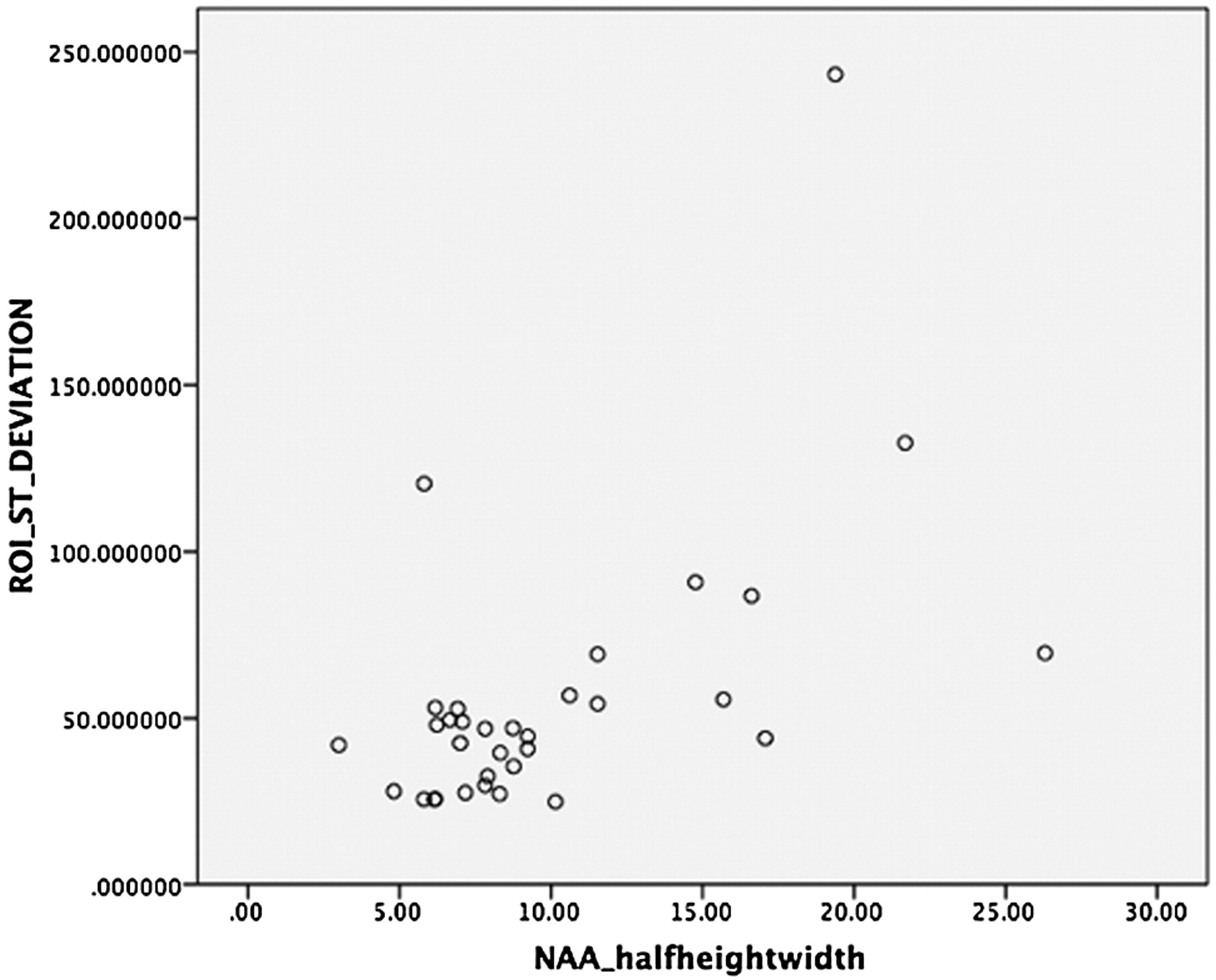
We investigated the relationship between spectral quality and the degree of susceptibility-induced phase distortion (ie, field inhomogeneity) by processing the phase maps into a 15-mm maximum intensity projection section to match the MR spectroscopy voxel dimensions (Fig 1). For each individual MR spectroscopy voxel on the registered phase map, the averaged SD in phase was correlated with the spectral peak width at half-height for NAA, Cr, and Cho (Fig 2). Pearson correlation coefficients were determined to relate spectral quality (metabolite line width) with field inhomogeneity (magnitude of the susceptibility-induced phase change) for a particular spatial location. These coefficients were 0.56 for NAA, 0.60 for Cr, and 0.60 for Cho (P = .001).

Fig 1.
  • Download figure
  • Open in new tab
  • Download powerpoint
Fig 1.

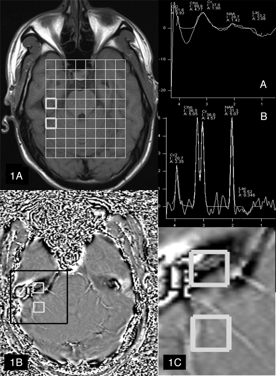
Representative data from a healthy subject. 1A, T1-weighted image showing placement of the multivoxel spectroscopy grid (top left) and (1B) SWI phase map. Spectrum A for the anterior voxel corresponding to brain tissue exhibiting large, susceptibility-induced phase heterogeneity. This spectrum is of nondiagnostic quality. Spectrum B is acquired from the more posterior voxel in which there is less phase shift compared with the anterior voxel and is of good diagnostic quality. 1C, Enlargement from (1B) showing increased phase heterogeneity within the anterior voxel.

Fig 2.
  • Download figure
  • Open in new tab
  • Download powerpoint
Fig 2.

Relationship between spectral quality and phase heterogeneity. Correlation between the line width at half-height for NAA and pixel SD for each MR spectroscopy voxel within the phase image (Pearson rank correlation coefficient, 0.56; P = .001).

Discussion

Magnetic field inhomogeneity identified on SWI-generated phase maps (indicated in Fig 2 by a increased SD of pixel values) correlates significantly with poor quality of spectroscopic data (widened metabolite peaks, poor water suppression, excessive baseline roll) obtained in both healthy tissue and contrast-enhancing tumor (Figs 1, 2, 3). Spectral quality may be improved by using the phase image to guide placement of the sampling voxel away from regions of poor field homogeneity in both single and multivoxel MR spectroscopy studies. In addition, voxel placement may be optimized by use of postcontrast SWI to identify areas of hemorrhage and contrast enhancement. Benefits of use of SWI data for MR spectroscopy voxel placement include higher-quality spectra, fewer repeated studies, faster study acquisition in critically ill and pediatric patients, and optimal use of scanner time.

Fig 3.
  • Download figure
  • Open in new tab
  • Download powerpoint
Fig 3.

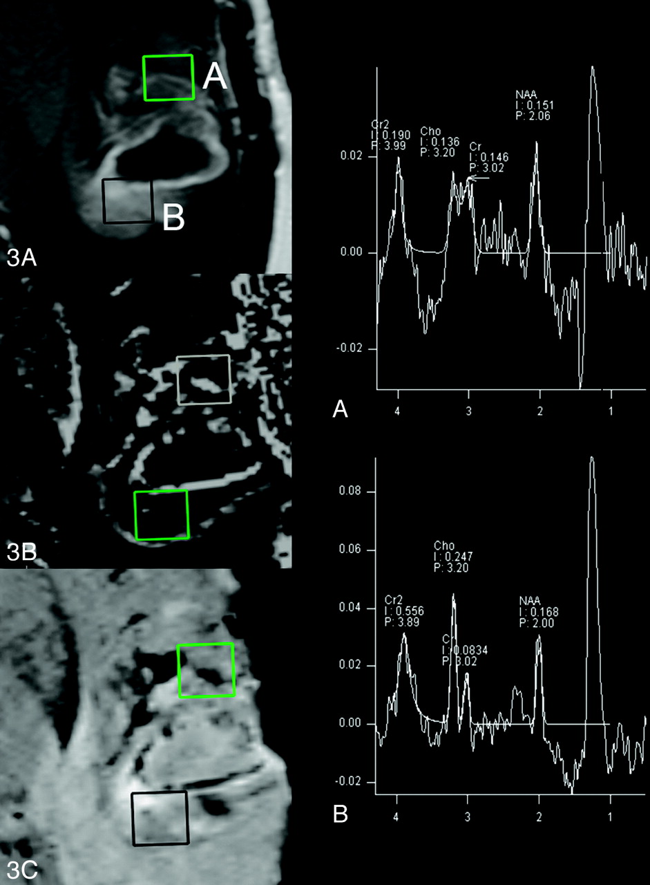
3A, T1-weighted postgadolinium image showing tumor enhancement and 2 representative sampling voxels A and B. Voxel A is placed in an area correlating to magnetic field heterogeneity on the phase map image (3B). This heterogeneity is secondary to the paramagnetic hemorrhagic tissue seen on the SWI image (3C). The MR spectrum from voxel A is shown top right. Voxel B has been placed in a region without phase heterogeneity (3B) and with contrast enhancement (3A, 3C) with resultant diagnostic spectrum (B, bottom right).

Acknowledgments

We would like to acknowledge associate professor Stephen Rose for his assistance with this study.

References

  1. ↵
    Lin A, Ross BD, Harris K, et al. Efficacy of proton magnetic resonance spectroscopy in neurological diagnosis and neurotherapeutic decision making. NeuroRx 2005;2:197–214
    Abstract/FREE Full Text
  2. ↵
    Di Costanzo A, Trojsi F, Tosetti M, et al. High-field proton MRS of human brain. Eur J Radiol 2003;48:146–53
    CrossRefPubMed
  3. ↵
    Drost DJ, Riddle WR, Clarke GD, et al. Proton magnetic resonance spectroscopy in the brain: report of AAPM MR Task Group #9. Med Physics 2002;29:2177–97
    CrossRefPubMed
  4. ↵
    Rauscher A, Sedlacik J, Barth M, et al. Magnetic susceptibility sensitive MR phase imaging of the human brain. AJNR Am J Neuroradiol 2005;26:736–42
    Abstract/FREE Full Text
  • Received September 3, 2008.
  • Accepted after revision October 13, 2008.
  • Copyright © American Society of Neuroradiology
View Abstract
PreviousNext
Back to top

In this issue

American Journal of Neuroradiology: 30 (4)
American Journal of Neuroradiology
Vol. 30, Issue 4
April 2009
  • Table of Contents
  • Index by author
Advertisement
Print
Download PDF
Email Article

Thank you for your interest in spreading the word on American Journal of Neuroradiology.

NOTE: We only request your email address so that the person you are recommending the page to knows that you wanted them to see it, and that it is not junk mail. We do not capture any email address.

Enter multiple addresses on separate lines or separate them with commas.
Usefulness of Susceptibility-Weighted Imaging for Voxel Placement in MR Spectroscopy
(Your Name) has sent you a message from American Journal of Neuroradiology
(Your Name) thought you would like to see the American Journal of Neuroradiology web site.
CAPTCHA
This question is for testing whether or not you are a human visitor and to prevent automated spam submissions.
Cite this article
L. Josey, A. Al Sayyari, R. Buckley, A. Coulthard
Usefulness of Susceptibility-Weighted Imaging for Voxel Placement in MR Spectroscopy
American Journal of Neuroradiology Apr 2009, 30 (4) 752-754; DOI: 10.3174/ajnr.A1403

Citation Manager Formats

  • BibTeX
  • Bookends
  • EasyBib
  • EndNote (tagged)
  • EndNote 8 (xml)
  • Medlars
  • Mendeley
  • Papers
  • RefWorks Tagged
  • Ref Manager
  • RIS
  • Zotero
0 Responses
Respond to this article
Share
Bookmark this article
Usefulness of Susceptibility-Weighted Imaging for Voxel Placement in MR Spectroscopy
L. Josey, A. Al Sayyari, R. Buckley, A. Coulthard
American Journal of Neuroradiology Apr 2009, 30 (4) 752-754; DOI: 10.3174/ajnr.A1403
del.icio.us logo Twitter logo Facebook logo Mendeley logo
  • Tweet Widget
  • Facebook Like
  • Google Plus One
Purchase

Jump to section

  • Article
    • Abstract
    • Technique
    • Discussion
    • Acknowledgments
    • References
  • Figures & Data
  • Info & Metrics
  • Responses
  • References
  • PDF

Related Articles

  • No related articles found.
  • PubMed
  • Google Scholar

Cited By...

  • No citing articles found.
  • Crossref (1)
  • Google Scholar

This article has been cited by the following articles in journals that are participating in Crossref Cited-by Linking.

  • Fast T2*‐weighted MRI of the prostate at 3 tesla
    Rulon L. Hardman, Fadi El‐Merhi, Adam J. Jung, Steve Ware, Ian M. Thompson, Harry T. Friel, Qi Peng
    Journal of Magnetic Resonance Imaging 2011 33 4

More in this TOC Section

  • Progression of Microstructural Damage in Spinocerebellar Ataxia Type 2: A Longitudinal DTI Study
  • Usefulness of Quantitative Susceptibility Mapping for the Diagnosis of Parkinson Disease
  • Evaluating the Effects of White Matter Multiple Sclerosis Lesions on the Volume Estimation of 6 Brain Tissue Segmentation Methods
Show more Brain

Similar Articles

Advertisement

Indexed Content

  • Current Issue
  • Accepted Manuscripts
  • Article Preview
  • Past Issues
  • Editorials
  • Editors Choice
  • Fellow Journal Club
  • Letters to the Editor

Cases

  • Case Collection
  • Archive - Case of the Week
  • Archive - Case of the Month
  • Archive - Classic Case

Special Collections

  • Special Collections

Resources

  • News and Updates
  • Turn around Times
  • Submit a Manuscript
  • Author Policies
  • Manuscript Submission Guidelines
  • Evidence-Based Medicine Level Guide
  • Publishing Checklists
  • Graphical Abstract Preparation
  • Imaging Protocol Submission
  • Submit a Case
  • Become a Reviewer/Academy of Reviewers
  • Get Peer Review Credit from Publons

Multimedia

  • AJNR Podcast
  • AJNR SCANtastic
  • Video Articles

About Us

  • About AJNR
  • Editorial Board
  • Not an AJNR Subscriber? Join Now
  • Alerts
  • Feedback
  • Advertise with us
  • Librarian Resources
  • Permissions
  • Terms and Conditions

American Society of Neuroradiology

  • Not an ASNR Member? Join Now

© 2025 by the American Society of Neuroradiology All rights, including for text and data mining, AI training, and similar technologies, are reserved.
Print ISSN: 0195-6108 Online ISSN: 1936-959X

Powered by HighWire