Advertisement

Main menu

  • Home
  • Content
    • Current Issue
    • Accepted Manuscripts
    • Article Preview
    • Past Issue Archive
    • AJNR Case Collection
    • Case of the Week Archive
    • Classic Case Archive
    • Case of the Month Archive
  • Special Collections
    • Spinal CSF Leak Articles (Jan 2020-June 2024)
    • 2024 AJNR Journal Awards
    • Most Impactful AJNR Articles
  • Multimedia
    • AJNR Podcast
    • AJNR Scantastics
    • Video Articles
  • For Authors
    • Submit a Manuscript
    • Author Policies
    • Fast publishing of Accepted Manuscripts
    • Graphical Abstract Preparation
    • Manuscript Submission Guidelines
    • Imaging Protocol Submission
    • Submit a Case for the Case Collection
  • About Us
    • About AJNR
    • Editorial Board
  • More
    • Become a Reviewer/Academy of Reviewers
    • Subscribers
    • Permissions
    • Alerts
    • Feedback
    • Advertisers
    • ASNR Home
  • Other Publications
    • ajnr

User menu

  • Alerts
  • Log in
  • Log out

Search

  • Advanced search
American Journal of Neuroradiology

American Journal of Neuroradiology

ASHNR American Society of Functional Neuroradiology ASHNR American Society of Pediatric Neuroradiology ASSR
  • Alerts
  • Log in
  • Log out

Advanced Search

  • Home
  • Content
    • Current Issue
    • Accepted Manuscripts
    • Article Preview
    • Past Issue Archive
    • AJNR Case Collection
    • Case of the Week Archive
    • Classic Case Archive
    • Case of the Month Archive
  • Special Collections
    • Spinal CSF Leak Articles (Jan 2020-June 2024)
    • 2024 AJNR Journal Awards
    • Most Impactful AJNR Articles
  • Multimedia
    • AJNR Podcast
    • AJNR Scantastics
    • Video Articles
  • For Authors
    • Submit a Manuscript
    • Author Policies
    • Fast publishing of Accepted Manuscripts
    • Graphical Abstract Preparation
    • Manuscript Submission Guidelines
    • Imaging Protocol Submission
    • Submit a Case for the Case Collection
  • About Us
    • About AJNR
    • Editorial Board
  • More
    • Become a Reviewer/Academy of Reviewers
    • Subscribers
    • Permissions
    • Alerts
    • Feedback
    • Advertisers
    • ASNR Home
  • Follow AJNR on Twitter
  • Visit AJNR on Facebook
  • Follow AJNR on Instagram
  • Join AJNR on LinkedIn
  • RSS Feeds

Welcome to the new AJNR, Updated Hall of Fame, and more. Read the full announcements.


AJNR is seeking candidates for the position of Associate Section Editor, AJNR Case Collection. Read the full announcement.


OtherHEAD & NECK

Transdural Spread of Glioblastoma through the Foramen Ovale with Presentation as a Masticator Space Mass

R. Kwak and D. Shatzkes
American Journal of Neuroradiology April 2009, 30 (4) 808-810; DOI: https://doi.org/10.3174/ajnr.A1349
R. Kwak
  • Find this author on Google Scholar
  • Find this author on PubMed
  • Search for this author on this site
D. Shatzkes
  • Find this author on Google Scholar
  • Find this author on PubMed
  • Search for this author on this site
  • Article
  • Figures & Data
  • Info & Metrics
  • Responses
  • References
  • PDF
Loading

Abstract

SUMMARY: Direct extension of a glioblastoma through the dura at the skull base is an uncommon occurrence. We report an unusual case of recurrent glioblastoma that spread transdurally through the foramen ovale and presented primarily as a masticator space mass. There was mandibular denervation and a relative paucity of intracranial disease.

Despite the aggressive nature of glioblastoma multiforme (GBM), extracranial spread is not a common occurrence. With improving treatment options and survival times, reports of extracranial recurrence of GBM have increased. Most commonly these metastases are to the lungs, lymph nodes, liver, and bones.1 There have been many fewer reports of glioblastoma spreading primarily through the skull base. We report an exceptional case of GBM spreading extracranially through a natural skull base foramen and presenting primarily as a masticator space mass.

Case Report

A 53-year-old right-handed man without significant medical history initially presented in October of 2006 with headaches, right-sided hemiparesis, and episodes of odd episodic smells. He was found to have a left temporal lobe mass, which was resected via a left pterional craniotomy and was found to be a grade IV astrocytoma with infiltration of the dura and leptomeninges. He received chemoradiation and 11 cycles of temozolomide (Temodar) monotherapy.

Approximately 15 months later, he experienced hearing loss and chronic drainage from his left ear. Review of systems was also positive for left jaw and tongue numbness. His physical examination revealed left temporal hollowing and slight left facial asymmetry. There was dense numbness over the central left V3 distribution. Other cranial nerves were intact.

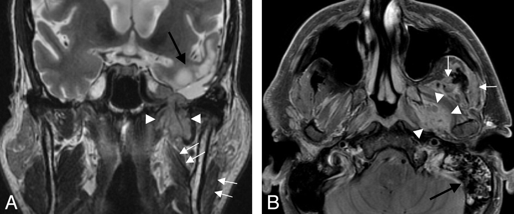
MR imaging performed in February 2008 demonstrated abnormal soft-tissue extending transcranially through the widened foramen ovale into the infratemporal fossa (Fig 1). Additionally, findings of subacute denervation involving the muscles of mastication were noted, including atrophy and hyperintensity on T2-weighted imaging. Intracranially, there were expected postoperative changes related to the prior pterional craniotomy, remote from the skull base foramina and without significant soft-tissue mass at the craniotomy site.

Fig 1.
  • Download figure
  • Open in new tab
  • Download powerpoint
Fig 1.

A 53-year-old man with history of glioblastoma presented with chronic otomastoiditis and mandibular nerve denervation. A, Coronal T2-weighted MR image demonstrates a transcranial mass extending through the left foramen ovale, with the largest component in the masticator space (white arrowheads). Subacute denervation of the left medial pterygoid and masseter muscles is evidenced by their increased signal intensity and decreased volume (white arrows). Chronic postoperative and treatment-related changes are noted in the left middle cranial fossa and left temporal lobe (black arrow). B, Axial contrast-enhanced fat-suppressed T1-weighted MR image demonstrates the extracranial portion of the mass interposed between the left medial and lateral pterygoid muscles (white arrowheads). The left temporalis and masseter muscles demonstrate asymmetrically decreased volume (white arrows). Note the mass effect on the left eustachian tube orifice and abnormal signal within the left mastoid air cells, likely on an obstructive basis (black arrow).

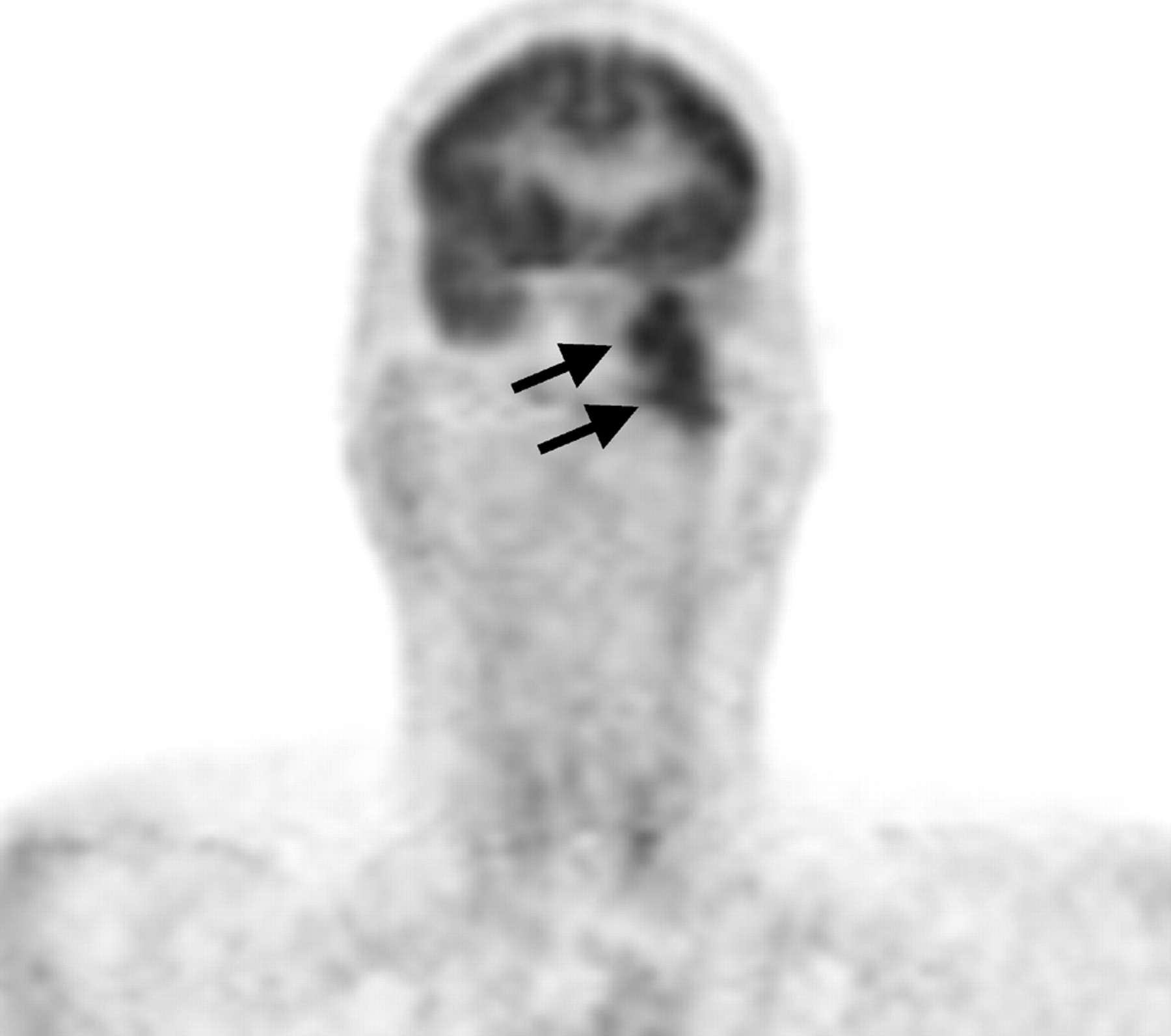
A positron-emission tomography (PET)/CT performed in March 2008 demonstrated the mass to be hypermetabolic with a maximum standard uptake value of 10.9–12.0 g/mL (Fig 2).

Fig 2.
  • Download figure
  • Open in new tab
  • Download powerpoint
Fig 2.

PET images in the coronal plane clearly show transcranial tumor extending from the floor of the left middle cranial fossa into the soft tissues of the left deep face (black arrows).

In April of 2008, the patient underwent transnasal and transmaxillary infratemporal fossa biopsies utilizing CT navigation. Light microscopy showed atypical cells with hyperchromatic nuclei and mitotic activity as well as areas of palisading necrosis. Cells stained strongly positive for glial fibrillary acidic protein, vimentin, and S100 and were negative for epithelial membrane antigen, synaptophysin, cytokeratins (AE1/AE3), and neurofilament. A diagnosis of recurrent GBM was made.

Discussion

There are many barriers to the spread of disease from the intracranial compartment. These include the dural coverings, the basement membranes around vessels of the central nervous system, the absence of true lymphatics in the intracranial compartment, and the early occlusion of compliant veins by tumor.1,2 For hematogenous dissemination to occur, the tumor cells must gain access to the intravascular space. Although there are data to suggest that glial tumors can perform the steps necessary to do this primarily,3 historically, it has been thought that extracranial dissemination of GBM occurred solely in the setting of a prior craniotomy, biopsy, or shunt placement.1,3 Such a procedure would, theoretically, provide tumor cells a mechanical entry point into the vascular or extracranial lymphatic system (or a direct route into the peritoneum in the setting of a ventriculoperitoneal shunt). Many reports and exhaustive reviews have described remote extracranial metastases in the setting of a prior surgical intervention.1

There has been a smaller number of case reports documenting remote extracranial metastases in patients who have never had any prior violation of the dura other than radiation therapy. Local invasion of the dura and, subsequently, the dural venous sinuses, is thought to play a role in many of these cases. In fact, at postmortem, many of these patients are found to have tumor that has entered the venous sinuses.3,4

Transdural spread of GBM in patients without prior dural defects or at sites distant from a craniotomy defect is a rare occurrence with few reported cases. In their review, Kawano et al5 described 3 routes for extradural extension of gliomas: 1) through the perivascular or dural slit at the middle cranial fossa, which is facilitated by intracranial hypertension; 2) along the cranial or spinal nerves; and 3) via the direct destruction of the dura mater. Although the most common sites for extradural spread with direct involvement of bone are the cerebral convexities and the middle cranial fossae, these occurrences are exceedingly rare.3

We were able to find 12 reported cases of glioblastoma spreading spontaneously through the skull base. They are summarized in the Table. Our case is unique in that there was minimal intracranial/intradural disease and isolated extension through the native foramen ovale. This resulted in a large masticator space mass and denervation in the distribution of the mandibular nerve. Due to the rarity of such a presentation, we were initially forced to expand our differential diagnosis to include pathology arising primarily within the masticator space.

View this table:
  • View inline
  • View popup

Spontaneous extradural spread of glioblastoma through the skull base

In summary, the extracranial spread of a glioblastoma, particularly through an intact dura, is a rare occurrence. We reviewed the different mechanisms by which glioblastomas are thought to spread and presented a unique case of recurrent tumor that extended through foramen ovale, resulting in a large volume of extradural tumor and secondary denervation.

References

  1. ↵
    Pasquier B, Pasquier D, N'Golet A, et al. Extraneural metastases of astrocytomas and glioblastomas: clinicopathological study of two cases and review of literature. Cancer 1980;45:112–25
    CrossRefPubMed
  2. ↵
    Wallace CJ, Forsyth PA, Edwards DR. Lymph node metastases from glioblastoma multiforme. AJNR Am J Neuroradiol 1996;17:1929–31
    Abstract
  3. ↵
    Liwnicz B, Rubinstein L. The pathways of extraneural spread in metastasizing gliomas. Hum Pathol 1979;10:453–67
    CrossRefPubMed
  4. ↵
    Kleinschmidt-Demasters BK. Diffuse bone marrow metastasis from glioblastoma multiforme: the role of dural invasion. Hum Pathol 1996;27:197–201
    CrossRefPubMed
  5. ↵
    Kawano N, Yada K, Ogawa Y, et al. Spontaneous transdural extension of malignant astrocytoma. J Neurosurg 1977;47:766–70
    PubMed
  6. Sanerkin NG. Transdural spread of glioblastoma multiforme. J Pathol Bacteriol 1962;84:228–33
    CrossRefPubMed
  7. Nager GT. Gliomas involving the temporal bone: clinical and pathological aspects. Laryngoscope 1967;77:454–88
    PubMed
  8. Hoyt WF, Piovanetti E, Malumud N, et al. Cranio-orbital involvement in glioblastoma multiforme. Neurochirurgia (Stuttg) 1972;15:1–8
    PubMed
  9. Aoyama I, Makita Y, Nabeshima S, et al. Extradural nasal and orbital extension of glioblastoma multiforme without previous surgical intervention. Surg Neurol 1980;14:343–47
    PubMed
  10. Shuangshoti S, Kasantikul V, Suwanwela N. Spontaneous penetration of dura mater and bone by glioblastoma multiforme. J Surg Oncol 1987;36:36–44
    CrossRefPubMed
  11. Russell and Rubinstein's Pathology of Tumors of the Nervous System. Baltimore: Williams & Wilkins;1989 :245
  12. Lampl Y, Eshel Y, Gilad R, et al. Glioblastoma multiforme with bone metastases and cauda equina syndrome. J Neurooncol 1990;8:167–72
    PubMed
  13. Pompili A, Calvosa F, Caroli F, et al. The transdural extension of gliomas. J Neurooncol 1993;15:67–74
    CrossRefPubMed
  14. Horiuchi T, Osawa M, Itoh N, et al. Extradural extension of glioblastoma multiforme into the oral cavity: case report. Surg Neurol 1996;46:42–6
    CrossRefPubMed
  15. Rainov NG, Holzhausen HJ, Meyer H, et al. Local invasivity of glioblastoma multiforme with destruction of skull bone: case report and review of the literature. Neurosurg Rev 1996;19:183–88
    CrossRefPubMed
  • Received August 3, 2008.
  • Accepted after revision August 30, 2008.
  • Copyright © American Society of Neuroradiology
View Abstract
PreviousNext
Back to top

In this issue

American Journal of Neuroradiology: 30 (4)
American Journal of Neuroradiology
Vol. 30, Issue 4
April 2009
  • Table of Contents
  • Index by author
Advertisement
Print
Download PDF
Email Article

Thank you for your interest in spreading the word on American Journal of Neuroradiology.

NOTE: We only request your email address so that the person you are recommending the page to knows that you wanted them to see it, and that it is not junk mail. We do not capture any email address.

Enter multiple addresses on separate lines or separate them with commas.
Transdural Spread of Glioblastoma through the Foramen Ovale with Presentation as a Masticator Space Mass
(Your Name) has sent you a message from American Journal of Neuroradiology
(Your Name) thought you would like to see the American Journal of Neuroradiology web site.
CAPTCHA
This question is for testing whether or not you are a human visitor and to prevent automated spam submissions.
Cite this article
R. Kwak, D. Shatzkes
Transdural Spread of Glioblastoma through the Foramen Ovale with Presentation as a Masticator Space Mass
American Journal of Neuroradiology Apr 2009, 30 (4) 808-810; DOI: 10.3174/ajnr.A1349

Citation Manager Formats

  • BibTeX
  • Bookends
  • EasyBib
  • EndNote (tagged)
  • EndNote 8 (xml)
  • Medlars
  • Mendeley
  • Papers
  • RefWorks Tagged
  • Ref Manager
  • RIS
  • Zotero
0 Responses
Respond to this article
Share
Bookmark this article
Transdural Spread of Glioblastoma through the Foramen Ovale with Presentation as a Masticator Space Mass
R. Kwak, D. Shatzkes
American Journal of Neuroradiology Apr 2009, 30 (4) 808-810; DOI: 10.3174/ajnr.A1349
del.icio.us logo Twitter logo Facebook logo Mendeley logo
  • Tweet Widget
  • Facebook Like
  • Google Plus One
Purchase

Jump to section

  • Article
    • Abstract
    • Case Report
    • Discussion
    • References
  • Figures & Data
  • Info & Metrics
  • Responses
  • References
  • PDF

Related Articles

  • No related articles found.
  • PubMed
  • Google Scholar

Cited By...

  • Unusual extracranial spread of glioblastoma
  • Crossref (9)
  • Google Scholar

This article has been cited by the following articles in journals that are participating in Crossref Cited-by Linking.

  • The Skull Base in the Evaluation of Sinonasal Disease
    Steve E.J. Connor
    Neuroimaging Clinics of North America 2015 25 4
  • Endocranial Lesions
    Surjith Vattoth, Roger Steven DeLappe, Philip R. Chapman
    Seminars in Ultrasound, CT and MRI 2013 34 5
  • Unusual Case of Transdural Extension of Glioblastoma Multiforme in the Maxillofacial Region and a Review of the Literature
    Shahin Taghikhan, Brian M. Woo, Yu-Hung Kuo
    Journal of Oral and Maxillofacial Surgery 2019 77 11
  • Advances in the Study of the Middle Cranial Fossa through Cutting Edge Neuroimaging Techniques
    Juan A. Juanes Méndez, Pablo Ruisoto, Juan C. Paniagua, Alberto Prats
    Journal of Medical Systems 2018 42 2
  • Glioblastoma metastases: a literature review and a description of six clinical observations
    S. A. Goryaynov, A. A. Potapov, M. A. Ignatenko, V. Yu. Zhukov, S. V. Protskiy, N. A. Zakharova, V. A. Okhlopkov, L. V. Shishkina
    Voprosy neirokhirurgii imeni N.N. Burdenko 2015 79 2
  • Unusual extracranial spread of glioblastoma
    Ricardo J Fernández-de Thomas, Joel E Pellot Cestero, Cesar M Carballo Cuello, Orlando De Jesus
    BMJ Case Reports 2021 14 8
  • Glioblastoma multiforme with transdural extension to the external auditory canal via the tegmen tympani—Clinical report with review of the literature
    Arba Cecia, Emal Lesha, David G. Laird, Elsa Nico, Kaan Yagmurlu, Bruce L. Fetterman, L. Madison Michael
    Eye & ENT Research 2025
  • Primary cavernous sinus germinoma with atypical extension pattern: a case report and review of the literature
    Kazuki Sakakura, Takao Tsurubuchi, Tomohiko Masumoto, Ai Muroi, Eiichi Ishikawa, Akira Matsumura
    Child's Nervous System 2019 35 9
  • Transdural Skull Base Infiltration by Glioblastoma: Case Report and Review of the Literature
    Michael Thrull, Khaled Atasi, Lennart-Maximilian Boese, Mahmut Cakar, Ullrich Heller, Nils Jansen, Leoni-Christine Menzel, Hassan Omaimen, Katharina Theis, Damir Karacic, Diyan Dimov, Roland Coras, Randolf Klingebiel, Abr o Rapoport
    Case Reports in Otolaryngology 2023 2023

More in this TOC Section

  • WHO Classification Update: Nasal&Skull Base Tumors
  • Peritumoral Signal in Vestibular Schwannomas
  • Chondrosarcoma vs Synovial Chondromatosis: Imaging
Show more HEAD & NECK

Similar Articles

Advertisement

Indexed Content

  • Current Issue
  • Accepted Manuscripts
  • Article Preview
  • Past Issues
  • Editorials
  • Editors Choice
  • Fellow Journal Club
  • Letters to the Editor

Cases

  • Case Collection
  • Archive - Case of the Week
  • Archive - Case of the Month
  • Archive - Classic Case

Special Collections

  • Special Collections

Resources

  • News and Updates
  • Turn around Times
  • Submit a Manuscript
  • Author Policies
  • Manuscript Submission Guidelines
  • Evidence-Based Medicine Level Guide
  • Publishing Checklists
  • Graphical Abstract Preparation
  • Imaging Protocol Submission
  • Submit a Case
  • Become a Reviewer/Academy of Reviewers
  • Get Peer Review Credit from Publons

Multimedia

  • AJNR Podcast
  • AJNR SCANtastic
  • Video Articles

About Us

  • About AJNR
  • Editorial Board
  • Not an AJNR Subscriber? Join Now
  • Alerts
  • Feedback
  • Advertise with us
  • Librarian Resources
  • Permissions
  • Terms and Conditions

American Society of Neuroradiology

  • Not an ASNR Member? Join Now

© 2025 by the American Society of Neuroradiology All rights, including for text and data mining, AI training, and similar technologies, are reserved.
Print ISSN: 0195-6108 Online ISSN: 1936-959X

Powered by HighWire