Advertisement

Main menu

  • Home
  • Content
    • Current Issue
    • Accepted Manuscripts
    • Article Preview
    • Past Issue Archive
    • AJNR Case Collection
    • Case of the Week Archive
    • Classic Case Archive
    • Case of the Month Archive
  • Special Collections
    • Spinal CSF Leak Articles (Jan 2020-June 2024)
    • 2024 AJNR Journal Awards
    • Most Impactful AJNR Articles
  • Multimedia
    • AJNR Podcast
    • AJNR Scantastics
    • Video Articles
  • For Authors
    • Submit a Manuscript
    • Author Policies
    • Fast publishing of Accepted Manuscripts
    • Graphical Abstract Preparation
    • Manuscript Submission Guidelines
    • Imaging Protocol Submission
    • Submit a Case for the Case Collection
  • About Us
    • About AJNR
    • Editorial Board
  • More
    • Become a Reviewer/Academy of Reviewers
    • Subscribers
    • Permissions
    • Alerts
    • Feedback
    • Advertisers
    • ASNR Home
  • Other Publications
    • ajnr

User menu

  • Alerts
  • Log in

Search

  • Advanced search
American Journal of Neuroradiology

American Journal of Neuroradiology

ASHNR American Society of Functional Neuroradiology ASHNR American Society of Pediatric Neuroradiology ASSR
  • Alerts
  • Log in

Advanced Search

  • Home
  • Content
    • Current Issue
    • Accepted Manuscripts
    • Article Preview
    • Past Issue Archive
    • AJNR Case Collection
    • Case of the Week Archive
    • Classic Case Archive
    • Case of the Month Archive
  • Special Collections
    • Spinal CSF Leak Articles (Jan 2020-June 2024)
    • 2024 AJNR Journal Awards
    • Most Impactful AJNR Articles
  • Multimedia
    • AJNR Podcast
    • AJNR Scantastics
    • Video Articles
  • For Authors
    • Submit a Manuscript
    • Author Policies
    • Fast publishing of Accepted Manuscripts
    • Graphical Abstract Preparation
    • Manuscript Submission Guidelines
    • Imaging Protocol Submission
    • Submit a Case for the Case Collection
  • About Us
    • About AJNR
    • Editorial Board
  • More
    • Become a Reviewer/Academy of Reviewers
    • Subscribers
    • Permissions
    • Alerts
    • Feedback
    • Advertisers
    • ASNR Home
  • Follow AJNR on Twitter
  • Visit AJNR on Facebook
  • Follow AJNR on Instagram
  • Join AJNR on LinkedIn
  • RSS Feeds

Welcome to the new AJNR, Updated Hall of Fame, and more. Read the full announcements.


AJNR is seeking candidates for the position of Associate Section Editor, AJNR Case Collection. Read the full announcement.


OtherHEAD & NECK

Primary Retropharyngeal Synovial Sarcoma

H. Shi, S. Wang, P. Wang and Q. Yu
American Journal of Neuroradiology April 2009, 30 (4) 811-812; DOI: https://doi.org/10.3174/ajnr.A1333
H. Shi
  • Find this author on Google Scholar
  • Find this author on PubMed
  • Search for this author on this site
S. Wang
  • Find this author on Google Scholar
  • Find this author on PubMed
  • Search for this author on this site
P. Wang
  • Find this author on Google Scholar
  • Find this author on PubMed
  • Search for this author on this site
Q. Yu
  • Find this author on Google Scholar
  • Find this author on PubMed
  • Search for this author on this site
  • Article
  • Figures & Data
  • Info & Metrics
  • Responses
  • References
  • PDF
Loading

Abstract

SUMMARY: Synovial sarcoma is the third most common histologic type of extremity soft tissue sarcoma. However, primary retropharyngeal synovial sarcoma is extremely rare. We present a case of primary retropharyngeal synovial sarcoma in a 60-year-old woman. The radiographic imaging of our present cases was characteristic of a heterogeneously enhancing mass.

Synovial sarcoma, a mesenchymal spindle-cell tumor, arises most commonly in the deep soft tissues of the extremities (especially around the knee), and frequently adjacent to the joints or tendon sheaths. The retropharyngeal space is a rare site for this tumor. We describe a case of synovial sarcoma located in the retropharyngeal space, focusing on the CT imaging findings.

Case Report

Clinical History and Physical Examination Findings

In August 2006, a 60-year-old woman with difficulties in swallowing and nausea for 3 months was referred to our hospital for evaluation. A clinical examination revealed a submucosal bulging of the left posterior pharyngeal wall. The overlying mucosa seemed to be hypervascular but was not ulcerated. There were no pathologic lymph nodes in the neck.

Radiographic Findings

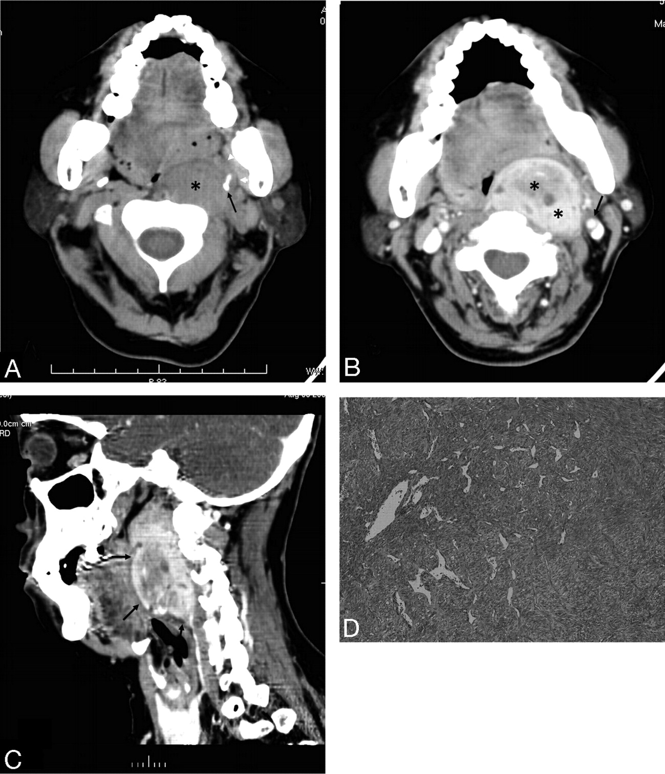
CT scans of the head and neck showed a large, well-circumscribed, solid mass in the left retropharyngeal space, which was isoattenuated to the muscle (Fig 1A). The styloid process was anterolaterally displaced by the mass. After infusion of the contrast material, the mass showed strong heterogeneous enhancement (Fig 1B). The mass displaced the parapharyngeal fat anteriorly, pharyngeal mucosal space medially, and internal carotid artery laterally. A multiplanar reformatted image showed a clearly limited oval mass in the retropharyngeal space extending from the first to the fifth vertebrae (Fig 1C).

  Fig 1.
  • Download figure
  • Open in new tab
  • Download powerpoint
Fig 1.

A 60-year-old woman with a bulging mass in the left retropharyngeal space. A, Axial CT scan shows a large, well-circumscribed, solid mass in the left retropharyngeal space (black asterisk). The mass displaces the parapharyngeal fat anteriorly (white arrowheads). Note the styloid process, which is anterolaterally displaced by the mass (black arrow). B, Contrast-enhanced axial CT scan shows a well-marginated strong heterogeneous enhancement mass in the left retropharyngeal space (black asterisks). The mass displaces the parapharyngeal fat anteriorly, the pharyngeal mucosal space medially, and the internal carotid artery laterally (black arrow). C, A multiplanar reformatted image shows a clearly limited oval mass in the retropharyngeal space extending from the first to the fifth vertebrae (black arrows). D, Low-power photomicrograph (original magnification, ×20) reveals a proliferation of oval to spindle-shaped cells arranged in sheets and fascicles.

Histologic Findings

The patient underwent a left cervicotomy, in which the lesion was enucleated. During surgery, the tumor appeared tan, circumscribed, and adherent to the anterior common vertebral ligament. The gross specimen, measuring 5.0 × 3.5 × 6.0 cm, was encapsulated. The histologic examination revealed a proliferation of oval to spindle-shaped cells arranged in sheets and fascicles (Fig 1D). On immunohistochemical analysis, tumor cells were positive for keratins, vimentin, and epithelial membrane antigen, and negative for S-100, desmin. The histologic diagnosis was monophasic synovial sarcoma.

Two years after initial surgery, the patient is free of disease, without signs of local recurrence and distant metastases, and functioning adequately.

Discussion

Synovial sarcoma is a mesenchymal spindle-cell tumor that displays variable epithelial differentiation including glandular formation and has a specific chromosomal translocation t(X; 18) (p11; q11). Synovial sarcoma accounts for 5% to 10% of soft tissue sarcomas.1 The reported annual incidence is 2.57 per 100,000.2 They are reported from birth to age 89 years but occur mainly between the ages of 15 and 40 years3 and are more common in males. Synovial sarcoma is unrelated to the synovium, and less than 5% of these sarcomas originate within a joint or bursa. More than 80% of these tumors arise in the deep soft tissue of the extremities (especially around the knee), and the tumor frequently arises adjacent to joints or tendon sheaths. Approximately 5% of these tumors arise in the head and neck region; however, any site can be affected.4,5 In the case of our patient, the tumor was located within the left suprahyoid portion of the retropharyngeal space.

The most common radiologic finding of primary synovial sarcoma has been described to be a heterogeneously enhancing soft tissue mass, with well-defined margins and without calcifications on CT.6 In the case of our patient, the tumor concurred with previous reports.

The retropharyngeal space is a distinct space along the midline of the extracranial portion of the head and neck that extends from the skull base to the upper mediastinum. It is bounded by the middle and deep layers of the deep cervical fascia. Lymph nodes and fat are found in the suprahyoid portion of the retropharyngeal space, whereas only fat is present in the infrahyoid portion. In the case of our patient, the tumor was thought to arise from a retropharyngeal node, probably the lateral group, which is located at the anterolateral aspect of the prevertebral muscle.

Disease processes in the retropharyngeal space are relatively uncommon; however, they assume greater significance because of the proximity of the retropharyngeal space to the airway and because of the inability to examine this space clinically.7-9 The nodal pattern of suprahyoid retropharyngeal space involvement is identified on CT or MR images when the center of the lesion is anterior to the prevertebral musculature, posteromedial to the parapharyngeal space, and medial to the carotid artery. The CT differential diagnosis of retropharyngeal space abnormalities is limited and includes malignant tumors and inflammatory diseases. Malignant tumors account for most abnormalities in the retropharyngeal space mass, and nodal metastases from head and neck squamous cell carcinoma represent the most common form of involvement. Malignant nodal involvement of the suprahyoid retropharyngeal space was diagnosed when unilateral or bilateral discrete nodal masses of 1 cm or larger were present, or when nodes of any size contained central inhomogeneity on CT scans. Metastatic disease in the retropharyngeal space can result in nodal necrosis.10 On occasion, nodal necrosis may be confused with an abscess or suppurative lymphadenitis. Necrotic nodes often maintain a clear nodal margin with no signs of surrounding inflammation. Causes of inflammatory diseases in the retropharyngeal space include cellulitis, suppurative adenopathy, and abscess. Except for lipomas, most low-attenuation massive lesions in the retropharyngeal space should be considered large abscesses that will extend rapidly into the lower neck and ultimately to the mediastinum; their existence and extension should be diagnosed correctly and promptly by sectional images.11

In general, synovial sarcoma is treated surgically with or without adjuvant radiation. Up to 50% of synovial sarcomas recur, usually within 2 years.12 Some 40% metastasize, commonly to the lungs and bone and also to the regional lymph nodes. Prognosis does not differ between monophasic and biphasic tumors or in relationship to immunophenotype.13

To the best of our knowledge, only 1 case of retropharyngeal synovial sarcoma has been reported. Radiologists should be familiar with synovial sarcoma in the different sites, and synovial sarcoma should be considered in the differential diagnosis of masses involving the retropharyngeal space.

References

  1. ↵
    Kransdorf MJ. Malignant soft tissue tumors in a large referral population: distribution of diagnoses by age, sex, and location. AJR Am J Roentgenol 1955;164:129–34
  2. ↵
    Scully SP, Temple H, Harrelson J. Synovial sarcoma of the foot and ankle. Clin Orthop Relat Res 1999;364:220–26
    CrossRefPubMed
  3. ↵
    Ryan JR, Baker LH, Benjamin RS. The natural history of metastatic synovial sarcoma: Experience of the Southwest Oncology Group. Clin Orthop Relat Res 1982;164:257–60
    PubMed
  4. ↵
    Argani P, Faria PA, Epstein JI, et al. Primary renal synovial sarcoma: molecular and morphologic delineation of an entity previously included among embryonal sarcomas of the kidney. Am J Surg Pathol 2000;24:1087–96
    CrossRefPubMed
  5. ↵
    Fisher C. Synovial sarcoma. Am Diagn Pathol 1998;2:401–21
    CrossRef
  6. ↵
    Duran-Mendicuti A, Costello P, Vargas SO. Primary synovial sarcoma of the chest: radiographic and clinicopathologic correlation. J Thorac Imaging. 2003;18:87–93
    CrossRefPubMed
  7. ↵
    Nyberg DA, Jeffrey RB, Brant-Zawadzki M, et al. Computed tomography of cervical infection. J Comput Assist Tomogr 1985;9:288–96
    CrossRefPubMed
  8. Wong YK, Novotny GM. Retropharyngeal space: a review of anatomy, pathology, and clinical presentation. J Otolaryngol 1978;7:528–36
    PubMed
  9. ↵
    Mancuso AA, Harnsberger HR, Muraki AS, et al. Computed tomography of cervical and retropharyngeal lymph nodes: normal anatomy, variants of normal, and application in staging head and neck cancer. Part II: pathology. Radiology 1983;148:715–23
    PubMed
  10. ↵
    Chong VF, Fan YF, Khoo JB. Retropharyngeal lymphadenopathy in nasopharyngeal carcinoma. Eur J Radiol 1995;21:100–05
    CrossRefPubMed
  11. ↵
    Yokinori K, Tomohiro O. The pharyngeal mucosal, parapharyngeal, and retropharyngeal spaces. Oral Radiol 2003;19:106–18
  12. ↵
    Fletcher CD, Unni KK, Mertens F, eds. World Health Organization Classification of Tumours. Pathology and Genetics of Tumours of Soft Tissue and Bone. Lyon, France: IARC Press;2002 :200–04
  13. ↵
    Antonescu CR, Kawai A, Leung DH, et al. Strong association of SYT-SSX fusion type and morphologic epithelial differentiation in synovial sarcoma. Diagn Mol Pathol 2000;9:1–8
    CrossRefPubMed
  • Received August 9, 2008.
  • Accepted after revision August 25, 2008.
  • Copyright © American Society of Neuroradiology
View Abstract
PreviousNext
Back to top

In this issue

American Journal of Neuroradiology: 30 (4)
American Journal of Neuroradiology
Vol. 30, Issue 4
April 2009
  • Table of Contents
  • Index by author
Advertisement
Print
Download PDF
Email Article

Thank you for your interest in spreading the word on American Journal of Neuroradiology.

NOTE: We only request your email address so that the person you are recommending the page to knows that you wanted them to see it, and that it is not junk mail. We do not capture any email address.

Enter multiple addresses on separate lines or separate them with commas.
Primary Retropharyngeal Synovial Sarcoma
(Your Name) has sent you a message from American Journal of Neuroradiology
(Your Name) thought you would like to see the American Journal of Neuroradiology web site.
CAPTCHA
This question is for testing whether or not you are a human visitor and to prevent automated spam submissions.
Cite this article
H. Shi, S. Wang, P. Wang, Q. Yu
Primary Retropharyngeal Synovial Sarcoma
American Journal of Neuroradiology Apr 2009, 30 (4) 811-812; DOI: 10.3174/ajnr.A1333

Citation Manager Formats

  • BibTeX
  • Bookends
  • EasyBib
  • EndNote (tagged)
  • EndNote 8 (xml)
  • Medlars
  • Mendeley
  • Papers
  • RefWorks Tagged
  • Ref Manager
  • RIS
  • Zotero
0 Responses
Respond to this article
Share
Bookmark this article
Primary Retropharyngeal Synovial Sarcoma
H. Shi, S. Wang, P. Wang, Q. Yu
American Journal of Neuroradiology Apr 2009, 30 (4) 811-812; DOI: 10.3174/ajnr.A1333
del.icio.us logo Twitter logo Facebook logo Mendeley logo
  • Tweet Widget
  • Facebook Like
  • Google Plus One
Purchase

Jump to section

  • Article
    • Abstract
    • Case Report
    • Discussion
    • References
  • Figures & Data
  • Info & Metrics
  • Responses
  • References
  • PDF

Related Articles

  • No related articles found.
  • PubMed
  • Google Scholar

Cited By...

  • No citing articles found.
  • Crossref (3)
  • Google Scholar

This article has been cited by the following articles in journals that are participating in Crossref Cited-by Linking.

  • Soft Tissue Tumors of the Head and Neck: Imaging-based Review of the WHO Classification
    Ahmed Abdel Razek, Benjamin Y. Huang
    RadioGraphics 2011 31 7
  • Update on MR Imaging of Soft Tissue Tumors of Head and Neck
    Justin D. Rodriguez, A. Morgan Selleck, Ahmed Abdel Khalek Abdel Razek, Benjamin Y. Huang
    Magnetic Resonance Imaging Clinics of North America 2022 30 1
  • A rare case of retropharyngeal liposarcoma: a rare location of a rare diagnosis
    Wajiha Arshad, Shahzaib Maqbool, Javeria A Kiany, Ali Raza, Umer Farooq, Qasim Ali, Ka Y Lee
    Journal of Surgical Case Reports 2023 2023 3

More in this TOC Section

  • WHO Classification Update: Nasal&Skull Base Tumors
  • Peritumoral Signal in Vestibular Schwannomas
  • Chondrosarcoma vs Synovial Chondromatosis: Imaging
Show more HEAD & NECK

Similar Articles

Advertisement

Indexed Content

  • Current Issue
  • Accepted Manuscripts
  • Article Preview
  • Past Issues
  • Editorials
  • Editors Choice
  • Fellow Journal Club
  • Letters to the Editor

Cases

  • Case Collection
  • Archive - Case of the Week
  • Archive - Case of the Month
  • Archive - Classic Case

Special Collections

  • Special Collections

Resources

  • News and Updates
  • Turn around Times
  • Submit a Manuscript
  • Author Policies
  • Manuscript Submission Guidelines
  • Evidence-Based Medicine Level Guide
  • Publishing Checklists
  • Graphical Abstract Preparation
  • Imaging Protocol Submission
  • Submit a Case
  • Become a Reviewer/Academy of Reviewers
  • Get Peer Review Credit from Publons

Multimedia

  • AJNR Podcast
  • AJNR SCANtastic
  • Video Articles

About Us

  • About AJNR
  • Editorial Board
  • Not an AJNR Subscriber? Join Now
  • Alerts
  • Feedback
  • Advertise with us
  • Librarian Resources
  • Permissions
  • Terms and Conditions

American Society of Neuroradiology

  • Not an ASNR Member? Join Now

© 2025 by the American Society of Neuroradiology All rights, including for text and data mining, AI training, and similar technologies, are reserved.
Print ISSN: 0195-6108 Online ISSN: 1936-959X

Powered by HighWire