Advertisement

Main menu

  • Home
  • Content
    • Current Issue
    • Accepted Manuscripts
    • Article Preview
    • Past Issue Archive
    • AJNR Case Collection
    • Case of the Week Archive
    • Classic Case Archive
    • Case of the Month Archive
  • Special Collections
    • Spinal CSF Leak Articles (Jan 2020-June 2024)
    • 2024 AJNR Journal Awards
    • Most Impactful AJNR Articles
  • Multimedia
    • AJNR Podcast
    • AJNR Scantastics
    • Video Articles
  • For Authors
    • Submit a Manuscript
    • Author Policies
    • Fast publishing of Accepted Manuscripts
    • Graphical Abstract Preparation
    • Manuscript Submission Guidelines
    • Imaging Protocol Submission
    • Submit a Case for the Case Collection
  • About Us
    • About AJNR
    • Editorial Board
  • More
    • Become a Reviewer/Academy of Reviewers
    • Subscribers
    • Permissions
    • Alerts
    • Feedback
    • Advertisers
    • ASNR Home
  • Other Publications
    • ajnr

User menu

  • Alerts
  • Log in

Search

  • Advanced search
American Journal of Neuroradiology

American Journal of Neuroradiology

ASHNR American Society of Functional Neuroradiology ASHNR American Society of Pediatric Neuroradiology ASSR
  • Alerts
  • Log in

Advanced Search

  • Home
  • Content
    • Current Issue
    • Accepted Manuscripts
    • Article Preview
    • Past Issue Archive
    • AJNR Case Collection
    • Case of the Week Archive
    • Classic Case Archive
    • Case of the Month Archive
  • Special Collections
    • Spinal CSF Leak Articles (Jan 2020-June 2024)
    • 2024 AJNR Journal Awards
    • Most Impactful AJNR Articles
  • Multimedia
    • AJNR Podcast
    • AJNR Scantastics
    • Video Articles
  • For Authors
    • Submit a Manuscript
    • Author Policies
    • Fast publishing of Accepted Manuscripts
    • Graphical Abstract Preparation
    • Manuscript Submission Guidelines
    • Imaging Protocol Submission
    • Submit a Case for the Case Collection
  • About Us
    • About AJNR
    • Editorial Board
  • More
    • Become a Reviewer/Academy of Reviewers
    • Subscribers
    • Permissions
    • Alerts
    • Feedback
    • Advertisers
    • ASNR Home
  • Follow AJNR on Twitter
  • Visit AJNR on Facebook
  • Follow AJNR on Instagram
  • Join AJNR on LinkedIn
  • RSS Feeds

Welcome to the new AJNR, Updated Hall of Fame, and more. Read the full announcements.


AJNR is seeking candidates for the position of Associate Section Editor, AJNR Case Collection. Read the full announcement.


Getting new auth cookie, if you see this message a lot, tell someone!
OtherHEAD & NECK

Apparent Diffusion Coefficient Values of Middle Ear Cholesteatoma Differ from Abscess and Cholesteatoma Admixed Infection

S. Thiriat, S. Riehm, S. Kremer, E. Martin and F. Veillon
American Journal of Neuroradiology June 2009, 30 (6) 1123-1126; DOI: https://doi.org/10.3174/ajnr.A1473
S. Thiriat
  • Find this author on Google Scholar
  • Find this author on PubMed
  • Search for this author on this site
S. Riehm
  • Find this author on Google Scholar
  • Find this author on PubMed
  • Search for this author on this site
S. Kremer
  • Find this author on Google Scholar
  • Find this author on PubMed
  • Search for this author on this site
E. Martin
  • Find this author on Google Scholar
  • Find this author on PubMed
  • Search for this author on this site
F. Veillon
  • Find this author on Google Scholar
  • Find this author on PubMed
  • Search for this author on this site
  • Article
  • Figures & Data
  • Info & Metrics
  • Responses
  • References
  • PDF
Loading

Article Figures & Data

Figures

  • Tables
  • Fig 1.
    • Download figure
    • Open in new tab
    • Download powerpoint
    Fig 1.

    A, ADC value cartography and region of interest to calculate ADC value. B, DW image.

  • Fig 2.
    • Download figure
    • Open in new tab
    • Download powerpoint
    Fig 2.

    A, corresponding calculated ADC value on ADC cartography (0.917 × 10⁻³ mm2/s). B, DW image of a group 1 patient.

  • Fig 3.
    • Download figure
    • Open in new tab
    • Download powerpoint
    Fig 3.

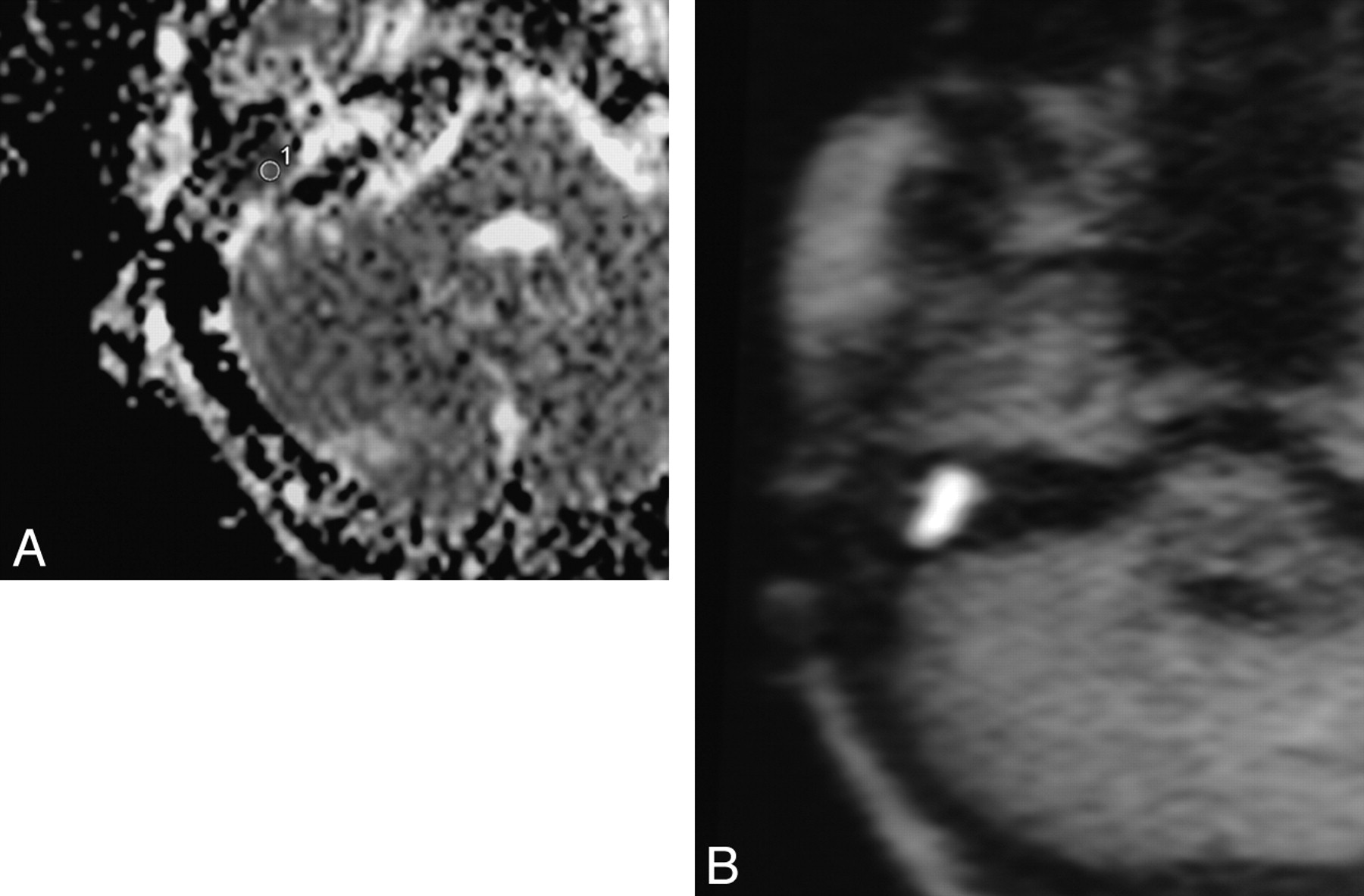
    A, corresponding calculated ADC value on ADC cartography (0.774 × 10⁻³ mm2/s). B, DW image of a group 2 patient.

  • Fig 4.
    • Download figure
    • Open in new tab
    • Download powerpoint
    Fig 4.

    A, corresponding calculated ADC value on ADC cartography (0.570 × 10⁻³ mm2/s). B, DW image of a group 3 patient.

Tables

  • Figures
    • View popup
    Table 1:

    ADC values for the 3 different surgical groups

    Patient No.ADC Value (×10−3 mm2/s)
    Group 1Group 2Group 3
    10.8470.6280.571
    21.0540.7740.568
    31.0460.6300.107
    40.917
    51.047
    60.790
    70.871
    80.698
    90.863
    • Note:—ADC indicates apparent diffusion coefficient.

    • View popup
    Table 2:

    ADC values calculated twice to test the reliability of the measurements

    Patient No./Surgical GroupADC Value (×10−3 mm2/s) First CalculationADC Value (×10−3 mm2/s) Second Calculation
    1/g10.8470.866
    2/g11.0541.015
    3/g11.0461.026
    4/g10.9170.805
    5/g11.0471.054
    6/g10.7900.857
    7/g10.8710.832
    8/g10.6980.696
    9/g10.8630.824
    1/g20.6280.668
    2/g20.7740.670
    3/g20.6300.621
    1/g30.5710.531
    2/g30.5680.576
    3/g30.1070.075
    • Note:—g1 indicates group 1; g2, group 2; g3, group 3.

    • View popup
    Table 3:

    Diagnosis made by DWI alone vs DWI and calculated ADC values

    Patient No./Surgical GroupDiagnosis Made by DWI AloneDiagnosis Inferred by DWI with Calculated ADC Values
    1/g1G1G1
    2/g1G1G1
    3/g1G1G1
    4/g1G1G1
    5/g1G1G1
    6/g1G1G1 or 2
    7/g1G1G1
    8/g1G1G2
    9/g1G1G1
    1/g2G1G2
    2/g2G1G1 or 2
    3/g2G1 or 2G2
    1/g3G2G3
    2/g3G1 or 2G3
    3/g3G3G3
    • Note:—DWI indicates diffusion-weighted imaging; G1, diagnosed as a group 1 patient; G2, diagnosed as a group 2 patient; G3, diagnosed as a group 3 patient.

PreviousNext
Back to top

In this issue

American Journal of Neuroradiology: 30 (6)
American Journal of Neuroradiology
Vol. 30, Issue 6
June 2009
  • Table of Contents
  • Index by author
Advertisement
Print
Download PDF
Email Article

Thank you for your interest in spreading the word on American Journal of Neuroradiology.

NOTE: We only request your email address so that the person you are recommending the page to knows that you wanted them to see it, and that it is not junk mail. We do not capture any email address.

Enter multiple addresses on separate lines or separate them with commas.
Apparent Diffusion Coefficient Values of Middle Ear Cholesteatoma Differ from Abscess and Cholesteatoma Admixed Infection
(Your Name) has sent you a message from American Journal of Neuroradiology
(Your Name) thought you would like to see the American Journal of Neuroradiology web site.
CAPTCHA
This question is for testing whether or not you are a human visitor and to prevent automated spam submissions.
Cite this article
S. Thiriat, S. Riehm, S. Kremer, E. Martin, F. Veillon
Apparent Diffusion Coefficient Values of Middle Ear Cholesteatoma Differ from Abscess and Cholesteatoma Admixed Infection
American Journal of Neuroradiology Jun 2009, 30 (6) 1123-1126; DOI: 10.3174/ajnr.A1473

Citation Manager Formats

  • BibTeX
  • Bookends
  • EasyBib
  • EndNote (tagged)
  • EndNote 8 (xml)
  • Medlars
  • Mendeley
  • Papers
  • RefWorks Tagged
  • Ref Manager
  • RIS
  • Zotero
0 Responses
Respond to this article
Share
Bookmark this article
Apparent Diffusion Coefficient Values of Middle Ear Cholesteatoma Differ from Abscess and Cholesteatoma Admixed Infection
S. Thiriat, S. Riehm, S. Kremer, E. Martin, F. Veillon
American Journal of Neuroradiology Jun 2009, 30 (6) 1123-1126; DOI: 10.3174/ajnr.A1473
del.icio.us logo Twitter logo Facebook logo Mendeley logo
  • Tweet Widget
  • Facebook Like
  • Google Plus One
Purchase

Jump to section

  • Article
    • Abstract
    • Materials and Methods
    • Results
    • Discussion
    • Conclusions
    • References
  • Figures & Data
  • Info & Metrics
  • Responses
  • References
  • PDF

Related Articles

  • No related articles found.
  • PubMed
  • Google Scholar

Cited By...

  • Diffusion Analysis of Intracranial Epidermoid, Head and Neck Epidermal Inclusion Cyst, and Temporal Bone Cholesteatoma
  • Improved Assessment of Middle Ear Recurrent Cholesteatomas Using a Fusion of Conventional CT and Non-EPI-DWI MRI
  • Facial extention of ear pathology: infected cholesteatoma causing a parotid abscess
  • MR Imaging Features of Acute Mastoiditis and Their Clinical Relevance
  • The Diagnostic Accuracy of Non-Echo-Planar Diffusion-Weighted Imaging in the Detection of Residual and/or Recurrent Cholesteatoma of the Temporal Bone
  • Detection of Middle Ear Cholesteatoma by Diffusion-Weighted MR Imaging: Multishot Echo-Planar Imaging Compared with Single-Shot Echo-Planar Imaging
  • Crossref (41)
  • Google Scholar

This article has been cited by the following articles in journals that are participating in Crossref Cited-by Linking.

  • Diffusion-Weighted Imaging in Meningioma: Prediction of Tumor Grade and Association with Histopathological Parameters
    Alexey Surov, Sebastian Gottschling, Christian Mawrin, Julian Prell, Rolf Peter Spielmann, Andreas Wienke, Eckhard Fiedler
    Translational Oncology 2015 8 6
  • Contemporary Non–Echo-planar Diffusion-weighted Imaging of Middle Ear Cholesteatomas
    Fernando Más-Estellés, Manuel Mateos-Fernández, Blanca Carrascosa-Bisquert, Fernando Facal de Castro, Iciar Puchades-Román, Constantino Morera-Pérez
    RadioGraphics 2012 32 4
  • Apparent Diffusion Coefficients for Detection of Postoperative Middle Ear Cholesteatoma on Non–Echo-planar Diffusion-weighted Images
    Ravi K. Lingam, Pushpa Khatri, Jonathan Hughes, Arvind Singh
    Radiology 2013 269 2
  • The Diagnostic Accuracy of Non-Echo-Planar Diffusion-Weighted Imaging in the Detection of Residual and/or Recurrent Cholesteatoma of the Temporal Bone
    M.H.G. Dremmen, P.A.M. Hofman, J.R. Hof, R.J. Stokroos, A.A. Postma
    American Journal of Neuroradiology 2012 33 3
  • Detection of Middle Ear Cholesteatoma by Diffusion-Weighted MR Imaging: Multishot Echo-Planar Imaging Compared with Single-Shot Echo-Planar Imaging
    K. Yamashita, T. Yoshiura, A. Hiwatashi, H. Kamano, T. Dashjamts, S. Shibata, A. Tamae, H. Honda
    American Journal of Neuroradiology 2011 32 10
  • Diffusion-weighted MRI for evaluating perianal fistula activity: Feasibility study
    Takeshi Yoshizako, Akihiko Wada, Taro Takahara, Thomas C. Kwee, Megumi Nakamura, Koji Uchida, Shinji Hara, Peter R. Luijten, Hajime Kitagaki
    European Journal of Radiology 2012 81 9
  • Predictive validity of MRI in detecting and following cholesteatoma
    Milan Profant, Katarína Sláviková, Zuzana Kabátová, Peter Slezák, Iveta Waczulíková
    European Archives of Oto-Rhino-Laryngology 2012 269 3
  • Non echo planar, diffusion-weighted magnetic resonance imaging (periodically rotated overlapping parallel lines with enhanced reconstruction sequence) compared with echo planar imaging for the detection of middle-ear cholesteatoma
    A V Kasbekar, D J Scoffings, B Kenway, J Cross, N Donnelly, S W K Lloyd, D Moffat, P R Axon
    The Journal of Laryngology & Otology 2011 125 4
  • MR Imaging Features of Acute Mastoiditis and Their Clinical Relevance
    R. Saat, A.H. Laulajainen-Hongisto, G. Mahmood, L.J. Lempinen, A.A. Aarnisalo, A.T. Markkola, J.P. Jero
    American Journal of Neuroradiology 2015 36 2
  • Performance of TGSE BLADE DWI compared with RESOLVE DWI in the diagnosis of cholesteatoma
    Yaru Sheng, Rujian Hong, Yan Sha, Zhongshuai Zhang, Kun Zhou, Caixia Fu
    BMC Medical Imaging 2020 20 1

More in this TOC Section

  • NI-RADS for HEAD&NECK Cancer Recurrence
  • WHO Classification Update: Nasal&Skull Base Tumors
  • Peritumoral Signal in Vestibular Schwannomas
Show more HEAD & NECK

Similar Articles

Advertisement

Indexed Content

  • Current Issue
  • Accepted Manuscripts
  • Article Preview
  • Past Issues
  • Editorials
  • Editors Choice
  • Fellow Journal Club
  • Letters to the Editor

Cases

  • Case Collection
  • Archive - Case of the Week
  • Archive - Case of the Month
  • Archive - Classic Case

Special Collections

  • Special Collections

Resources

  • News and Updates
  • Turn around Times
  • Submit a Manuscript
  • Author Policies
  • Manuscript Submission Guidelines
  • Evidence-Based Medicine Level Guide
  • Publishing Checklists
  • Graphical Abstract Preparation
  • Imaging Protocol Submission
  • Submit a Case
  • Become a Reviewer/Academy of Reviewers
  • Get Peer Review Credit from Publons

Multimedia

  • AJNR Podcast
  • AJNR SCANtastic
  • Video Articles

About Us

  • About AJNR
  • Editorial Board
  • Not an AJNR Subscriber? Join Now
  • Alerts
  • Feedback
  • Advertise with us
  • Librarian Resources
  • Permissions
  • Terms and Conditions

American Society of Neuroradiology

  • Not an ASNR Member? Join Now

© 2025 by the American Society of Neuroradiology All rights, including for text and data mining, AI training, and similar technologies, are reserved.
Print ISSN: 0195-6108 Online ISSN: 1936-959X

Powered by HighWire