Advertisement

Main menu

  • Home
  • Content
    • Current Issue
    • Accepted Manuscripts
    • Article Preview
    • Past Issue Archive
    • Video Articles
    • AJNR Case Collection
    • Case of the Week Archive
    • Case of the Month Archive
    • Classic Case Archive
  • Special Collections
    • AJNR Awards
    • Low-Field MRI
    • Alzheimer Disease
    • ASNR Foundation Special Collection
    • Photon-Counting CT
    • View All
  • Multimedia
    • AJNR Podcasts
    • AJNR SCANtastic
    • Trainee Corner
    • MRI Safety Corner
    • Imaging Protocols
  • For Authors
    • Submit a Manuscript
    • Submit a Video Article
    • Submit an eLetter to the Editor/Response
    • Manuscript Submission Guidelines
    • Statistical Tips
    • Fast Publishing of Accepted Manuscripts
    • Graphical Abstract Preparation
    • Imaging Protocol Submission
    • Author Policies
  • About Us
    • About AJNR
    • Editorial Board
    • Editorial Board Alumni
  • More
    • Become a Reviewer/Academy of Reviewers
    • Subscribers
    • Permissions
    • Alerts
    • Feedback
    • Advertisers
    • ASNR Home

User menu

  • Alerts
  • Log in

Search

  • Advanced search
American Journal of Neuroradiology

American Journal of Neuroradiology

ASHNR American Society of Functional Neuroradiology ASHNR American Society of Pediatric Neuroradiology ASSR
  • Alerts
  • Log in

Advanced Search

  • Home
  • Content
    • Current Issue
    • Accepted Manuscripts
    • Article Preview
    • Past Issue Archive
    • Video Articles
    • AJNR Case Collection
    • Case of the Week Archive
    • Case of the Month Archive
    • Classic Case Archive
  • Special Collections
    • AJNR Awards
    • Low-Field MRI
    • Alzheimer Disease
    • ASNR Foundation Special Collection
    • Photon-Counting CT
    • View All
  • Multimedia
    • AJNR Podcasts
    • AJNR SCANtastic
    • Trainee Corner
    • MRI Safety Corner
    • Imaging Protocols
  • For Authors
    • Submit a Manuscript
    • Submit a Video Article
    • Submit an eLetter to the Editor/Response
    • Manuscript Submission Guidelines
    • Statistical Tips
    • Fast Publishing of Accepted Manuscripts
    • Graphical Abstract Preparation
    • Imaging Protocol Submission
    • Author Policies
  • About Us
    • About AJNR
    • Editorial Board
    • Editorial Board Alumni
  • More
    • Become a Reviewer/Academy of Reviewers
    • Subscribers
    • Permissions
    • Alerts
    • Feedback
    • Advertisers
    • ASNR Home
  • Follow AJNR on Twitter
  • Visit AJNR on Facebook
  • Follow AJNR on Instagram
  • Join AJNR on LinkedIn
  • RSS Feeds

AJNR Awards, New Junior Editors, and more. Read the latest AJNR updates

Research ArticleHead and Neck Imaging

CT Assessment of Woodworkers’ Nasal Adenocarcinomas Confirms the Origin in the Olfactory Cleft

T. Georgel, R. Jankowski, P. Henrot, C. Baumann, S. Kacha, B. Grignon, B. Toussaint, P. Graff, M.C. Kaminsky, L. Geoffrois and J.M. Vignaud
American Journal of Neuroradiology August 2009, 30 (7) 1440-1444; DOI: https://doi.org/10.3174/ajnr.A1648
T. Georgel
  • Find this author on Google Scholar
  • Find this author on PubMed
  • Search for this author on this site
R. Jankowski
  • Find this author on Google Scholar
  • Find this author on PubMed
  • Search for this author on this site
P. Henrot
  • Find this author on Google Scholar
  • Find this author on PubMed
  • Search for this author on this site
C. Baumann
  • Find this author on Google Scholar
  • Find this author on PubMed
  • Search for this author on this site
S. Kacha
  • Find this author on Google Scholar
  • Find this author on PubMed
  • Search for this author on this site
B. Grignon
  • Find this author on Google Scholar
  • Find this author on PubMed
  • Search for this author on this site
B. Toussaint
  • Find this author on Google Scholar
  • Find this author on PubMed
  • Search for this author on this site
P. Graff
  • Find this author on Google Scholar
  • Find this author on PubMed
  • Search for this author on this site
M.C. Kaminsky
  • Find this author on Google Scholar
  • Find this author on PubMed
  • Search for this author on this site
L. Geoffrois
  • Find this author on Google Scholar
  • Find this author on PubMed
  • Search for this author on this site
J.M. Vignaud
  • Find this author on Google Scholar
  • Find this author on PubMed
  • Search for this author on this site
  • Article
  • Figures & Data
  • Info & Metrics
  • Responses
  • References
  • PDF
Loading

Article Figures & Data

Figures

  • Tables
  • Fig 1.
    • Download figure
    • Open in new tab
    • Download powerpoint
    Fig 1.

    Anatomic terminology.

  • Fig 2.
    • Download figure
    • Open in new tab
    • Download powerpoint
    Fig 2.

    Measurement of widths on a coronal scan passing through the crista galli and the posterior half of both ocular globes. The midline is defined by the xy line joining the bases of the crista galli and the vomer bone on the nasal floor. A perpendicular line zt is drawn tangent to the inferior limits of both ocular globes, cutting xy in O and the nasal septum line in C. The OC segment is equal to zero when the nasal septum is straight on the midline and is measured as positive when the C point is displaced opposite to the pathologic side and as negative in reverse conditions. The AE segment on the zt line measures the nasoethmoidal cavity total width and is decomposed into the following subsegments: AB for the width of the healthy ethmoidal labyrinth, BC for the width of the healthy olfactory cleft, CD for the width of the olfactory cleft on the pathologic side (in patients, the CD segment measures the median opacity without recognizable bony lamella, located between the nasal septum and the squeezed ethmoidal labyrinth on the pathologic side), and DE for the width of the ethmoidal labyrinth on the pathologic side (in patients, the DE segment measures the lateral opacity squeezed onto the orbital wall, in which ethmoidal bony lamellas are still recognizable).

  • Fig 3.
    • Download figure
    • Open in new tab
    • Download powerpoint
    Fig 3.

    Measurement of the ECL angle between the midline xy and the conchal lamina. The conchal lamina is the upper part of the ethmoidal turbinate wall (Fig 1). A line lm is drawn tangent and parallel to the conchal lamina on the pathologic side to measure the ECL angle. In patients, the line lm also represents the border between the median opacity without recognizable bony lamella and the lateral opacity squeezed onto the orbital wall in which ethmoidal bony lamellas are still recognizable.

  • Fig 4.
    • Download figure
    • Open in new tab
    • Download powerpoint
    Fig 4.

    CT scan demonstrates significant enlargement of the olfactory cleft in the adenocarcinoma (ADC) group compared with NSP and healthy sinus controls (HSC) (P < .05). A, OC, bulging of the nasal septum across the midline. B, CD, width of the pathologic olfactory cleft. C, DE, width of the ethmoidal labyrinth on the pathologic side. D, ECL, angle between the midline xy and the conchal lamina. NS indicates not statistically significant.

  • Fig 5.
    • Download figure
    • Open in new tab
    • Download powerpoint
    Fig 5.

    Endoscopic surgical view of a woodworkers’ adenocarcinoma originating in left olfactory cleft. IT indicates inferior turbinate; MT, middle turbinate; NS, nasal septum; T, tumor into the olfactory cleft.
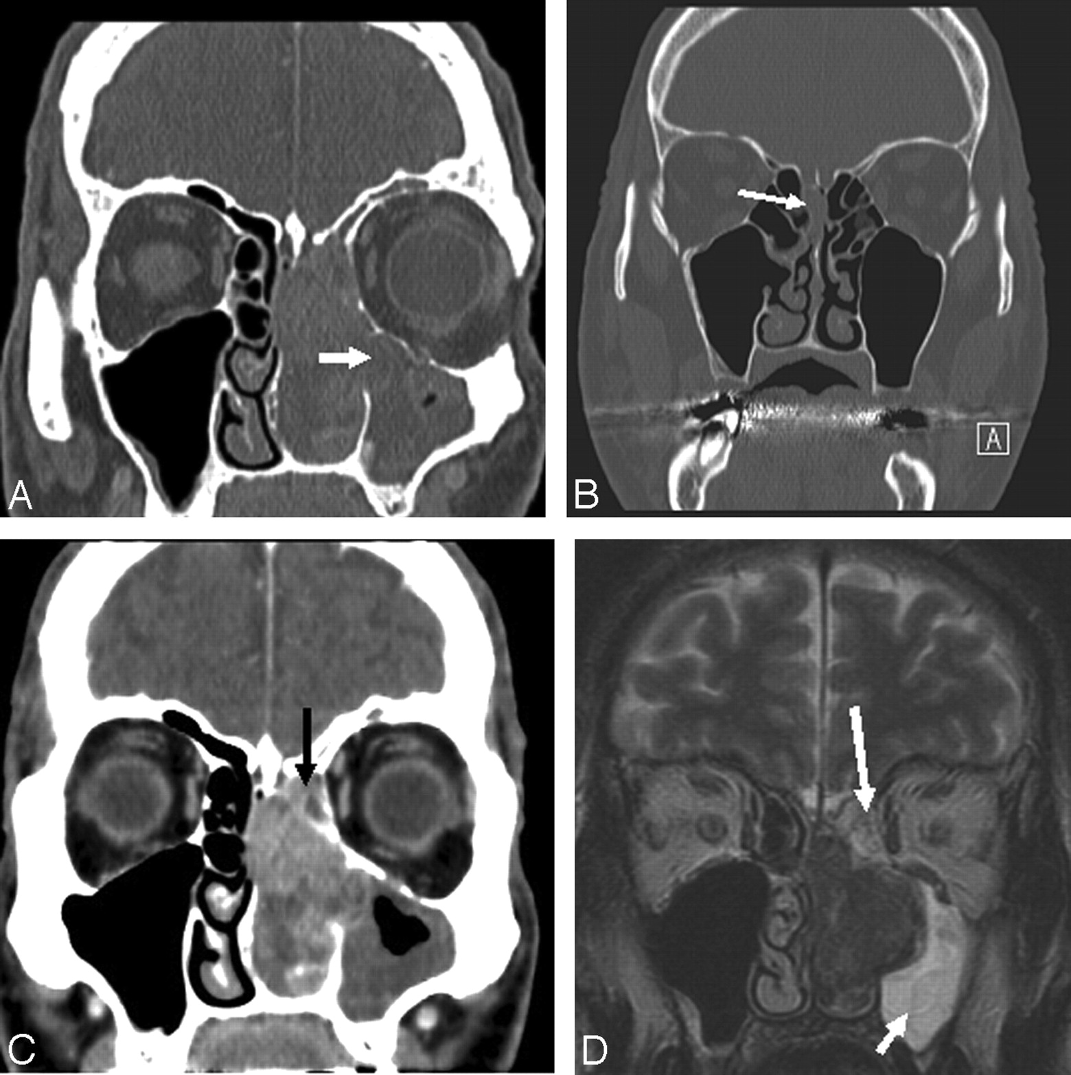
  • Fig 6.
    • Download figure
    • Open in new tab
    • Download powerpoint
    Fig 6.

    More examples of woodworkers’ adenocarcinomas. A, The inferior part of the ethmoidal labyrinth is reduced to a thin opaque layer with the tumor penetrating into the maxillary sinus through the fontanelle area (arrow). B, CT scan of an anosmic woodworker, which led to the early diagnosis of a small adenocarcinoma of the right olfactory cleft (arrow). The right olfactory cleft is opacified with a slight bulging of the corresponding nasal septum to the left and of the conchal lamina to the right. Note that there is still a small space filled with air under the cribriform plate and between the conchal lamina and the septum in the left olfactory cleft. C, The slow-growing tumor originating in the olfactory cleft (below the cribriform plate) sequestrates a small triangle (arrow) of ethmoidal cells under the ethmoidal roof (which lies more cranially) by lateralizing (not invading) the conchal lamina. D, MR image confirms the origin in the olfactory cleft with retention in the squeezed ethmoidal labyrinth (long arrow) (note the triangle of ethmoidal cells under the ethmoidal roof) and maxillary sinus (short arrow).

Tables

  • Figures
  • Descriptive results*

    VariableWoodworkers’ Adenocarcinomas (n = 27)NSP (n = 30)Healthy Controls (n = 33)
    OC4.58 ± 3.050.71 ± 1.070.51 ± 0.98
    (−0, 1–13.7)(−2.1–2.3)(−1.2–2)
    CD15.12 ± 4.523.61 ± 0.453.28 ± 0.68
    (8.6–25.7)(2.8–4.6)(1.4–4.6)
    DE7.18 ± 2.6814.82 ± 0.8813.23 ± 1.47
    (3.2–14.2)(11.4–15.3)(9.8–17.5)
    AE31.03 ± 4.9329.04 ± 2.4729.4 ± 2.12
    (16–40.9)(23.7–35.1)(25.3–34.3)
    ECL (degrees)39.76 ± 13.830.03 ± 2.250.45 ± 2.13
    (14.9–74.6)(−5–3)(−5–5)
    • Note:—NSP indicates nasosinusal polyposis; OC, width between midline and septal bulging or deviation; CD, width of olfactory cleft on pathologic side; DE, ethmoidal labyrinth width; AE, nasoethmoidal cavity total width; ECL, angle between midline and conchal lamina.

    • * Mean ± SD, range.

PreviousNext
Back to top

In this issue

American Journal of Neuroradiology: 30 (7)
American Journal of Neuroradiology
Vol. 30, Issue 7
August 2009
  • Table of Contents
  • Index by author
Advertisement
Print
Download PDF
Email Article

Thank you for your interest in spreading the word on American Journal of Neuroradiology.

NOTE: We only request your email address so that the person you are recommending the page to knows that you wanted them to see it, and that it is not junk mail. We do not capture any email address.

Enter multiple addresses on separate lines or separate them with commas.
CT Assessment of Woodworkers’ Nasal Adenocarcinomas Confirms the Origin in the Olfactory Cleft
(Your Name) has sent you a message from American Journal of Neuroradiology
(Your Name) thought you would like to see the American Journal of Neuroradiology web site.
CAPTCHA
This question is for testing whether or not you are a human visitor and to prevent automated spam submissions.
Cite this article
T. Georgel, R. Jankowski, P. Henrot, C. Baumann, S. Kacha, B. Grignon, B. Toussaint, P. Graff, M.C. Kaminsky, L. Geoffrois, J.M. Vignaud
CT Assessment of Woodworkers’ Nasal Adenocarcinomas Confirms the Origin in the Olfactory Cleft
American Journal of Neuroradiology Aug 2009, 30 (7) 1440-1444; DOI: 10.3174/ajnr.A1648

Citation Manager Formats

  • BibTeX
  • Bookends
  • EasyBib
  • EndNote (tagged)
  • EndNote 8 (xml)
  • Medlars
  • Mendeley
  • Papers
  • RefWorks Tagged
  • Ref Manager
  • RIS
  • Zotero
0 Responses
Respond to this article
Share
Bookmark this article
CT Assessment of Woodworkers’ Nasal Adenocarcinomas Confirms the Origin in the Olfactory Cleft
T. Georgel, R. Jankowski, P. Henrot, C. Baumann, S. Kacha, B. Grignon, B. Toussaint, P. Graff, M.C. Kaminsky, L. Geoffrois, J.M. Vignaud
American Journal of Neuroradiology Aug 2009, 30 (7) 1440-1444; DOI: 10.3174/ajnr.A1648
del.icio.us logo Twitter logo Facebook logo Mendeley logo
  • Tweet Widget
  • Facebook Like
  • Google Plus One
Purchase

Jump to section

  • Article
    • Abstract
    • Materials and Methods
    • Results
    • Discussion
    • References
  • Figures & Data
  • Info & Metrics
  • Responses
  • References
  • PDF

Related Articles

  • No related articles found.
  • PubMed
  • Google Scholar

Cited By...

  • Sinonasal imaging
  • Crossref
  • Google Scholar

This article has not yet been cited by articles in journals that are participating in Crossref Cited-by Linking.

More in this TOC Section

  • Hydrops Herniation into the Semicircular Canals
  • ASL Sensitivity for Head and Neck Paraganglioma
  • Post SRS Peritumoral Hyperintense Signal of VSs
Show more HEAD AND NECK IMAGING

Similar Articles

Advertisement

Indexed Content

  • Current Issue
  • Accepted Manuscripts
  • Article Preview
  • Past Issues
  • Editorials
  • Editor's Choice
  • Fellows' Journal Club
  • Letters to the Editor
  • Video Articles

Cases

  • Case Collection
  • Archive - Case of the Week
  • Archive - Case of the Month
  • Archive - Classic Case

More from AJNR

  • Trainee Corner
  • Imaging Protocols
  • MRI Safety Corner
  • Book Reviews

Multimedia

  • AJNR Podcasts
  • AJNR Scantastics

Resources

  • Turnaround Time
  • Submit a Manuscript
  • Submit a Video Article
  • Submit an eLetter to the Editor/Response
  • Manuscript Submission Guidelines
  • Statistical Tips
  • Fast Publishing of Accepted Manuscripts
  • Graphical Abstract Preparation
  • Imaging Protocol Submission
  • Evidence-Based Medicine Level Guide
  • Publishing Checklists
  • Author Policies
  • Become a Reviewer/Academy of Reviewers
  • News and Updates

About Us

  • About AJNR
  • Editorial Board
  • Editorial Board Alumni
  • Alerts
  • Permissions
  • Not an AJNR Subscriber? Join Now
  • Advertise with Us
  • Librarian Resources
  • Feedback
  • Terms and Conditions
  • AJNR Editorial Board Alumni

American Society of Neuroradiology

  • Not an ASNR Member? Join Now

© 2025 by the American Society of Neuroradiology All rights, including for text and data mining, AI training, and similar technologies, are reserved.
Print ISSN: 0195-6108 Online ISSN: 1936-959X

Powered by HighWire