Advertisement

Main menu

  • Home
  • Content
    • Current Issue
    • Accepted Manuscripts
    • Article Preview
    • Past Issue Archive
    • AJNR Case Collection
    • Case of the Week Archive
    • Classic Case Archive
    • Case of the Month Archive
  • Special Collections
    • Spinal CSF Leak Articles (Jan 2020-June 2024)
    • 2024 AJNR Journal Awards
    • Most Impactful AJNR Articles
  • Multimedia
    • AJNR Podcast
    • AJNR Scantastics
    • Video Articles
  • For Authors
    • Submit a Manuscript
    • Author Policies
    • Fast publishing of Accepted Manuscripts
    • Graphical Abstract Preparation
    • Manuscript Submission Guidelines
    • Imaging Protocol Submission
    • Submit a Case for the Case Collection
  • About Us
    • About AJNR
    • Editorial Board
  • More
    • Become a Reviewer/Academy of Reviewers
    • Subscribers
    • Permissions
    • Alerts
    • Feedback
    • Advertisers
    • ASNR Home
  • Other Publications
    • ajnr

User menu

  • Alerts
  • Log in

Search

  • Advanced search
American Journal of Neuroradiology

American Journal of Neuroradiology

ASHNR American Society of Functional Neuroradiology ASHNR American Society of Pediatric Neuroradiology ASSR
  • Alerts
  • Log in

Advanced Search

  • Home
  • Content
    • Current Issue
    • Accepted Manuscripts
    • Article Preview
    • Past Issue Archive
    • AJNR Case Collection
    • Case of the Week Archive
    • Classic Case Archive
    • Case of the Month Archive
  • Special Collections
    • Spinal CSF Leak Articles (Jan 2020-June 2024)
    • 2024 AJNR Journal Awards
    • Most Impactful AJNR Articles
  • Multimedia
    • AJNR Podcast
    • AJNR Scantastics
    • Video Articles
  • For Authors
    • Submit a Manuscript
    • Author Policies
    • Fast publishing of Accepted Manuscripts
    • Graphical Abstract Preparation
    • Manuscript Submission Guidelines
    • Imaging Protocol Submission
    • Submit a Case for the Case Collection
  • About Us
    • About AJNR
    • Editorial Board
  • More
    • Become a Reviewer/Academy of Reviewers
    • Subscribers
    • Permissions
    • Alerts
    • Feedback
    • Advertisers
    • ASNR Home
  • Follow AJNR on Twitter
  • Visit AJNR on Facebook
  • Follow AJNR on Instagram
  • Join AJNR on LinkedIn
  • RSS Feeds

Welcome to the new AJNR, Updated Hall of Fame, and more. Read the full announcements.


AJNR is seeking candidates for the position of Associate Section Editor, AJNR Case Collection. Read the full announcement.


Research ArticleBrain
Open Access

Normal Aging in the Basal Ganglia Evaluated by Eigenvalues of Diffusion Tensor Imaging

Q. Wang, X. Xu and M. Zhang
American Journal of Neuroradiology March 2010, 31 (3) 516-520; DOI: https://doi.org/10.3174/ajnr.A1862
Q. Wang
  • Find this author on Google Scholar
  • Find this author on PubMed
  • Search for this author on this site
X. Xu
  • Find this author on Google Scholar
  • Find this author on PubMed
  • Search for this author on this site
M. Zhang
  • Find this author on Google Scholar
  • Find this author on PubMed
  • Search for this author on this site
  • Article
  • Figures & Data
  • Info & Metrics
  • Responses
  • References
  • PDF
Loading

Article Figures & Data

Figures

  • Tables
  • Fig 1.
    • Download figure
    • Open in new tab
    • Download powerpoint
    Fig 1.

    Depictions of regions of interest used in this study. A, T2-weighted trace image. B and C, T2-weighted images. A, Regions of interest are drawn in the head of the caudate nucleus (regions of interest 1 and 2), the putamen (regions of interest 3 and 4), and the globus pallidus (regions of interest 5 and 6). B, Regions of interest are drawn in the prefrontal white matter (regions of interest 1 and 2), the genu (region of interest 3), and the splenium (region of interest 4) of the corpus callosum. C, Regions of interest are drawn in regions of the poster limb of the internal capsule (regions of interest 1 and 2).
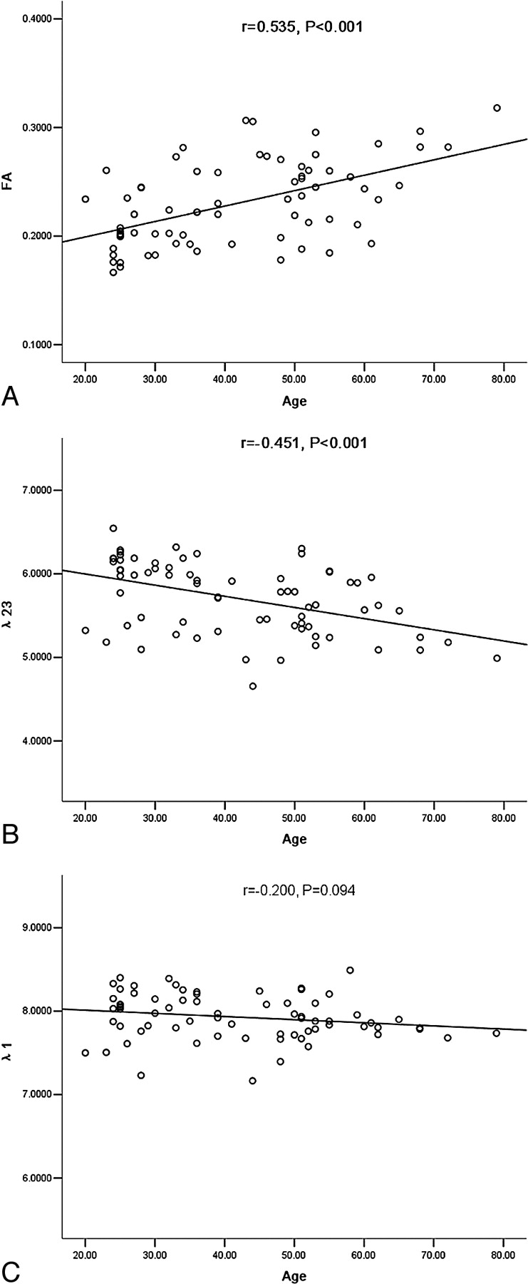
  • Fig 2.
    • Download figure
    • Open in new tab
    • Download powerpoint
    Fig 2.

    The scatterplots of FA (A) and eigenvalues λ23 (B) and λ1 (C) with aging in the putamen (λ1, λ23: × 10−4 mm2/s).

  • Fig 3.
    • Download figure
    • Open in new tab
    • Download powerpoint
    Fig 3.

    The color-coded FA (A), λ1 (B), λ2 (C), and λ3 (D) maps at the level of the basal ganglia (λ1, λ2, λ3: × 10−4 mm2/s).

Tables

  • Figures
  • Mean values and Pearson correlations between age and DTI metrics in the basal ganglia (MD, λ1, λ23 × 10−4 mm2/s)

    RegionMD (Mean ± SD)FA (Mean ± SD)λ1 (Mean ± SD)λ23 (Mean ± SD)
    Caudate6.88 ± 0.42, r = −0.319, P = .0070.24 ± 0.03, r = 0.178, P = .1388.57 ± 0.46, r = −0.299, P = .0116.03 ± 0.43, r = −0.312, P = .008
    Putamen6.45 ± 0.36, r = −0.410, P < .0010.23 ± 0.04, r = 0.535, P < .0017.93 ± 0.27, r = −0.200, P = .0945.71 ± 0.43, r = −0.451, P < .001
    Globus pallidus6.00 ± 0.49, r = −0.142, P = .2380.36 ± 0.04, r = 0.276, P = .0208.16 ± 0.42, r = −0.117, P = .3294.89 ± 0.45, r = −0.215, P = .072
    Prefrontal7.18 ± 0.51, r = 0.314, P = .0080.35 ± 0.05, r = −0.391, P = .0019.82 ± 0.65, r = 0.053, P = .6635.86 ± 0.56, r = 0.403, P < .001
    Genu6.98 ± 0.65, r = 0.319, P = .0070.65 ± 0.06, r = −0.307, P = .00912.97 ± 1.19, r = 0.139, P = .2493.98 ± 0.65, r = 0.353, P = .003
    Splenium6.77 ± 0.67, r = −0.153, P = .2020.75 ± 0.05, r = −0.008, P = .94614.16 ± 1.51, r = −0.156, P = .1943.06 ± 0.56, r = −0.066, P = .585
    Internal capsule6.43 ± 0.51, r = −0.185, P = .1230.69 ± 0.04, r = 0.013, P = .91412.56 ± 0.85, r = −0.231, P = .0533.36 ± 0.46, r = −0.090, P = .458
PreviousNext
Back to top

In this issue

American Journal of Neuroradiology: 31 (3)
American Journal of Neuroradiology
Vol. 31, Issue 3
1 Mar 2010
  • Table of Contents
  • Index by author
Advertisement
Print
Download PDF
Email Article

Thank you for your interest in spreading the word on American Journal of Neuroradiology.

NOTE: We only request your email address so that the person you are recommending the page to knows that you wanted them to see it, and that it is not junk mail. We do not capture any email address.

Enter multiple addresses on separate lines or separate them with commas.
Normal Aging in the Basal Ganglia Evaluated by Eigenvalues of Diffusion Tensor Imaging
(Your Name) has sent you a message from American Journal of Neuroradiology
(Your Name) thought you would like to see the American Journal of Neuroradiology web site.
CAPTCHA
This question is for testing whether or not you are a human visitor and to prevent automated spam submissions.
Cite this article
Q. Wang, X. Xu, M. Zhang
Normal Aging in the Basal Ganglia Evaluated by Eigenvalues of Diffusion Tensor Imaging
American Journal of Neuroradiology Mar 2010, 31 (3) 516-520; DOI: 10.3174/ajnr.A1862

Citation Manager Formats

  • BibTeX
  • Bookends
  • EasyBib
  • EndNote (tagged)
  • EndNote 8 (xml)
  • Medlars
  • Mendeley
  • Papers
  • RefWorks Tagged
  • Ref Manager
  • RIS
  • Zotero
0 Responses
Respond to this article
Share
Bookmark this article
Normal Aging in the Basal Ganglia Evaluated by Eigenvalues of Diffusion Tensor Imaging
Q. Wang, X. Xu, M. Zhang
American Journal of Neuroradiology Mar 2010, 31 (3) 516-520; DOI: 10.3174/ajnr.A1862
del.icio.us logo Twitter logo Facebook logo Mendeley logo
  • Tweet Widget
  • Facebook Like
  • Google Plus One
Purchase

Jump to section

  • Article
    • Abstract
    • Abbreviations
    • Materials and Methods
    • Results
    • Discussion
    • Conclusions
    • Footnotes
    • References
  • Figures & Data
  • Info & Metrics
  • Responses
  • References
  • PDF

Related Articles

  • No related articles found.
  • PubMed
  • Google Scholar

Cited By...

  • Automated Surface-Based Segmentation of Deep Gray Matter Regions Based on Diffusion Tensor Images Reveals Unique Age Trajectories Over the Healthy Lifespan
  • Variation in reported human head tissue electrical conductivity values
  • Simple Linear Regression Model Is Misleading When Used to Analyze Quantitative Diffusion Tensor Imaging Data That Include Young and Old Adults
  • Reply:
  • Crossref (39)
  • Google Scholar

This article has been cited by the following articles in journals that are participating in Crossref Cited-by Linking.

  • Variation in Reported Human Head Tissue Electrical Conductivity Values
    Hannah McCann, Giampaolo Pisano, Leandro Beltrachini
    Brain Topography 2019 32 5
  • The neural basis of central proprioceptive processing in older versus younger adults: An important sensory role for right putamen
    Daniel J. Goble, James P. Coxon, Annouchka Van Impe, Monique Geurts, Wim Van Hecke, Stefan Sunaert, Nicole Wenderoth, Stephan P. Swinnen
    Human Brain Mapping 2012 33 4
  • Meta-analyses of the n-back working memory task: fMRI evidence of age-related changes in prefrontal cortex involvement across the adult lifespan
    Zachary A. Yaple, W. Dale Stevens, Marie Arsalidou
    NeuroImage 2019 196
  • Prediction of individual subject&#039;s age across the human lifespan using diffusion tensor imaging: A machine learning approach
    Benson Mwangi, Khader M. Hasan, Jair C. Soares
    NeuroImage 2013 75
  • Multiple time scale complexity analysis of resting state FMRI
    Robert X. Smith, Lirong Yan, Danny J. J. Wang
    Brain Imaging and Behavior 2014 8 2
  • Aging in deep gray matter and white matter revealed by diffusional kurtosis imaging
    Nan-Jie Gong, Chun-Sing Wong, Chun-Chung Chan, Lam-Ming Leung, Yiu-Ching Chu
    Neurobiology of Aging 2014 35 10
  • MR‐based age‐related effects on the striatum, globus pallidus, and thalamus in healthy individuals across the adult lifespan
    Stephanie Tullo, Raihaan Patel, Gabriel A. Devenyi, Alyssa Salaciak, Saashi A. Bedford, Sarah Farzin, Nancy Wlodarski, Christine L. Tardif, John C. S. Breitner, M. Mallar Chakravarty
    Human Brain Mapping 2019 40 18
  • Chasing shadows: What determines DTI metrics in gray matter regions? An in vitro and in vivo study
    Aaron M. Rulseh, Jiří Keller, Jaroslav Tintěra, Milan Kožíšek, Josef Vymazal
    Journal of Magnetic Resonance Imaging 2013 38 5
  • Diffusion-Tensor Imaging of Thigh Muscles in Duchenne Muscular Dystrophy: Correlation of Apparent Diffusion Coefficient and Fractional Anisotropy Values With Fatty Infiltration
    Gui Dian Li, Ying Yin Liang, Ping Xu, Jian Ling, Ying Ming Chen
    American Journal of Roentgenology 2016 206 4
  • Lower Prefrontal and Hippocampal Volume and Diffusion Tensor Imaging Differences Reflect Structural and Functional Abnormalities in Abstinent Individuals with Alcohol Use Disorder
    Ashwini Kumar Pandey, Babak Assai Ardekani, Chella Kamarajan, Jian Zhang, David Balin Chorlian, Kelly Nicole‐Helen Byrne, Gayathri Pandey, Jacquelyn Leigh Meyers, Sivan Kinreich, Arthur Stimus, Bernice Porjesz
    Alcoholism: Clinical and Experimental Research 2018 42 10

More in this TOC Section

  • SWI or T2*: Which MRI Sequence to Use in the Detection of Cerebral Microbleeds? The Karolinska Imaging Dementia Study
  • Progression of Microstructural Damage in Spinocerebellar Ataxia Type 2: A Longitudinal DTI Study
  • Usefulness of Quantitative Susceptibility Mapping for the Diagnosis of Parkinson Disease
Show more Brain

Similar Articles

Advertisement

Indexed Content

  • Current Issue
  • Accepted Manuscripts
  • Article Preview
  • Past Issues
  • Editorials
  • Editors Choice
  • Fellow Journal Club
  • Letters to the Editor

Cases

  • Case Collection
  • Archive - Case of the Week
  • Archive - Case of the Month
  • Archive - Classic Case

Special Collections

  • Special Collections

Resources

  • News and Updates
  • Turn around Times
  • Submit a Manuscript
  • Author Policies
  • Manuscript Submission Guidelines
  • Evidence-Based Medicine Level Guide
  • Publishing Checklists
  • Graphical Abstract Preparation
  • Imaging Protocol Submission
  • Submit a Case
  • Become a Reviewer/Academy of Reviewers
  • Get Peer Review Credit from Publons

Multimedia

  • AJNR Podcast
  • AJNR SCANtastic
  • Video Articles

About Us

  • About AJNR
  • Editorial Board
  • Not an AJNR Subscriber? Join Now
  • Alerts
  • Feedback
  • Advertise with us
  • Librarian Resources
  • Permissions
  • Terms and Conditions

American Society of Neuroradiology

  • Not an ASNR Member? Join Now

© 2025 by the American Society of Neuroradiology All rights, including for text and data mining, AI training, and similar technologies, are reserved.
Print ISSN: 0195-6108 Online ISSN: 1936-959X

Powered by HighWire