Advertisement

Main menu

  • Home
  • Content
    • Current Issue
    • Accepted Manuscripts
    • Article Preview
    • Past Issue Archive
    • AJNR Case Collection
    • Case of the Week Archive
    • Classic Case Archive
    • Case of the Month Archive
  • Special Collections
    • Spinal CSF Leak Articles (Jan 2020-June 2024)
    • 2024 AJNR Journal Awards
    • Most Impactful AJNR Articles
  • Multimedia
    • AJNR Podcast
    • AJNR Scantastics
    • Video Articles
  • For Authors
    • Submit a Manuscript
    • Author Policies
    • Fast publishing of Accepted Manuscripts
    • Graphical Abstract Preparation
    • Manuscript Submission Guidelines
    • Imaging Protocol Submission
    • Submit a Case for the Case Collection
  • About Us
    • About AJNR
    • Editorial Board
  • More
    • Become a Reviewer/Academy of Reviewers
    • Subscribers
    • Permissions
    • Alerts
    • Feedback
    • Advertisers
    • ASNR Home
  • Other Publications
    • ajnr

User menu

  • Alerts
  • Log in

Search

  • Advanced search
American Journal of Neuroradiology

American Journal of Neuroradiology

ASHNR American Society of Functional Neuroradiology ASHNR American Society of Pediatric Neuroradiology ASSR
  • Alerts
  • Log in

Advanced Search

  • Home
  • Content
    • Current Issue
    • Accepted Manuscripts
    • Article Preview
    • Past Issue Archive
    • AJNR Case Collection
    • Case of the Week Archive
    • Classic Case Archive
    • Case of the Month Archive
  • Special Collections
    • Spinal CSF Leak Articles (Jan 2020-June 2024)
    • 2024 AJNR Journal Awards
    • Most Impactful AJNR Articles
  • Multimedia
    • AJNR Podcast
    • AJNR Scantastics
    • Video Articles
  • For Authors
    • Submit a Manuscript
    • Author Policies
    • Fast publishing of Accepted Manuscripts
    • Graphical Abstract Preparation
    • Manuscript Submission Guidelines
    • Imaging Protocol Submission
    • Submit a Case for the Case Collection
  • About Us
    • About AJNR
    • Editorial Board
  • More
    • Become a Reviewer/Academy of Reviewers
    • Subscribers
    • Permissions
    • Alerts
    • Feedback
    • Advertisers
    • ASNR Home
  • Follow AJNR on Twitter
  • Visit AJNR on Facebook
  • Follow AJNR on Instagram
  • Join AJNR on LinkedIn
  • RSS Feeds

Welcome to the new AJNR, Updated Hall of Fame, and more. Read the full announcements.


AJNR is seeking candidates for the position of Associate Section Editor, AJNR Case Collection. Read the full announcement.


Case ReportHead and Neck

Successful Sonography-Guided Fine-Needle Aspiration Biopsy of a 1-Millimeter-Diameter Papillary Thyroid Microcarcinoma

D.W. Kim, S.H. Kim and S.J. Jung
American Journal of Neuroradiology June 2010, 31 (6) 1082-1084; DOI: https://doi.org/10.3174/ajnr.A1850
D.W. Kim
  • Find this author on Google Scholar
  • Find this author on PubMed
  • Search for this author on this site
S.H. Kim
  • Find this author on Google Scholar
  • Find this author on PubMed
  • Search for this author on this site
S.J. Jung
  • Find this author on Google Scholar
  • Find this author on PubMed
  • Search for this author on this site
  • Article
  • Figures & Data
  • Info & Metrics
  • Responses
  • References
  • PDF
Loading

Abstract

SUMMARY: A PTMC is defined as a thyroid papillary cancer that measures ≤10 mm in the greatest diameter. The detection rate for a PTMC has increased because of the worldwide use of high-resolution sonography and US-FNAB. We report a case of a 1-mm-diameter PTMC confirmed at thyroidectomy for which the cytologic diagnosis had an important role in deciding the extent of surgery (ie, either a total thyroidectomy or a hemithyroidectomy).

Abbreviations

ATA
American Thyroid Association
HE
hematoxylin-eosin
PTC
papillary thyroid carcinoma
PTMC
papillary thyroid microcarcinoma
US-FNAB
sonography-guided fine-needle aspiration biopsy

AUS-FNAB is a widely used procedure for the cytological evaluation of thyroid nodules. However, US-FNAB of a thyroid nodule <10 mm in maximal diameter seems to be limited because of a poor clinical significance and social-medical-cost problem. We report a case of 1 mm diameter PTMC that was diagnosed on US-FNAB and this cytological diagnosis influenced the decision of thyroid surgery type.

Case Report

A 43-year-old woman who had recently complained of fatigue and neck discomfort was referred to the radiology department from the department of general surgery for sonographic evaluation of the thyroid. The patient showed normal values on a thyroid function test (T3, 1.3 nmol/L; free T4, 11.6 pmol/L; thyroid-stimulating hormone, 1.03 mU/L), and no specific abnormalities were detected on a physical examination of the neck. The patient had neither previously undergone a fine-needle biopsy for a thyroid lesion nor had a history of neck irradiation or surgery or a family history of a thyroid malignancy.

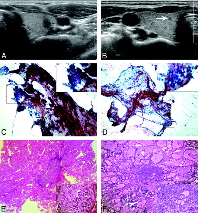
A thyroid nodule that showed marked hypoechogenicity, an ill-defined margin, and a taller-than-wide shape was identified in the left lobe on thyroid sonography. The nodule measured 5.5 mm in the maximal diameter and was considered to be highly suspicious for a malignancy (Fig 1 A). A hypoechoic thyroid nodule that measured 1.0 mm in maximal diameter was detected in the medial aspect of the opposite right midlobe on a simultaneous diagnostic sonography examination, and this nodule was considered as an “indeterminate” nodule for a malignancy (Fig 1B). In addition, several thyroid nodules that showed benign sonographic features were observed in both lobes.

Fig 1.
  • Download figure
  • Open in new tab
  • Download powerpoint
Fig 1.

A, A transverse sonogram shows a 5.5-mm-diameter solid thyroid nodule (calipers) with marked hypoechogenicity, an irregular margin, and a taller-than-wide shape located in the left thyroid lobe. B, A transverse sonogram shows a 1.0-mm-diameter solid thyroid nodule with hypoechogenicity in the right thyroid lobe (arrow). C, On aspiration cytology of left thyroid nodule, many 3D papillary or caplike structured clusters are noted. The characteristic nuclear features of papillary carcinoma, including fine and powdery chromatin and irregular nuclear membranes with nuclear grooves, are noted in most nuclei (Papanicolaou, original magnification ×100 [inset, ×400]). D, Aspiration cytology of the right thyroid nodule reveals a few small aggregates of follicular cells within some lymphocytes. The follicular cells reveal nuclear clearing and grooves suspicious for papillary carcinoma (Papanicolaou, original magnification ×100 [inset, ×400]). E, On higher magnification of left thyroid nodule, the papillary arrangement is well recognized and is considered a classic type of PTMC (HE, original magnification ×40 [inset, ×100]). F, Right thyroid nodule reveals a follicular variant of PTMC, which is composed of follicles having nuclear features of papillary carcinoma on a frozen-section slide (HE, original magnification ×10 [inset, ×100]).

A US-FNAB was performed by 1 investigator (D.W.K.) on the 2 nodules that showed a likelihood of being thyroid malignancies. Only 1 sampling was achieved for each thyroid nodule, without the administration of local anesthetic. A US-FNAB was not performed on several other thyroid nodules suggestive of being benign lesions on thyroid sonography because the selection of a nodule for US-FNAB was based primarily on the sonographic features rather than the nodule size. The patient had no significant pain or other complications following the US-FNAB procedure. On the basis of the cytology results, a nodule highly suspicious for a malignancy in the left lobe was identified as compatible with PTC (Fig 1C), and a 1-mm right-lobe nodule suspicious for a malignancy on thyroid sonography was also identified as suspicious for PTC (Fig 1D).

Although the primary PTC had a maximal diameter of 5.5 mm and an intraglandular location, the surgeon and the patient chose a total thyroidectomy rather than a left hemithyroidectomy on the basis of the cytology findings. On pathology, the tiny malignancy in the right thyroid lobe measured 1.5 mm in maximal diameter at gross and microscopic examinations. On the pathologic findings, PTMCs in left and right thyroid lobes were a classic type and follicular variant, respectively (Fig 1E, -F). These 2 PTMCs showed no capsular or nodal invasion in the perithyroidal areas. In addition, several thyroid nodules that had showed benign features on thyroid sonography in both lobes were determined to be nodular hyperplasia at microscopic findings. After thyroid surgery, the patient has undergone follow-up and has not had any problems related to the thyroid malignancy or the surgery.

Discussion

A PTMC is defined as a PTC <10 mm in the maximal diameter. The detection and incidence rates of PTMCs have increased recently with the worldwide use of thyroid sonography and US-FNABs, and there have been many reports related to the diagnostic efficacy of US-FNAB for PTMCs.1–6 The ATA guidelines and the Society of Radiologists in Sonography Consensus Statement suggest that, in general, only nodules >10 mm in diameter should be evaluated because these nodules have the potential to represent a clinically significant cancer.7,8 However, both the ATA and Society of Radiologists in Sonography Consensus Statement suggest performing US-FNAB on thyroid nodules ≥10 mm in maximal diameter when the nodules exhibit sonographic features associated with thyroid cancer. Recently, Roti et al9 reported that this incidence of bilaterality of a PTMC is 19%. This suggests that incomplete treatment would occur in 19% of patients if only hemithyroidectomy was performed. Therefore, we believe that an accurate evaluation of satellite malignancy in an opposite thyroid lobe through sonographic diagnosis and US-FNAB of the thyroid nodule is necessary.

The sampling technique for US-FNAB of small thyroid nodules <5 mm in maximal diameter is the same as that for thyroid nodules >5 mm in maximal diameter. After the needle tip is placed in the appropriate area of the target nodule, the sampling begins by using the “mixed sampling technique,”6,10 in which the needle is moved up and down for a few seconds only by movement of the operator's wrist and without initial aspiration under sonographic guidance. When the hub of the needle is filled with the proper amount of material, the syringe-needle unit should be rapidly withdrawn. However, if no aspirates are obtained after a few seconds of capillary sampling, negative pressure can gradually be added by use of the first finger of the right hand. This to-and-fro needle movement should be continued during the entire sampling procedure. Only 1 sampling is achieved in most of the US-FNABs for thyroid nodules by using 1 needle puncture and without local anesthesia.

The smear technique is as follows: The operator expels the contents of the needle hub onto 1 or 2 slides for smear preparation after filling the empty syringe with room air. Two-to-4 slides are then obtained by duplicating the smear. Finally, a special technique is used in which the operator flips the plastic hub of the needle onto the slide for effective cell acquisition of the remaining material in the needle hub.10

One of the investigators (D.W.K.) has performed US-FNAB for small thyroid nodules <5 mm in maximal diameter when a nodule has shown the likelihood of a malignancy on diagnostic thyroid sonography.6 In particular, US-FNAB has been performed for small thyroid nodules regardless of the nodule size when it is necessary to determine the type of thyroid surgery (a total thyroidectomy or hemithyroidectomy), if the primary thyroid malignancy is a PTC and is <10 mm in maximal diameter. Our present patient had a small thyroid nodule in the left thyroid lobe that was considered highly suspicious for a malignancy as well as a tiny nodule in the right thyroid lobe that was considered indeterminate for a malignancy on thyroid sonography. The patient underwent a total thyroidectomy based on cytologic findings after a successful US-FNAB for each nodule.

In conclusion, we report a case of a 1-mm-diameter PTMC that was diagnosed by cytology after US-FNAB and confirmed after thyroidectomy. The use of US-FNAB for a 1-mm-diameter thyroid nodule influenced the decision regarding the most appropriate type of thyroid surgery technique.

References

  1. 1.↵
    1. Yokozawa T,
    2. Miyauchi A,
    3. Kuma K,
    4. et al
    . Accurate and simple method of diagnosing thyroid nodules: the modified technique of ultrasound-guided fine needle aspiration biopsy. Thyroid 1995;5:141–45
    PubMed
  2. 2.↵
    1. Leenhardt L,
    2. Hejblum G,
    3. Farnc B,
    4. et al
    . Indications and limits of ultrasound-guided cytology in the management of nonpalpable thyroid nodules. J Clin Endocrinol Metab 1999;84:242–48
  3. 3.↵
    1. Kim SJ,
    2. Kim EK,
    3. Park CS,
    4. et al
    . Ultrasound-guided fine-needle aspiration biopsy in nonpalpable thyroid nodules: is it useful in infracentimetric nodules? Yonsei Med J 2003;44:635–40
    PubMed
  4. 4.↵
    1. Nam-Goong IS,
    2. Kim HY,
    3. Gong GY,
    4. et al
    . Ultrasonography-guided fine-needle aspiration of thyroid incidentaloma: correlation with pathological findings. J Clin Endocrinol 2004;60:21–28
  5. 5.↵
    1. Burman KD
    . Editorial: micropapillary thyroid cancer—should we aspirate all nodules regardless of size? J Clin Endocrinol Metab 2006;91:2043–46
    CrossRefPubMed
  6. 6.↵
    1. Kim DW,
    2. Lee EJ,
    3. Kim SH,
    4. et al
    . Ultrasound-guided fine-needle aspiration biopsy of thyroid nodules: comparison in efficacy according to nodule size. Thyroid 2009;19:27–31
    CrossRefPubMed
  7. 7.↵
    1. Gharib H,
    2. Papini E,
    3. Valcavi R,
    4. et al
    . American Association of Clinical Endocrinologists and Associazione Medici Endorinologi medical guidelines for clinical practice for the diagnosis and management of thyroid nodules. Endocr Pract 2006;12:63–102
    PubMed
  8. 8.↵
    1. Frates MC,
    2. Benson CB,
    3. Charboneau JW,
    4. et al
    . Management of thyroid nodules detected at US: Society of Radiologists in Ultrasound consensus conference statement. Radiology 2005;237:794–800
    CrossRefPubMed
  9. 9.↵
    1. Roti E,
    2. Rossi R,
    3. Tranforini G,
    4. et al
    . Clinical and histological characteristics of papillary thyroid microcarcinoma: results of retrospective study in 243 patients. J Clin Endocrinol Metab 2006;91:2171–78
    CrossRefPubMed
  10. 10.↵
    1. Park KR,
    2. Kim DW,
    3. Rho MH,
    4. et al
    . Ultrasonography-guided fine-needle aspiration biopsy of thyroid nodule: effective technique and a peculiar smear method [In Korean]. J Korean Radiol Soc 2006;55:543–49
  • Received April 14, 2009.
  • Accepted after revision July 25, 2009.
  • Copyright © American Society of Neuroradiology
View Abstract
PreviousNext
Back to top

In this issue

American Journal of Neuroradiology: 31 (6)
American Journal of Neuroradiology
Vol. 31, Issue 6
1 Jun 2010
  • Table of Contents
  • Index by author
Advertisement
Print
Download PDF
Email Article

Thank you for your interest in spreading the word on American Journal of Neuroradiology.

NOTE: We only request your email address so that the person you are recommending the page to knows that you wanted them to see it, and that it is not junk mail. We do not capture any email address.

Enter multiple addresses on separate lines or separate them with commas.
Successful Sonography-Guided Fine-Needle Aspiration Biopsy of a 1-Millimeter-Diameter Papillary Thyroid Microcarcinoma
(Your Name) has sent you a message from American Journal of Neuroradiology
(Your Name) thought you would like to see the American Journal of Neuroradiology web site.
CAPTCHA
This question is for testing whether or not you are a human visitor and to prevent automated spam submissions.
Cite this article
D.W. Kim, S.H. Kim, S.J. Jung
Successful Sonography-Guided Fine-Needle Aspiration Biopsy of a 1-Millimeter-Diameter Papillary Thyroid Microcarcinoma
American Journal of Neuroradiology Jun 2010, 31 (6) 1082-1084; DOI: 10.3174/ajnr.A1850

Citation Manager Formats

  • BibTeX
  • Bookends
  • EasyBib
  • EndNote (tagged)
  • EndNote 8 (xml)
  • Medlars
  • Mendeley
  • Papers
  • RefWorks Tagged
  • Ref Manager
  • RIS
  • Zotero
0 Responses
Respond to this article
Share
Bookmark this article
Successful Sonography-Guided Fine-Needle Aspiration Biopsy of a 1-Millimeter-Diameter Papillary Thyroid Microcarcinoma
D.W. Kim, S.H. Kim, S.J. Jung
American Journal of Neuroradiology Jun 2010, 31 (6) 1082-1084; DOI: 10.3174/ajnr.A1850
del.icio.us logo Twitter logo Facebook logo Mendeley logo
  • Tweet Widget
  • Facebook Like
  • Google Plus One
Purchase

Jump to section

  • Article
    • Abstract
    • Abbreviations
    • Case Report
    • Discussion
    • References
  • Figures & Data
  • Info & Metrics
  • Responses
  • References
  • PDF

Related Articles

  • No related articles found.
  • PubMed
  • Google Scholar

Cited By...

  • An Exponential Growth in Incidence of Thyroid Cancer: Trends and Impact of CT Imaging
  • Biopsy of Tiny Thyroid Nodules: Not a Question of "Can We" but "Should We"
  • Crossref (11)
  • Google Scholar

This article has been cited by the following articles in journals that are participating in Crossref Cited-by Linking.

  • An Exponential Growth in Incidence of Thyroid Cancer: Trends and Impact of CT Imaging
    J. K. Hoang, K. R. Choudhury, J. D. Eastwood, R. M. Esclamado, G. H. Lyman, T. M. Shattuck, X. V. Nguyen
    American Journal of Neuroradiology 2014 35 4
  • Size and Ultrasound Features Affecting Results of Ultrasound‐Guided Fine‐Needle Aspiration of Thyroid Nodules
    YiJie Dong, MinJing Mao, WeiWei Zhan, JianQiao Zhou, Wei Zhou, JieJie Yao, YunYun Hu, Yan Wang, TingJun Ye
    Journal of Ultrasound in Medicine 2018 37 6
  • Thyroglobulin measurement in fine‐needle aspiration biopsy of metastatic lymph nodes after rhTSH stimulation
    Carlo Cappelli, Ilenia Pirola, Elvira De Martino, Elena Gandossi, Elena Cimino, Francesca Samoni, Barbara Agosti, Enrico Agabiti Rosei, Claudio Casella, Maurizio Castellano
    Head & Neck 2013 35 1
  • Diagnostic Value of Ultrasound-Guided Fine Needle Aspiration Biopsy in Malignant Thyroid Nodules: Utility for Micronodules
    Betul Unal, Cem Sezer
    Asian Pacific Journal of Cancer Prevention 2014 15 20
  • Examining the impact of several factors including COVID‐19 on thyroid fine‐needle aspiration biopsy
    Muzaffer Serdar Deniz, Merve Dindar
    Diagnostic Cytopathology 2024 52 1
  • Cervical Lymph Node Features Predictive of Suboptimal Adequacy During Ultrasound‐Guided Fine‐Needle Aspiration in Thyroid Cancer Patients
    Joshua Vic Chen, Tara A. Morgan, Chienying Liu, Elham Khanafshar, Hailey H. Choi
    Journal of Ultrasound in Medicine 2022 41 1
  • Diagnóstico incidental versus clínico del microcarcinoma papilar de tiroides. Pronóstico a largo plazo
    José Ruiz, Antonio Ríos, José Manuel Rodríguez, Miriam Paredes, Víctor Soriano, María Isabel Oviedo, Antonio Miguel Hernández, Pascual Parrilla
    Endocrinología, Diabetes y Nutrición 2020 67 5
  • Biopsy of Tiny Thyroid Nodules: Not a Question of “Can We” but “Should We”
    A.M. Saindane, P.A. Hudgins
    American Journal of Neuroradiology 2011 32 1
  • Incidental versus clinical diagnosis of papillary thyroid microcarcinoma. Long-term prognosis
    José Ruiz, Antonio Ríos, José Manuel Rodríguez, Miriam Paredes, Victor Soriano, María Isabel Oviedo, Antonio Miguel Hernández, Pascual Parrilla
    Endocrinología, Diabetes y Nutrición (English ed.) 2020 67 5
  • Reply to Letter to the Editor
    Carlo Cappelli
    Head & Neck 2013 35 1

More in this TOC Section

  • Correlation of Apparent Diffusion Coefficient at 3T with Prognostic Parameters of Retinoblastoma
  • Parathyroid Lesions: Characterization with Dual-Phase Arterial and Venous Enhanced CT of the Neck
  • MR Evaluation of Sinonasal Angiomatous Polyp
Show more Head and Neck

Similar Articles

Advertisement

Indexed Content

  • Current Issue
  • Accepted Manuscripts
  • Article Preview
  • Past Issues
  • Editorials
  • Editors Choice
  • Fellow Journal Club
  • Letters to the Editor

Cases

  • Case Collection
  • Archive - Case of the Week
  • Archive - Case of the Month
  • Archive - Classic Case

Special Collections

  • Special Collections

Resources

  • News and Updates
  • Turn around Times
  • Submit a Manuscript
  • Author Policies
  • Manuscript Submission Guidelines
  • Evidence-Based Medicine Level Guide
  • Publishing Checklists
  • Graphical Abstract Preparation
  • Imaging Protocol Submission
  • Submit a Case
  • Become a Reviewer/Academy of Reviewers
  • Get Peer Review Credit from Publons

Multimedia

  • AJNR Podcast
  • AJNR SCANtastic
  • Video Articles

About Us

  • About AJNR
  • Editorial Board
  • Not an AJNR Subscriber? Join Now
  • Alerts
  • Feedback
  • Advertise with us
  • Librarian Resources
  • Permissions
  • Terms and Conditions

American Society of Neuroradiology

  • Not an ASNR Member? Join Now

© 2025 by the American Society of Neuroradiology All rights, including for text and data mining, AI training, and similar technologies, are reserved.
Print ISSN: 0195-6108 Online ISSN: 1936-959X

Powered by HighWire