Advertisement

Main menu

  • Home
  • Content
    • Current Issue
    • Accepted Manuscripts
    • Article Preview
    • Past Issue Archive
    • AJNR Case Collection
    • Case of the Week Archive
    • Classic Case Archive
    • Case of the Month Archive
  • Special Collections
    • Spinal CSF Leak Articles (Jan 2020-June 2024)
    • 2024 AJNR Journal Awards
    • Most Impactful AJNR Articles
  • Multimedia
    • AJNR Podcast
    • AJNR Scantastics
    • Video Articles
  • For Authors
    • Submit a Manuscript
    • Author Policies
    • Fast publishing of Accepted Manuscripts
    • Graphical Abstract Preparation
    • Manuscript Submission Guidelines
    • Imaging Protocol Submission
    • Submit a Case for the Case Collection
  • About Us
    • About AJNR
    • Editorial Board
  • More
    • Become a Reviewer/Academy of Reviewers
    • Subscribers
    • Permissions
    • Alerts
    • Feedback
    • Advertisers
    • ASNR Home
  • Other Publications
    • ajnr

User menu

  • Alerts
  • Log in

Search

  • Advanced search
American Journal of Neuroradiology

American Journal of Neuroradiology

ASHNR American Society of Functional Neuroradiology ASHNR American Society of Pediatric Neuroradiology ASSR
  • Alerts
  • Log in

Advanced Search

  • Home
  • Content
    • Current Issue
    • Accepted Manuscripts
    • Article Preview
    • Past Issue Archive
    • AJNR Case Collection
    • Case of the Week Archive
    • Classic Case Archive
    • Case of the Month Archive
  • Special Collections
    • Spinal CSF Leak Articles (Jan 2020-June 2024)
    • 2024 AJNR Journal Awards
    • Most Impactful AJNR Articles
  • Multimedia
    • AJNR Podcast
    • AJNR Scantastics
    • Video Articles
  • For Authors
    • Submit a Manuscript
    • Author Policies
    • Fast publishing of Accepted Manuscripts
    • Graphical Abstract Preparation
    • Manuscript Submission Guidelines
    • Imaging Protocol Submission
    • Submit a Case for the Case Collection
  • About Us
    • About AJNR
    • Editorial Board
  • More
    • Become a Reviewer/Academy of Reviewers
    • Subscribers
    • Permissions
    • Alerts
    • Feedback
    • Advertisers
    • ASNR Home
  • Follow AJNR on Twitter
  • Visit AJNR on Facebook
  • Follow AJNR on Instagram
  • Join AJNR on LinkedIn
  • RSS Feeds

Welcome to the new AJNR, Updated Hall of Fame, and more. Read the full announcements.


AJNR is seeking candidates for the position of Associate Section Editor, AJNR Case Collection. Read the full announcement.


Review ArticleReview Article
Open Access

Sphenoid Masses in Children: Radiologic Differential Diagnosis with Pathologic Correlation

Y.W. Lui, S.B. Dasari and R.J. Young
American Journal of Neuroradiology April 2011, 32 (4) 617-626; DOI: https://doi.org/10.3174/ajnr.A2144
Y.W. Lui
  • Find this author on Google Scholar
  • Find this author on PubMed
  • Search for this author on this site
S.B. Dasari
  • Find this author on Google Scholar
  • Find this author on PubMed
  • Search for this author on this site
R.J. Young
  • Find this author on Google Scholar
  • Find this author on PubMed
  • Search for this author on this site
  • Article
  • Figures & Data
  • Info & Metrics
  • Responses
  • References
  • PDF
Loading

Article Figures & Data

Figures

  • Fig 1.
    • Download figure
    • Open in new tab
    • Download powerpoint
    Fig 1.

    An imaging-guided algorithm.

  • Fig 2.
    • Download figure
    • Open in new tab
    • Download powerpoint
    Fig 2.

    An 18-year-old man with sphenoid mucocele. A, Axial T2WI demonstrates a fluid-filled and expanded sphenoid sinus. B, Sagittal noncontrast T1WI shows a fluid/debris level within the lesion. Relative hyperintense material on T1WI reflects proteinaceous fluid content. There is intracranial extension with loss of the cortical margin of the planum sphenoidale (arrow).
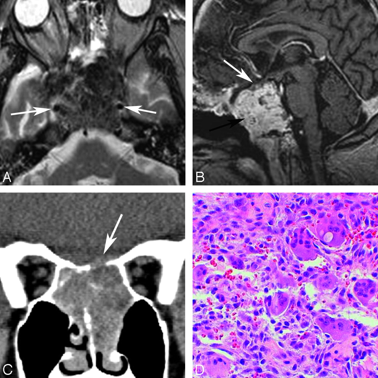
  • Fig 3.
    • Download figure
    • Open in new tab
    • Download powerpoint
    Fig 3.

    A 19-year-old woman with aneurysmal bone cyst. A and B, Gadolinium-enhanced sagittal T1WI (A) and axial T2WI (B) show multiple fluid/fluid levels in a complex multicystic mass centered on the sphenoid bone, expanding the bone and filling the sphenoid sinus. C, Photomicrograph shows blood-filled cavernous channels at low-power magnification with (HE staining, original magnification ×4). D, Higher power photomicrograph shows spindle cells in the vessel walls, reactive bone formation, and multinucleated giant cells typical of these lesions (HE staining, original magnification ×40).
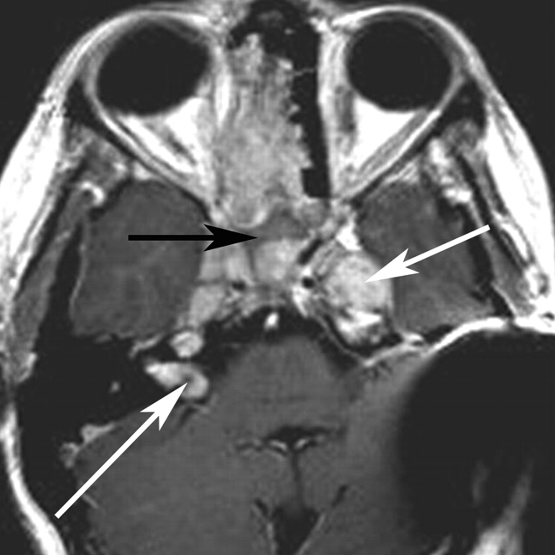
  • Fig 4.
    • Download figure
    • Open in new tab
    • Download powerpoint
    Fig 4.

    Two patients with giant cell lesions of the sphenoid bone. A, An 18-year-old man with sphenoid giant cell tumor: axial T2WI shows relative hypointense areas within a lesion centered on the sphenoid bone and obliterating the sphenoid sinus cavity. These hypointense areas may represent areas of old blood products and fibrotic change. The carotids are displaced laterally (arrows). B, Gadolinium-enhanced sagittal T1WI shows the lesion avidly enhancing (black arrow). The pituitary gland and sella are displaced cephalad (white arrow). C, A 27-year-old woman with giant cell reparative granuloma. Coronal reformatted image in a soft-tissue window from a noncontrast CT scan shows a soft-tissue mass centered on the sphenoid sinus with dehiscence of the planum sphenoidale (arrow). D, HE stain on the photomicrograph shows giant cells with multiple nuclei, fibrous tissue, and blood products characteristic of this reactive lesion (original magnification ×40).

  • Fig 5.
    • Download figure
    • Open in new tab
    • Download powerpoint
    Fig 5.

    A 10-year-old female patient with central skull base meningioma invading the paranasal sinuses. Axial postcontrast T1WI demonstrates an enhancing mass within the central skull base. The mass involves the ethmoid and sphenoid sinuses and was a biopsy-proved meningioma. Nonenhancing anterior portion of the sphenoid sinus is filled with secretions (black arrow). Multiple additional schwannomas are seen in the Meckel cave and the internal auditory canal (white arrows) in this patient with neurofibromatosis type 2. Although meningioma is common in adults, in pediatric patients, the lesion is much rarer and occurs in the setting of neurofibromatosis type 2 or prior radiation therapy.

  • Fig 6.
    • Download figure
    • Open in new tab
    • Download powerpoint
    Fig 6.

    A 13-year-old girl with fibrous dysplasia affecting the sphenoid bone. A, Bone window on CT shows classic ground-glass attenuation in the sphenoid bone. B, The sphenoid sinuses are small and demonstrate mucosal disease. The lesion is hypointense on T2WI. C, This MR imaging appearance is likely attributable to the fibrous stroma, illustrated on the photomicrograph with HE stain demonstrating woven bone embedded in a spindle cell stroma (original magnification ×4). Fibrous dysplasia can have extreme heterogeneity on MR imaging and can be a confusing lesion to diagnose. Correlation with CT should always be done.
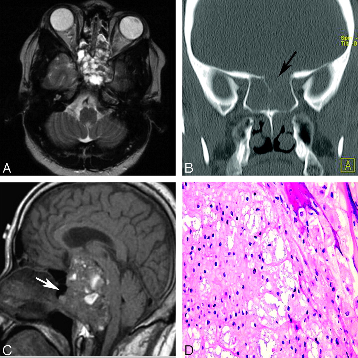
  • Fig 7.
    • Download figure
    • Open in new tab
    • Download powerpoint
    Fig 7.

    A 4-year-old girl with hypothalamic astrocytoma. A, Sagittal postcontrast T1WI shows a large enhancing mass extending from the suprasellar region. The lesion expands and fills the sella with remodeling of the sphenoid bone. The sphenoid sinus is small (black arrow). Postoperative changes and artifacts are present (white arrow). B and C, Compact and spongy areas are seen on photomicrographs of histopathologic specimens, correlating to solid and cystic components of the tumor (HE staining; original magnification ×40).

  • Fig 8.
    • Download figure
    • Open in new tab
    • Download powerpoint
    Fig 8.

    Two patients with chordoma involving the sphenoid bone. A, An 18-year-old male patient. Axial T2WI shows a multicystic lesion involving the central skull base, including the sphenoid bone, sphenoid sinus, posterior ethmoids, and left middle cranial fossa. Focal areas of T2 hyperintensity are seen. B, In the coronal reformatted image, a bone window shows dehiscence of the planum sphenoidale (arrow). C, An 11-year-old boy with chordoma. Sagittal noncontrast T1WI shows the lesion to nearly fill the sphenoid sinus. There is a small residual aerated portion of the sphenoid sinus cavity (arrow). D, Photomicrograph shows that neoplastic epithelioid cells form cords in a mucoid background (HE staining; original magnification ×40).
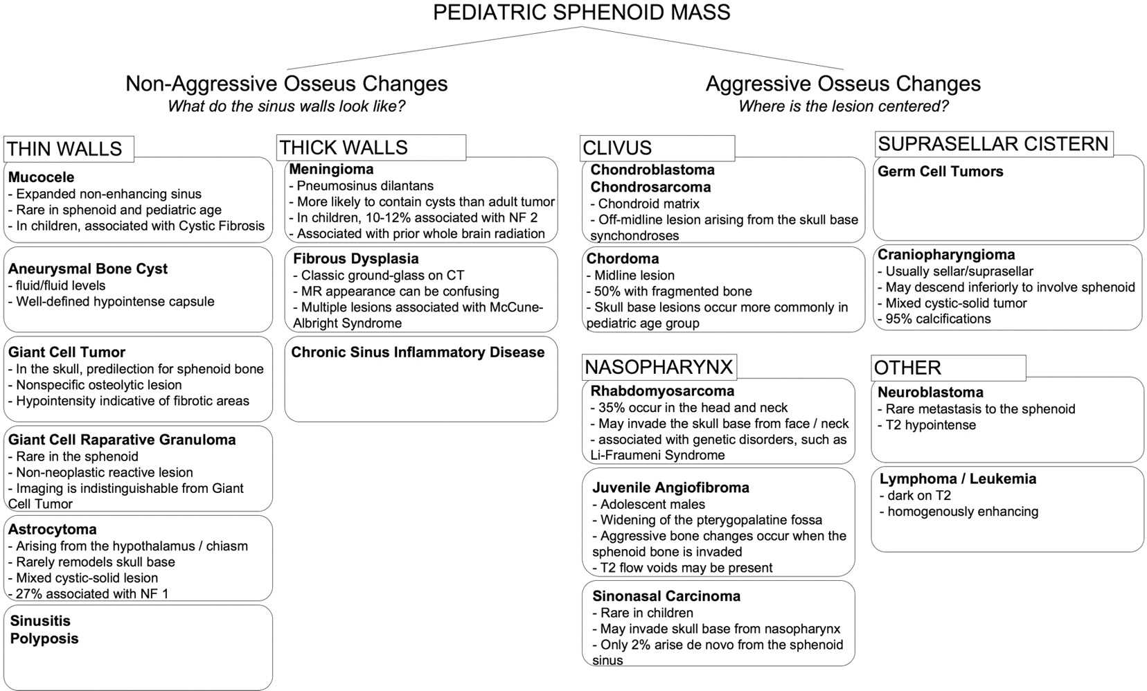
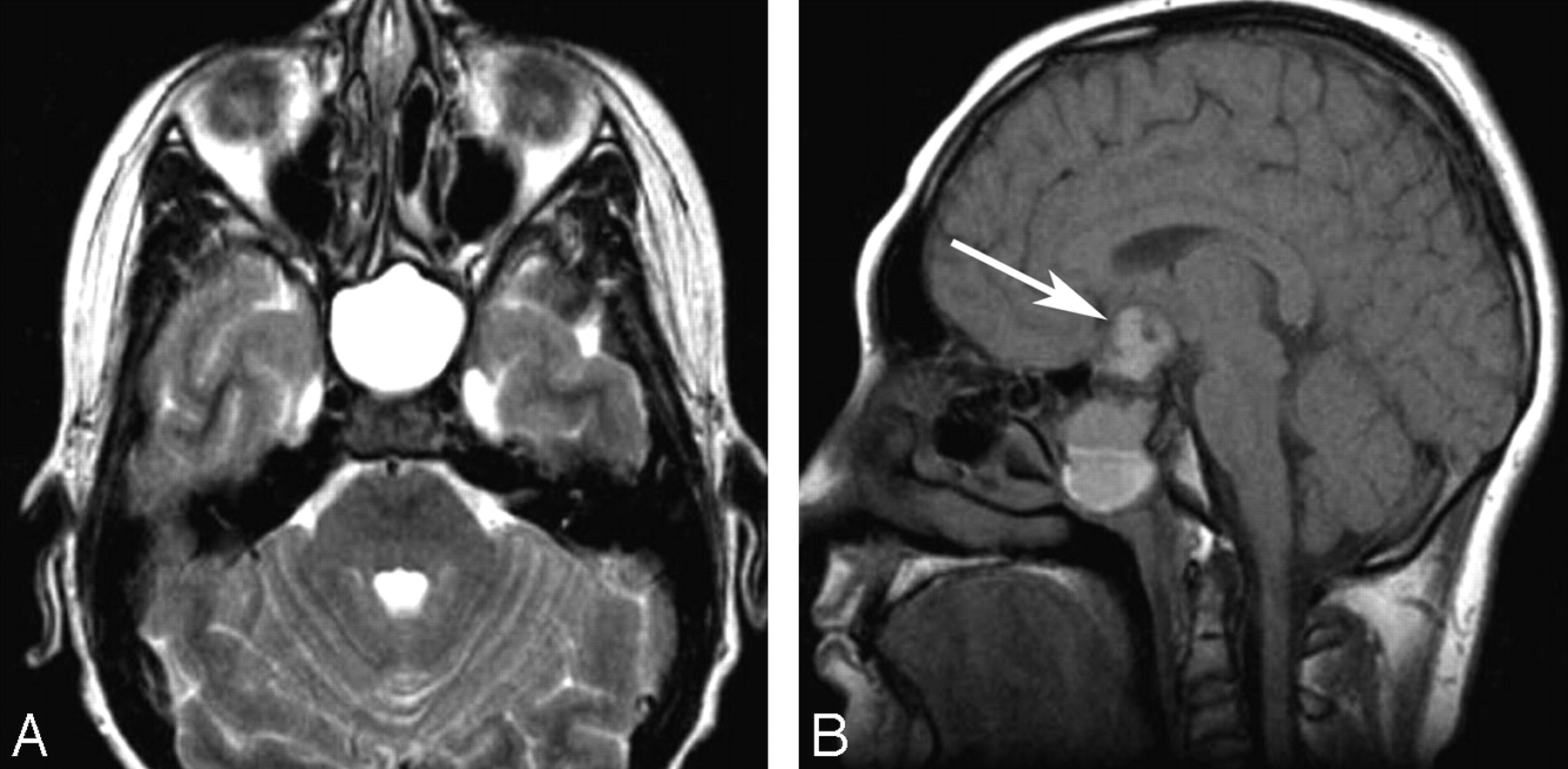
  • Fig 9.
    • Download figure
    • Open in new tab
    • Download powerpoint
    Fig 9.

    A 12-year-old boy with craniopharyngioma. A, Axial fluid-attenuated inversion recovery image shows the cystic portion of the tumor (black arrow) within the central sphenoid bone (white arrow). There is a fluid/fluid level. B, Photomicrograph of a histopathologic specimen shows findings characteristic of craniopharyngioma, with nests of anucleated squamous cells embedded in fibrous tissue and clefts consistent with cholesterol (HE staining; original magnification ×10).

  • Fig 10.
    • Download figure
    • Open in new tab
    • Download powerpoint
    Fig 10.

    A 16-year-old boy with rhabdomyosarcoma. A, Bone window from a noncontrast CT examination show opacification of the right-sided paranasal sinuses, including the right sphenoid sinus (arrow). There are areas of dehiscent bone involving the maxillary sinus walls. B, Soft-tissue window shows the lesion involving the right nasal cavity, right maxillary and sphenoid sinuses, right orbit, and right pterygopalatine fossa (white arrow). Note the normal fat within the left pterygopalatine fossa (black arrow).
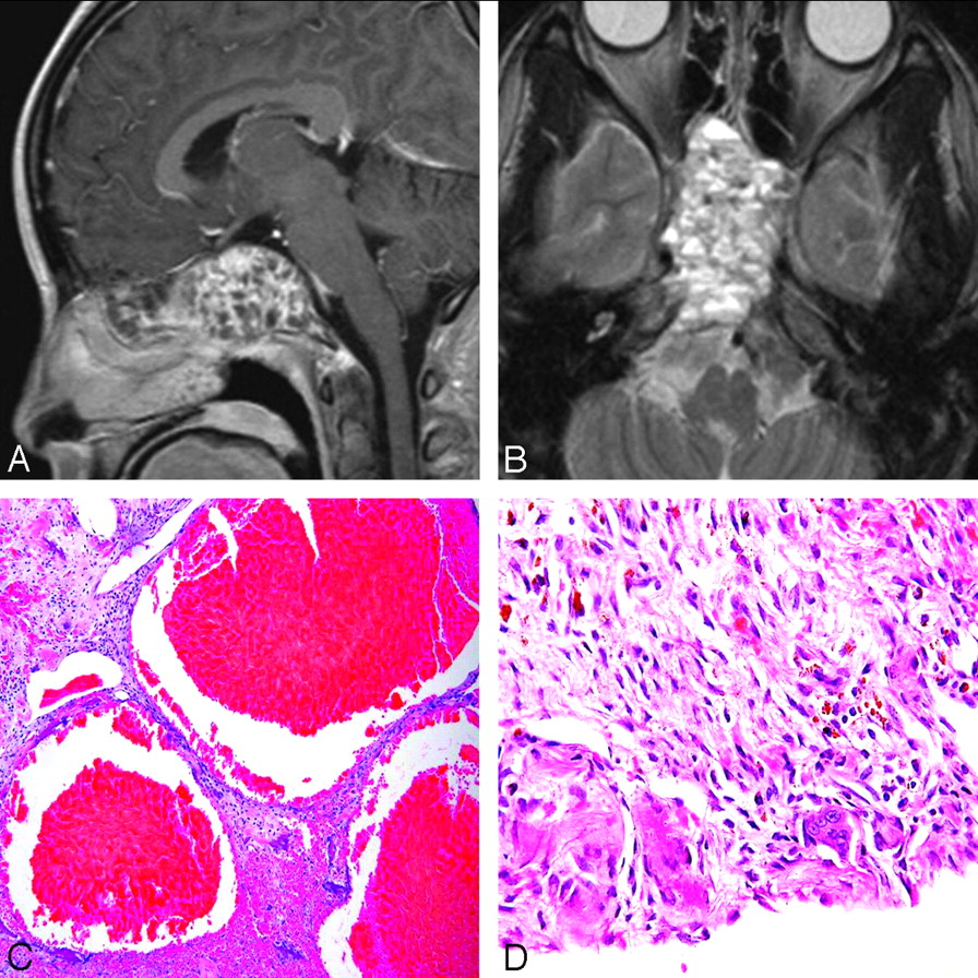
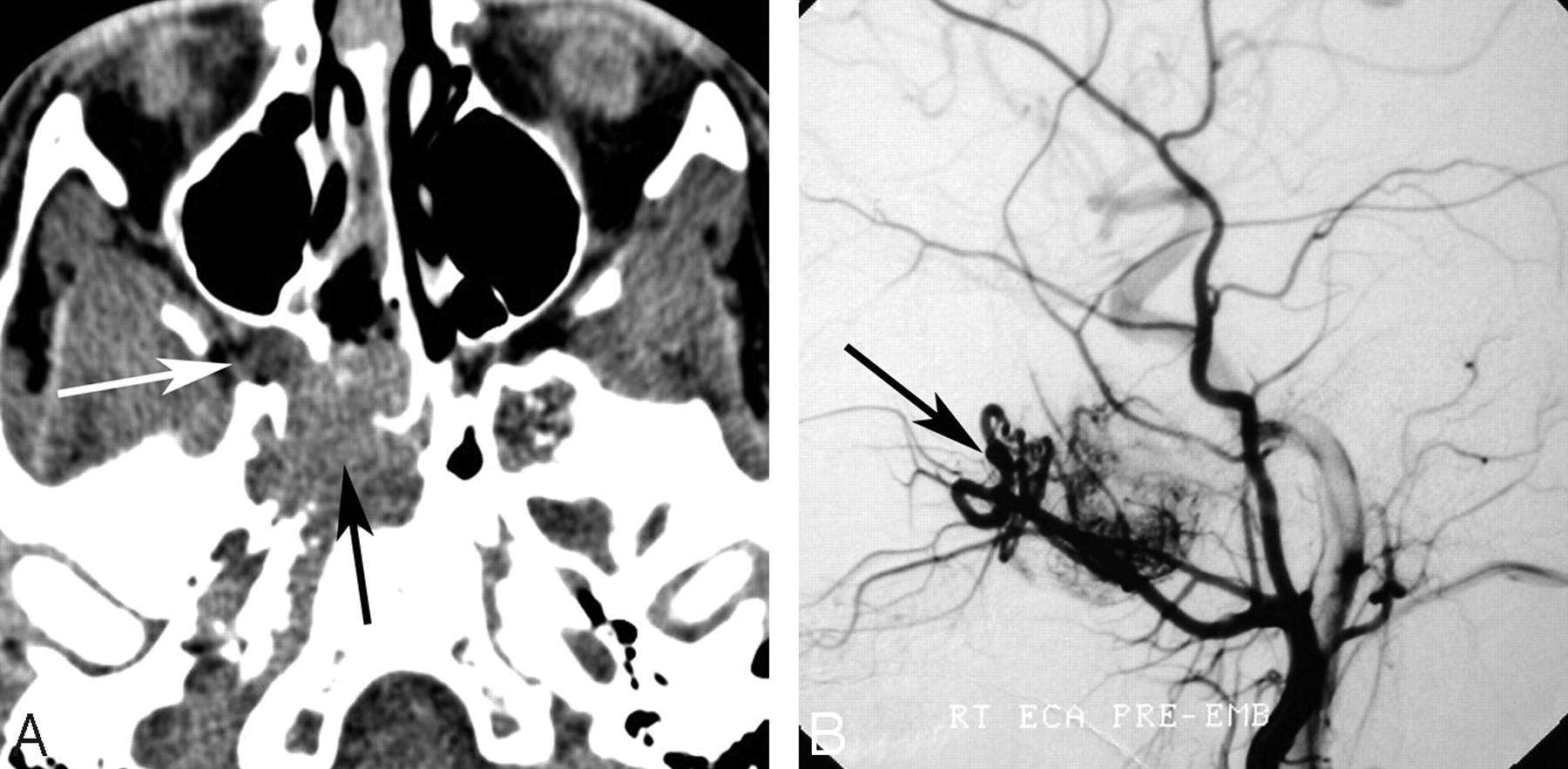
  • Fig 11.
    • Download figure
    • Open in new tab
    • Download powerpoint
    Fig 11.

    A 16-year-old boy with juvenile angiofibroma. A, Axial unenhanced CT scan demonstrates widening of the right sphenopalatine foramen with abnormal soft tissue obliterating the normal fat within the pterygopalatine fossa (white arrow). Soft tissue extends anteriorly into the posterior nasopharynx and posteriorly into the sphenoid bone and right sphenoid sinus (black arrow). B, Lateral projection from a right external carotid artery angiogram in the arterial phase demonstrates an attenuated tumor blush and an enlarged internal maxillary artery with tortuous vessels in the right pterygopalatine fossa (arrow).
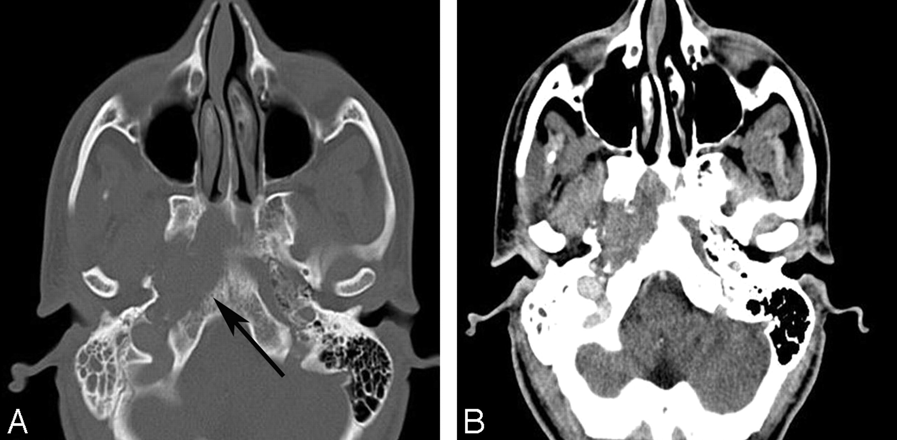
  • Fig 12.
    • Download figure
    • Open in new tab
    • Download powerpoint
    Fig 12.

    A 20-year-old man with sinonasal carcinoma. A, Bone window from an unenhanced CT scan shows destructive osseous changes (arrow). B, Soft tissue window at the same level shows an associated soft tissue mass in the right central skull base.

  • Fig 13.
    • Download figure
    • Open in new tab
    • Download powerpoint
    Fig 13.

    A 3-year-old girl with neuroblastoma. A, Sagittal postcontrast T1WI shows a mass centered on the sphenoid bone, an unusual location for a neuroblastoma metastasis, obliterating the sphenoid sinus. There is mass effect on the gyrus rectus. B, A multilobulated mass demonstrates characteristic relative hypointense signal intensity on an axial T2WI. The lesion has an intracranial component, extending along the anterior aspect of the middle cranial fossae bilaterally (arrows). C, The left adrenal primary is shown on coronal postcontrast T1WI of the abdomen.

PreviousNext
Back to top

In this issue

American Journal of Neuroradiology: 32 (4)
American Journal of Neuroradiology
Vol. 32, Issue 4
1 Apr 2011
  • Table of Contents
  • Index by author
Advertisement
Print
Download PDF
Email Article

Thank you for your interest in spreading the word on American Journal of Neuroradiology.

NOTE: We only request your email address so that the person you are recommending the page to knows that you wanted them to see it, and that it is not junk mail. We do not capture any email address.

Enter multiple addresses on separate lines or separate them with commas.
Sphenoid Masses in Children: Radiologic Differential Diagnosis with Pathologic Correlation
(Your Name) has sent you a message from American Journal of Neuroradiology
(Your Name) thought you would like to see the American Journal of Neuroradiology web site.
CAPTCHA
This question is for testing whether or not you are a human visitor and to prevent automated spam submissions.
Cite this article
Y.W. Lui, S.B. Dasari, R.J. Young
Sphenoid Masses in Children: Radiologic Differential Diagnosis with Pathologic Correlation
American Journal of Neuroradiology Apr 2011, 32 (4) 617-626; DOI: 10.3174/ajnr.A2144

Citation Manager Formats

  • BibTeX
  • Bookends
  • EasyBib
  • EndNote (tagged)
  • EndNote 8 (xml)
  • Medlars
  • Mendeley
  • Papers
  • RefWorks Tagged
  • Ref Manager
  • RIS
  • Zotero
0 Responses
Respond to this article
Share
Bookmark this article
Sphenoid Masses in Children: Radiologic Differential Diagnosis with Pathologic Correlation
Y.W. Lui, S.B. Dasari, R.J. Young
American Journal of Neuroradiology Apr 2011, 32 (4) 617-626; DOI: 10.3174/ajnr.A2144
del.icio.us logo Twitter logo Facebook logo Mendeley logo
  • Tweet Widget
  • Facebook Like
  • Google Plus One
Purchase

Jump to section

  • Article
    • Abstract
    • Abbreviations
    • Entities Typified by Nonaggressive Osseous Changes
    • Entities Typified by Aggressive Osseous Changes
    • Summary
    • References
  • Figures & Data
  • Info & Metrics
  • Responses
  • References
  • PDF

Related Articles

  • No related articles found.
  • PubMed
  • Google Scholar

Cited By...

  • Sphenoid mucocele with unusual panhypopituitarism
  • Crossref (23)
  • Google Scholar

This article has been cited by the following articles in journals that are participating in Crossref Cited-by Linking.

  • Clinical and Radiological Findings Suggesting Disorders Other Than Tolosa–Hunt Syndrome Among Ophthalmoplegic Patients: A Retrospective Analysis
    Chih‐Hsien Hung, Kuo‐Hsuan Chang, Yao‐Liang Chen, Yi‐Ming Wu, Chiou‐Lian Lai, Hong‐Shiu Chang, Rong‐Kuo Lyu, Yih‐Ru Wu, Chiung‐Mei Chen, Chin‐Chang Huang, Chun‐Che Chu, Chun‐Hung Chen, Long‐Sun Ro
    Headache: The Journal of Head and Face Pain 2015 55 2
  • The Skull Base in the Evaluation of Sinonasal Disease
    Steve E.J. Connor
    Neuroimaging Clinics of North America 2015 25 4
  • Spheno-Orbital Aneurysmal Bone Cyst in a 10-Month-Old Infant
    Elsa V. Arocho-Quinones, Scott Self, Mariko Suchi, Nathan T. Zwagerman, Sean M. Lew
    World Neurosurgery 2018 117
  • Sphenoid sinus pyocele after transsphenoidal approach for pituitary adenoma
    Mario Giordano, Venelin M. Gerganov, Wolfgang Draf, Rudolf Fahlbusch
    Pituitary 2012 15 2
  • Aneurysmal Bone Cyst of Sphenoid Bone and Clivus Misdiagnosed as Chordoma: A Case Report
    Fethi Emre Ustabasioglu, Cesur Samanci, Murat Asik, Inanc Yanik, Seyma Ozkanli, Onur Tutar, Zehra Isik Hasiloglu
    Brain Tumor Research and Treatment 2015 3 2
  • Sphenoid mucocele with unusual panhypopituitarism
    Saranya Devi, Anita Ganger, Sanjay Sharma, Rohit Saxena
    BMJ Case Reports 2016 2016
  • Intrasphenoidal Rathke Cleft Cyst
    Mahadevan S. Gowtham, Devaraj Sunilkumar, Andi S. Ramesh, Bheemanathi H. Srinivas, Dinesh Verma, Krishnan Nagarajan
    Journal of Pediatric Neurosciences 2021 16 4
  • Pure Endoscopic Endonasal Removal of Unusual Anterior Skull Base Aneursymal Bone Cyst Extending to the Frontal Lobe
    Ali Erdem Yildirim, Ibrahim Ekici, Emin Cagil, Denizhan Divanlioglu, Ahmed Deniz Belen
    Journal of Craniofacial Surgery 2015 26 3
  • A Boy with Acute Strabismus
    Federico Poropat, Giovanna Ventura, Flora M. Murru, Eva Orzan, Massimo Maschio
    The Journal of Pediatrics 2012 161 6
  • Radiographic Findings of a Well-Differentiated Sinonasal Neuroendocrine Neoplasm: Case Report and Review of the Literature
    Cui Ping Mao, Ming Zhang, Chen Niu, Min Li, Yuan Wang
    Ear, Nose & Throat Journal 2015 94 7

More in this TOC Section

  • Giant Cell Arteritis: Important Imaging Findings
  • DCE MRI in Spinal Disease Assessment
  • Cerebrovascular Anomalies in Fetal Imaging
Show more Review Article

Similar Articles

Advertisement

Indexed Content

  • Current Issue
  • Accepted Manuscripts
  • Article Preview
  • Past Issues
  • Editorials
  • Editors Choice
  • Fellow Journal Club
  • Letters to the Editor

Cases

  • Case Collection
  • Archive - Case of the Week
  • Archive - Case of the Month
  • Archive - Classic Case

Special Collections

  • Special Collections

Resources

  • News and Updates
  • Turn around Times
  • Submit a Manuscript
  • Author Policies
  • Manuscript Submission Guidelines
  • Evidence-Based Medicine Level Guide
  • Publishing Checklists
  • Graphical Abstract Preparation
  • Imaging Protocol Submission
  • Submit a Case
  • Become a Reviewer/Academy of Reviewers
  • Get Peer Review Credit from Publons

Multimedia

  • AJNR Podcast
  • AJNR SCANtastic
  • Video Articles

About Us

  • About AJNR
  • Editorial Board
  • Not an AJNR Subscriber? Join Now
  • Alerts
  • Feedback
  • Advertise with us
  • Librarian Resources
  • Permissions
  • Terms and Conditions

American Society of Neuroradiology

  • Not an ASNR Member? Join Now

© 2025 by the American Society of Neuroradiology All rights, including for text and data mining, AI training, and similar technologies, are reserved.
Print ISSN: 0195-6108 Online ISSN: 1936-959X

Powered by HighWire