Advertisement

Main menu

  • Home
  • Content
    • Current Issue
    • Accepted Manuscripts
    • Article Preview
    • Past Issue Archive
    • Video Articles
    • AJNR Case Collection
    • Case of the Week Archive
    • Case of the Month Archive
    • Classic Case Archive
  • Special Collections
    • AJNR Awards
    • Low-Field MRI
    • Alzheimer Disease
    • ASNR Foundation Special Collection
    • Photon-Counting CT
    • View All
  • Multimedia
    • AJNR Podcasts
    • AJNR SCANtastic
    • Trainee Corner
    • MRI Safety Corner
    • Imaging Protocols
  • For Authors
    • Submit a Manuscript
    • Submit a Video Article
    • Submit an eLetter to the Editor/Response
    • Manuscript Submission Guidelines
    • Statistical Tips
    • Fast Publishing of Accepted Manuscripts
    • Graphical Abstract Preparation
    • Imaging Protocol Submission
    • Author Policies
  • About Us
    • About AJNR
    • Editorial Board
    • Editorial Board Alumni
  • More
    • Become a Reviewer/Academy of Reviewers
    • Subscribers
    • Permissions
    • Alerts
    • Feedback
    • Advertisers
    • ASNR Home

User menu

  • Alerts
  • Log in

Search

  • Advanced search
American Journal of Neuroradiology

American Journal of Neuroradiology

ASHNR American Society of Functional Neuroradiology ASHNR American Society of Pediatric Neuroradiology ASSR
  • Alerts
  • Log in

Advanced Search

  • Home
  • Content
    • Current Issue
    • Accepted Manuscripts
    • Article Preview
    • Past Issue Archive
    • Video Articles
    • AJNR Case Collection
    • Case of the Week Archive
    • Case of the Month Archive
    • Classic Case Archive
  • Special Collections
    • AJNR Awards
    • Low-Field MRI
    • Alzheimer Disease
    • ASNR Foundation Special Collection
    • Photon-Counting CT
    • View All
  • Multimedia
    • AJNR Podcasts
    • AJNR SCANtastic
    • Trainee Corner
    • MRI Safety Corner
    • Imaging Protocols
  • For Authors
    • Submit a Manuscript
    • Submit a Video Article
    • Submit an eLetter to the Editor/Response
    • Manuscript Submission Guidelines
    • Statistical Tips
    • Fast Publishing of Accepted Manuscripts
    • Graphical Abstract Preparation
    • Imaging Protocol Submission
    • Author Policies
  • About Us
    • About AJNR
    • Editorial Board
    • Editorial Board Alumni
  • More
    • Become a Reviewer/Academy of Reviewers
    • Subscribers
    • Permissions
    • Alerts
    • Feedback
    • Advertisers
    • ASNR Home
  • Follow AJNR on Twitter
  • Visit AJNR on Facebook
  • Follow AJNR on Instagram
  • Join AJNR on LinkedIn
  • RSS Feeds

AJNR Awards, New Junior Editors, and more. Read the latest AJNR updates

Research ArticleBrain

Correlation between CT and Diffusion-Weighted Imaging of Acute Cerebral Ischemia in a Rat Model

H.S. Seo, D.G. Na, J.-h. Kim, K.W. Kim and K.-R. Son
American Journal of Neuroradiology April 2011, 32 (4) 728-733; DOI: https://doi.org/10.3174/ajnr.A2362
H.S. Seo
  • Find this author on Google Scholar
  • Find this author on PubMed
  • Search for this author on this site
D.G. Na
  • Find this author on Google Scholar
  • Find this author on PubMed
  • Search for this author on this site
J.-h. Kim
  • Find this author on Google Scholar
  • Find this author on PubMed
  • Search for this author on this site
K.W. Kim
  • Find this author on Google Scholar
  • Find this author on PubMed
  • Search for this author on this site
K.-R. Son
  • Find this author on Google Scholar
  • Find this author on PubMed
  • Search for this author on this site
  • Article
  • Figures & Data
  • Info & Metrics
  • Responses
  • References
  • PDF
Loading

Article Figures & Data

Figures

  • Tables
  • Fig 1.
    • Download figure
    • Open in new tab
    • Download powerpoint
    Fig 1.

    Measurement of the region of interest for CT attenuation in ischemic and normal brain tissue. The region of interest is positioned in the inferolateral part of the hemisphere on each CT or MR image, 5 mm ventral to the external auditory canal.

  • Fig 2.
    • Download figure
    • Open in new tab
    • Download powerpoint
    Fig 2.

    Longitudinal changes in acute ischemic lesions on CT, DWI, and the ADC map at each time point after MCA occlusion in a rat.

  • Fig 3.
    • Download figure
    • Open in new tab
    • Download powerpoint
    Fig 3.

    Time courses of rCT, rDWI, and rADC after MCA occlusion. The rCT decreases gradually and rDWI increases gradually with time (P < .01, respectively). However, rADC rapidly decreases at 1 hour after MCA occlusion, and rADC shows no substantial change after 3 hours (P =. 33).

  • Fig 4.
    • Download figure
    • Open in new tab
    • Download powerpoint
    Fig 4.

    Scatterplot of rCT and rDWI. Significant correlation occurs between rCT and rDWI in acute cerebral ischemia after 1, 3, 5, 7, or 9 hours of MCA occlusion. The rCT and rDWI are inversely correlated (P < .01, r = −0.592). The dotted line is the regression line for rCT and rDWI. One unit of rCT increase corresponds to 1.24 U of rDWI decrease.
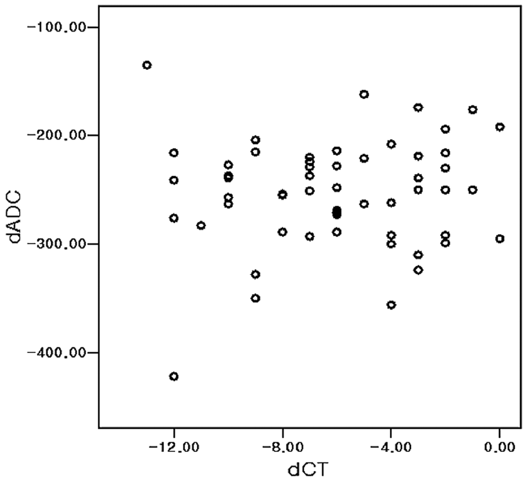
  • Fig 5.
    • Download figure
    • Open in new tab
    • Download powerpoint
    Fig 5.

    Scatterplot of dCT and dADC. No significant linear correlation occurs between dADC and dCT (P = .56) in acute cerebral ischemia after 1, 3, 5, 7, or 9 hours of MCA occlusion.

Tables

  • Figures
    • View popup
    Table 1:

    The mean values in CT, ADC, and DWI after MCA occlusiona

    HourrCTrDWIrADCdCTbdADCc
    10.94 ± 0.041.40 ± 0.070.62 ± 0.082.00 ± 1.48225.73 ± 52.60
    30.87 ± 0.051.60 ± 0.100.55 ± 0.064.09 ± 1.76268.18 ± 43.21
    (P < .01)(P < .01)(P = .04)(P = .01)(P = .06)
    50.81 ± 0.061.70 ± 0.130.56 ± 0.046.18 ± 2.44262.45 ± 28.54
    (P = .03)(P < .01)(P = .93)(P = .04)(P = .71)
    70.73 ± 0.071.71 ± 0.230.58 ± 0.068.91 ± 2.51250.55 ± 42.30
    (P < .01)(P = .82)(P = .09)(P < .01)(P = .35)
    90.71 ± 0.081.77 ± 0.280.57 ± 0.089.09 ± 2.39257.73 ± 73.12
    (P = .46)(P = .39)(P = .51)(P = .84)(P = .71)
    • a Data are mean ± SD. The figures in parentheses refer to the P values compared with the value of the previous time point in post hoc analysis of repeated-measure analysis.

    • b dCT is in Hounsfield units.

    • c dADC values are ×10−6 mm2/s.

    • View popup
    Table 2:

    The mean CNRs of CT attenuation, ADC value, and DWI signal intensity after MCA occlusiona

    HourCT (%)PbADC (%)PcDWI (%)Pd
    16.2 ± 4.4<.00138.5 ± 8.4.45339.9 ± 7.1<.001
    312.6 ± 4.5<.00144.8 ± 6.1<.00159.6 ± 9.8<.001
    518.9 ± 6.4<.00144.4 ± 4.0<.00169.9 ± 13.2<.001
    727.1 ± 6.5<.00141.8 ± 6.0.00471.2 ± 22.8<.001
    929.0 ± 8.0.00343.1 ± 7.9.00276.9 ± 28.2<.001
    • a Data are mean ± SD.

    • b CT attenuation versus ADC value.

    • c ADC value versus DWI signal intensity.

    • d DWI signal intensity versus CT attenuation.

PreviousNext
Back to top

In this issue

American Journal of Neuroradiology: 32 (4)
American Journal of Neuroradiology
Vol. 32, Issue 4
1 Apr 2011
  • Table of Contents
  • Index by author
Advertisement
Print
Download PDF
Email Article

Thank you for your interest in spreading the word on American Journal of Neuroradiology.

NOTE: We only request your email address so that the person you are recommending the page to knows that you wanted them to see it, and that it is not junk mail. We do not capture any email address.

Enter multiple addresses on separate lines or separate them with commas.
Correlation between CT and Diffusion-Weighted Imaging of Acute Cerebral Ischemia in a Rat Model
(Your Name) has sent you a message from American Journal of Neuroradiology
(Your Name) thought you would like to see the American Journal of Neuroradiology web site.
CAPTCHA
This question is for testing whether or not you are a human visitor and to prevent automated spam submissions.
Cite this article
H.S. Seo, D.G. Na, J.-h. Kim, K.W. Kim, K.-R. Son
Correlation between CT and Diffusion-Weighted Imaging of Acute Cerebral Ischemia in a Rat Model
American Journal of Neuroradiology Apr 2011, 32 (4) 728-733; DOI: 10.3174/ajnr.A2362

Citation Manager Formats

  • BibTeX
  • Bookends
  • EasyBib
  • EndNote (tagged)
  • EndNote 8 (xml)
  • Medlars
  • Mendeley
  • Papers
  • RefWorks Tagged
  • Ref Manager
  • RIS
  • Zotero
0 Responses
Respond to this article
Share
Bookmark this article
Correlation between CT and Diffusion-Weighted Imaging of Acute Cerebral Ischemia in a Rat Model
H.S. Seo, D.G. Na, J.-h. Kim, K.W. Kim, K.-R. Son
American Journal of Neuroradiology Apr 2011, 32 (4) 728-733; DOI: 10.3174/ajnr.A2362
del.icio.us logo Twitter logo Facebook logo Mendeley logo
  • Tweet Widget
  • Facebook Like
  • Google Plus One
Purchase

Jump to section

  • Article
    • Abstract
    • Abbreviations
    • Materials and Methods
    • Results
    • Discussion
    • Conclusions
    • Footnotes
    • References
  • Figures & Data
  • Info & Metrics
  • Responses
  • References
  • PDF

Related Articles

  • No related articles found.
  • PubMed
  • Google Scholar

Cited By...

  • No citing articles found.
  • Crossref
  • Google Scholar

This article has not yet been cited by articles in journals that are participating in Crossref Cited-by Linking.

More in this TOC Section

  • Statin Therapy Does Not Affect the Radiographic and Clinical Profile of Patients with TIA and Minor Stroke
  • Usefulness of Quantitative Susceptibility Mapping for the Diagnosis of Parkinson Disease
  • White Matter Alterations in the Brains of Patients with Active, Remitted, and Cured Cushing Syndrome: A DTI Study
Show more Brain

Similar Articles

Advertisement

Indexed Content

  • Current Issue
  • Accepted Manuscripts
  • Article Preview
  • Past Issues
  • Editorials
  • Editor's Choice
  • Fellows' Journal Club
  • Letters to the Editor
  • Video Articles

Cases

  • Case Collection
  • Archive - Case of the Week
  • Archive - Case of the Month
  • Archive - Classic Case

More from AJNR

  • Trainee Corner
  • Imaging Protocols
  • MRI Safety Corner
  • Book Reviews

Multimedia

  • AJNR Podcasts
  • AJNR Scantastics

Resources

  • Turnaround Time
  • Submit a Manuscript
  • Submit a Video Article
  • Submit an eLetter to the Editor/Response
  • Manuscript Submission Guidelines
  • Statistical Tips
  • Fast Publishing of Accepted Manuscripts
  • Graphical Abstract Preparation
  • Imaging Protocol Submission
  • Evidence-Based Medicine Level Guide
  • Publishing Checklists
  • Author Policies
  • Become a Reviewer/Academy of Reviewers
  • News and Updates

About Us

  • About AJNR
  • Editorial Board
  • Editorial Board Alumni
  • Alerts
  • Permissions
  • Not an AJNR Subscriber? Join Now
  • Advertise with Us
  • Librarian Resources
  • Feedback
  • Terms and Conditions
  • AJNR Editorial Board Alumni

American Society of Neuroradiology

  • Not an ASNR Member? Join Now

© 2025 by the American Society of Neuroradiology All rights, including for text and data mining, AI training, and similar technologies, are reserved.
Print ISSN: 0195-6108 Online ISSN: 1936-959X

Powered by HighWire