Advertisement

Main menu

  • Home
  • Content
    • Current Issue
    • Accepted Manuscripts
    • Article Preview
    • Past Issue Archive
    • Video Articles
    • AJNR Case Collection
    • Case of the Week Archive
    • Case of the Month Archive
    • Classic Case Archive
  • Special Collections
    • AJNR Awards
    • Low-Field MRI
    • Alzheimer Disease
    • ASNR Foundation Special Collection
    • Photon-Counting CT
    • View All
  • Multimedia
    • AJNR Podcasts
    • AJNR SCANtastic
    • Trainee Corner
    • MRI Safety Corner
    • Imaging Protocols
  • For Authors
    • Submit a Manuscript
    • Submit a Video Article
    • Submit an eLetter to the Editor/Response
    • Manuscript Submission Guidelines
    • Statistical Tips
    • Fast Publishing of Accepted Manuscripts
    • Graphical Abstract Preparation
    • Imaging Protocol Submission
    • Author Policies
  • About Us
    • About AJNR
    • Editorial Board
    • Editorial Board Alumni
  • More
    • Become a Reviewer/Academy of Reviewers
    • Subscribers
    • Permissions
    • Alerts
    • Feedback
    • Advertisers
    • ASNR Home

User menu

  • Alerts
  • Log in

Search

  • Advanced search
American Journal of Neuroradiology

American Journal of Neuroradiology

ASHNR American Society of Functional Neuroradiology ASHNR American Society of Pediatric Neuroradiology ASSR
  • Alerts
  • Log in

Advanced Search

  • Home
  • Content
    • Current Issue
    • Accepted Manuscripts
    • Article Preview
    • Past Issue Archive
    • Video Articles
    • AJNR Case Collection
    • Case of the Week Archive
    • Case of the Month Archive
    • Classic Case Archive
  • Special Collections
    • AJNR Awards
    • Low-Field MRI
    • Alzheimer Disease
    • ASNR Foundation Special Collection
    • Photon-Counting CT
    • View All
  • Multimedia
    • AJNR Podcasts
    • AJNR SCANtastic
    • Trainee Corner
    • MRI Safety Corner
    • Imaging Protocols
  • For Authors
    • Submit a Manuscript
    • Submit a Video Article
    • Submit an eLetter to the Editor/Response
    • Manuscript Submission Guidelines
    • Statistical Tips
    • Fast Publishing of Accepted Manuscripts
    • Graphical Abstract Preparation
    • Imaging Protocol Submission
    • Author Policies
  • About Us
    • About AJNR
    • Editorial Board
    • Editorial Board Alumni
  • More
    • Become a Reviewer/Academy of Reviewers
    • Subscribers
    • Permissions
    • Alerts
    • Feedback
    • Advertisers
    • ASNR Home
  • Follow AJNR on Twitter
  • Visit AJNR on Facebook
  • Follow AJNR on Instagram
  • Join AJNR on LinkedIn
  • RSS Feeds

AJNR Awards, New Junior Editors, and more. Read the latest AJNR updates

Research ArticleSpine Imaging and Spine Image-Guided Interventions

Parallel Imaging of the Cervical Spine at 3T: Optimized Trade-Off between Speed and Image Quality

J. Fruehwald-Pallamar, P. Szomolanyi, N. Fakhrai, A. Lunzer, M. Weber, M.M. Thurnher, M. Pallamar, S. Trattnig, D. Prayer and I.M. Noebauer-Huhmann
American Journal of Neuroradiology November 2012, 33 (10) 1867-1874; DOI: https://doi.org/10.3174/ajnr.A3101
J. Fruehwald-Pallamar
aFrom the Subdivision of Neuroradiology and Musculoskeletal Radiology (J.F.-P., N.F., M.M.T., D.P., I.M.N.-H.), Department of Radiology
  • Find this author on Google Scholar
  • Find this author on PubMed
  • Search for this author on this site
P. Szomolanyi
dCenter of High-Field MRI (P.S., M.P., S.T., I.M.N.-H.,), Department of Radiology; Medical University of Vienna, Vienna, Austria
eDepartment of Imaging Methods (P.S.), Institute of Measurement Science, Slovak Academy of Sciences, Bratislava, Slovakia.
  • Find this author on Google Scholar
  • Find this author on PubMed
  • Search for this author on this site
N. Fakhrai
aFrom the Subdivision of Neuroradiology and Musculoskeletal Radiology (J.F.-P., N.F., M.M.T., D.P., I.M.N.-H.), Department of Radiology
  • Find this author on Google Scholar
  • Find this author on PubMed
  • Search for this author on this site
A. Lunzer
bUniversity Clinic of Orthopaedic Surgery (A.L.)
  • Find this author on Google Scholar
  • Find this author on PubMed
  • Search for this author on this site
M. Weber
cDepartment of Radiology (M.W.)
  • Find this author on Google Scholar
  • Find this author on PubMed
  • Search for this author on this site
M.M. Thurnher
aFrom the Subdivision of Neuroradiology and Musculoskeletal Radiology (J.F.-P., N.F., M.M.T., D.P., I.M.N.-H.), Department of Radiology
  • Find this author on Google Scholar
  • Find this author on PubMed
  • Search for this author on this site
M. Pallamar
dCenter of High-Field MRI (P.S., M.P., S.T., I.M.N.-H.,), Department of Radiology; Medical University of Vienna, Vienna, Austria
  • Find this author on Google Scholar
  • Find this author on PubMed
  • Search for this author on this site
S. Trattnig
dCenter of High-Field MRI (P.S., M.P., S.T., I.M.N.-H.,), Department of Radiology; Medical University of Vienna, Vienna, Austria
  • Find this author on Google Scholar
  • Find this author on PubMed
  • Search for this author on this site
D. Prayer
aFrom the Subdivision of Neuroradiology and Musculoskeletal Radiology (J.F.-P., N.F., M.M.T., D.P., I.M.N.-H.), Department of Radiology
  • Find this author on Google Scholar
  • Find this author on PubMed
  • Search for this author on this site
I.M. Noebauer-Huhmann
dCenter of High-Field MRI (P.S., M.P., S.T., I.M.N.-H.,), Department of Radiology; Medical University of Vienna, Vienna, Austria
  • Find this author on Google Scholar
  • Find this author on PubMed
  • Search for this author on this site
  • Article
  • Figures & Data
  • Supplemental
  • Info & Metrics
  • Responses
  • References
  • PDF
Loading

Article Figures & Data

Figures

  • Tables
  • Fig 1.
    • Download figure
    • Open in new tab
    • Download powerpoint
    Fig 1.

    Region-of-interest measurements in the phantom.

  • Fig 2.
    • Download figure
    • Open in new tab
    • Download powerpoint
    Fig 2.

    The region-of-interest measurements in a volunteer.

  • Fig 3.
    • Download figure
    • Open in new tab
    • Download powerpoint
    Fig 3.

    The mean nonuniformity of the 10 volunteers for each sequence.
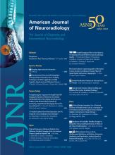
  • Fig 4.
    • Download figure
    • Open in new tab
    • Download powerpoint
    Fig 4.

    T1-weighted sequences (60-year-old female patient) showing degenerative changes, hemangioma at T1. A, T1, original sequence. B, Two averages. C, One average. D, Acceleration factor 2. E, Acceleration factor 3.
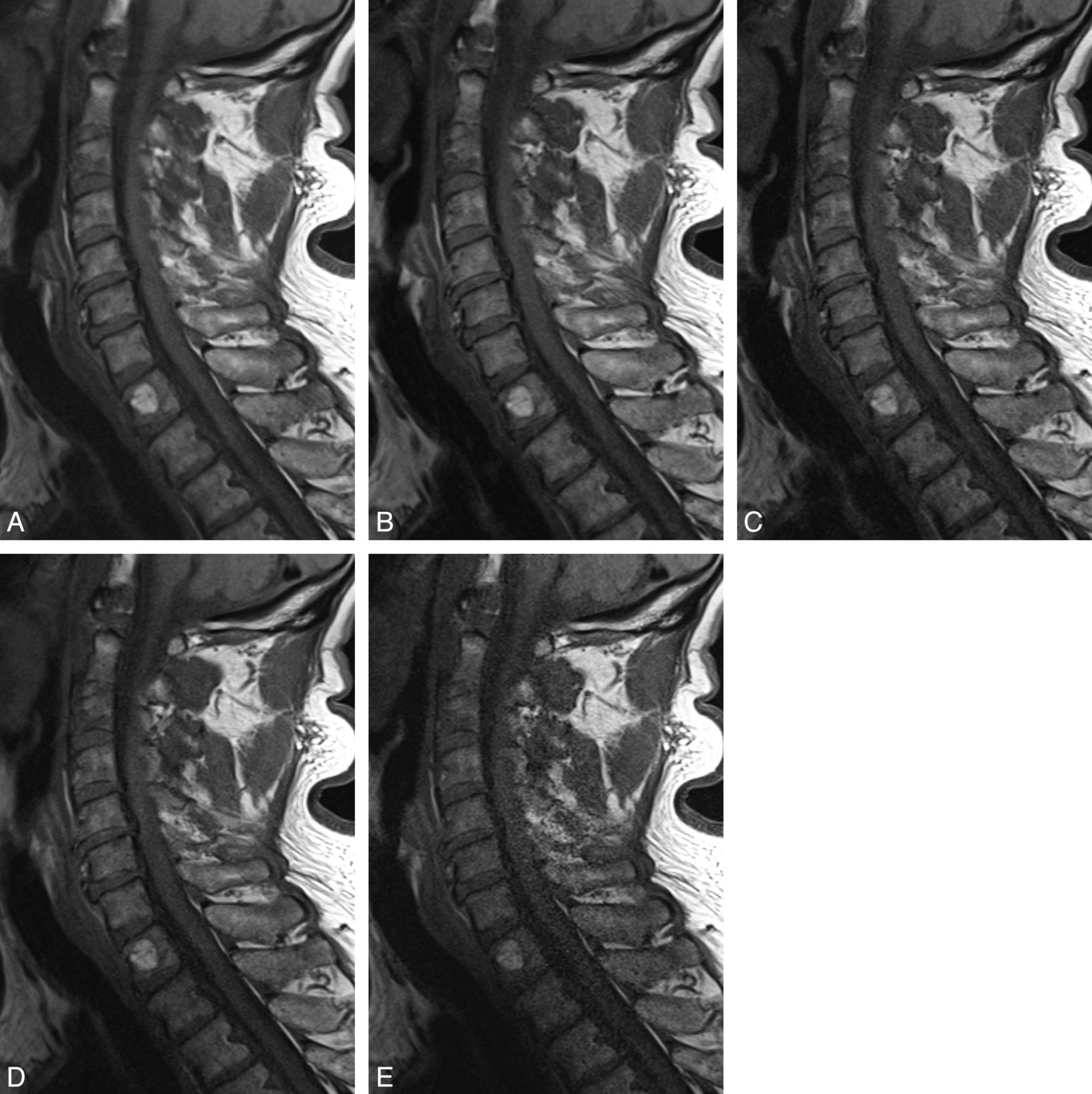
  • Fig 5.
    • Download figure
    • Open in new tab
    • Download powerpoint
    Fig 5.

    T2-weighted sequences (53-year-old female volunteer) showing a small hernia of the C5/6 disk. A, T2, original sequence. B, One average. C, Acceleration factor 2. D, Acceleration factor 3.

  • Fig 6.
    • Download figure
    • Open in new tab
    • Download powerpoint
    Fig 6.

    Axial T2* GRE sequences (46-year-old female patient) showing posterolateral disk herniation with compression of the spinal cord. A, T2* GRE, original sequence. B, Acceleration factor 2. C, Acceleration factor 3.

  • Fig 7.
    • Download figure
    • Open in new tab
    • Download powerpoint
    Fig 7.

    T2TIRM sequences (45-year-old male patient) showing slight edema of C5 due to disk degeneration and minor endplate edema of C5 (Modic type I). A, T2TIRM, original sequence. B, Acceleration factor 2. C, Acceleration factor 3.

Tables

  • Figures
  • Imaging parameters and examination times

    SequenceSagittal T1WISagittal T2WISagittal T2TIRMAxial T2* GRE
    TR (ms)600300030001300
    TE (ms)101093214
    Flip angle160°180°140°30°
    Section thickness (mm)3333
    Gap (mm)3.33.33.33.3
    NEX3211
    ETL533171
    Matrix (reconstruction)384 × 306320 × 320640 × 640512 × 512
    Matrix (acquisition)384 × 260320 × 256320 × 240256 × 256
    FOV191 × 240240 × 240240 × 240160 × 160
    Time (min/sec)
        Nonaccelerated (original)03:1003:0603:2408:19
            2 Averages (% reduction)02:06 (33.7%)
            1 average01:03 (67%)01:30 (54%)
    Acc 201:36 (49.5%)01:31 (53.4%)01:48 (47%)04:24 (47%)
    Acc 301:04 (66.4%)01:07 (66%)01:15 (63%)03:06 (63%)
    Acc 401:00 (68.8%)00:55 (67.6%)00:57 (72%)02:27 (74.5%)
    • Note:—ETL indicates echo-train length; Acc, acceleration factor.

PreviousNext
Back to top

In this issue

American Journal of Neuroradiology: 33 (10)
American Journal of Neuroradiology
Vol. 33, Issue 10
1 Nov 2012
  • Table of Contents
  • Index by author
Advertisement
Print
Download PDF
Email Article

Thank you for your interest in spreading the word on American Journal of Neuroradiology.

NOTE: We only request your email address so that the person you are recommending the page to knows that you wanted them to see it, and that it is not junk mail. We do not capture any email address.

Enter multiple addresses on separate lines or separate them with commas.
Parallel Imaging of the Cervical Spine at 3T: Optimized Trade-Off between Speed and Image Quality
(Your Name) has sent you a message from American Journal of Neuroradiology
(Your Name) thought you would like to see the American Journal of Neuroradiology web site.
CAPTCHA
This question is for testing whether or not you are a human visitor and to prevent automated spam submissions.
Cite this article
J. Fruehwald-Pallamar, P. Szomolanyi, N. Fakhrai, A. Lunzer, M. Weber, M.M. Thurnher, M. Pallamar, S. Trattnig, D. Prayer, I.M. Noebauer-Huhmann
Parallel Imaging of the Cervical Spine at 3T: Optimized Trade-Off between Speed and Image Quality
American Journal of Neuroradiology Nov 2012, 33 (10) 1867-1874; DOI: 10.3174/ajnr.A3101

Citation Manager Formats

  • BibTeX
  • Bookends
  • EasyBib
  • EndNote (tagged)
  • EndNote 8 (xml)
  • Medlars
  • Mendeley
  • Papers
  • RefWorks Tagged
  • Ref Manager
  • RIS
  • Zotero
0 Responses
Respond to this article
Share
Bookmark this article
Parallel Imaging of the Cervical Spine at 3T: Optimized Trade-Off between Speed and Image Quality
J. Fruehwald-Pallamar, P. Szomolanyi, N. Fakhrai, A. Lunzer, M. Weber, M.M. Thurnher, M. Pallamar, S. Trattnig, D. Prayer, I.M. Noebauer-Huhmann
American Journal of Neuroradiology Nov 2012, 33 (10) 1867-1874; DOI: 10.3174/ajnr.A3101
del.icio.us logo Twitter logo Facebook logo Mendeley logo
  • Tweet Widget
  • Facebook Like
  • Google Plus One
Purchase

Jump to section

  • Article
    • Abstract
    • ABBREVIATIONS:
    • Materials and Methods
    • Results
    • Discussion
    • Conclusions
    • Footnotes
    • References
  • Figures & Data
  • Supplemental
  • Info & Metrics
  • Responses
  • References
  • PDF

Related Articles

  • PubMed
  • Google Scholar

Cited By...

  • Diffusion Kurtosis Imaging of neonatal Spinal Cord in clinical routine
  • Clinical Utility of a Novel Ultrafast T2-Weighted Sequence for Spine Imaging
  • Crossref
  • Google Scholar

This article has not yet been cited by articles in journals that are participating in Crossref Cited-by Linking.

More in this TOC Section

  • Management Outcomes For VO Spine Biopsy
  • Characteristics of SIH Type I Culprit Lesions
  • Advanced Imaging of Type 2 Spinal CSF Leaks
Show more Spine Imaging and Spine Image-Guided Interventions

Similar Articles

Advertisement

Indexed Content

  • Current Issue
  • Accepted Manuscripts
  • Article Preview
  • Past Issues
  • Editorials
  • Editor's Choice
  • Fellows' Journal Club
  • Letters to the Editor
  • Video Articles

Cases

  • Case Collection
  • Archive - Case of the Week
  • Archive - Case of the Month
  • Archive - Classic Case

More from AJNR

  • Trainee Corner
  • Imaging Protocols
  • MRI Safety Corner
  • Book Reviews

Multimedia

  • AJNR Podcasts
  • AJNR Scantastics

Resources

  • Turnaround Time
  • Submit a Manuscript
  • Submit a Video Article
  • Submit an eLetter to the Editor/Response
  • Manuscript Submission Guidelines
  • Statistical Tips
  • Fast Publishing of Accepted Manuscripts
  • Graphical Abstract Preparation
  • Imaging Protocol Submission
  • Evidence-Based Medicine Level Guide
  • Publishing Checklists
  • Author Policies
  • Become a Reviewer/Academy of Reviewers
  • News and Updates

About Us

  • About AJNR
  • Editorial Board
  • Editorial Board Alumni
  • Alerts
  • Permissions
  • Not an AJNR Subscriber? Join Now
  • Advertise with Us
  • Librarian Resources
  • Feedback
  • Terms and Conditions
  • AJNR Editorial Board Alumni

American Society of Neuroradiology

  • Not an ASNR Member? Join Now

© 2025 by the American Society of Neuroradiology All rights, including for text and data mining, AI training, and similar technologies, are reserved.
Print ISSN: 0195-6108 Online ISSN: 1936-959X

Powered by HighWire