Advertisement

Main menu

  • Home
  • Content
    • Current Issue
    • Accepted Manuscripts
    • Article Preview
    • Past Issue Archive
    • Video Articles
    • AJNR Case Collection
    • Case of the Week Archive
    • Case of the Month Archive
    • Classic Case Archive
  • Special Collections
    • AJNR Awards
    • Low-Field MRI
    • Alzheimer Disease
    • ASNR Foundation Special Collection
    • Photon-Counting CT
    • View All
  • Multimedia
    • AJNR Podcasts
    • AJNR SCANtastic
    • Trainee Corner
    • MRI Safety Corner
    • Imaging Protocols
  • For Authors
    • Submit a Manuscript
    • Submit a Video Article
    • Submit an eLetter to the Editor/Response
    • Manuscript Submission Guidelines
    • Statistical Tips
    • Fast Publishing of Accepted Manuscripts
    • Graphical Abstract Preparation
    • Imaging Protocol Submission
    • Author Policies
  • About Us
    • About AJNR
    • Editorial Board
    • Editorial Board Alumni
  • More
    • Become a Reviewer/Academy of Reviewers
    • Subscribers
    • Permissions
    • Alerts
    • Feedback
    • Advertisers
    • ASNR Home

User menu

  • Alerts
  • Log in

Search

  • Advanced search
American Journal of Neuroradiology

American Journal of Neuroradiology

ASHNR American Society of Functional Neuroradiology ASHNR American Society of Pediatric Neuroradiology ASSR
  • Alerts
  • Log in

Advanced Search

  • Home
  • Content
    • Current Issue
    • Accepted Manuscripts
    • Article Preview
    • Past Issue Archive
    • Video Articles
    • AJNR Case Collection
    • Case of the Week Archive
    • Case of the Month Archive
    • Classic Case Archive
  • Special Collections
    • AJNR Awards
    • Low-Field MRI
    • Alzheimer Disease
    • ASNR Foundation Special Collection
    • Photon-Counting CT
    • View All
  • Multimedia
    • AJNR Podcasts
    • AJNR SCANtastic
    • Trainee Corner
    • MRI Safety Corner
    • Imaging Protocols
  • For Authors
    • Submit a Manuscript
    • Submit a Video Article
    • Submit an eLetter to the Editor/Response
    • Manuscript Submission Guidelines
    • Statistical Tips
    • Fast Publishing of Accepted Manuscripts
    • Graphical Abstract Preparation
    • Imaging Protocol Submission
    • Author Policies
  • About Us
    • About AJNR
    • Editorial Board
    • Editorial Board Alumni
  • More
    • Become a Reviewer/Academy of Reviewers
    • Subscribers
    • Permissions
    • Alerts
    • Feedback
    • Advertisers
    • ASNR Home
  • Follow AJNR on Twitter
  • Visit AJNR on Facebook
  • Follow AJNR on Instagram
  • Join AJNR on LinkedIn
  • RSS Feeds

AJNR Awards, New Junior Editors, and more. Read the latest AJNR updates

Getting new auth cookie, if you see this message a lot, tell someone!
Research ArticleBrain
Open Access

MR Imaging–Based Correction for Partial Volume Effect Improves Detectability of Intractable Epileptogenic Foci on Iodine 123 Iomazenil Brain SPECT Images: An Extended Study with a Larger Sample Size

H. Kato, K. Matsuda, K. Baba, E. Shimosegawa, K. Isohashi, M. Imaizumi and J. Hatazawa
American Journal of Neuroradiology December 2012, 33 (11) 2088-2094; DOI: https://doi.org/10.3174/ajnr.A3121
H. Kato
aFrom the Department of Nuclear Medicine and Tracer Kinetics (H.K., E.S., K.I., J.H.), Osaka University Graduate School of Medicine, Osaka, Japan
  • Find this author on Google Scholar
  • Find this author on PubMed
  • Search for this author on this site
K. Matsuda
bNational Epilepsy Center (K.M., K.B.), Shizuoka Institute of Epilepsy and Neurological Disorders, Shizuoka, Japan
  • Find this author on Google Scholar
  • Find this author on PubMed
  • Search for this author on this site
K. Baba
bNational Epilepsy Center (K.M., K.B.), Shizuoka Institute of Epilepsy and Neurological Disorders, Shizuoka, Japan
  • Find this author on Google Scholar
  • Find this author on PubMed
  • Search for this author on this site
E. Shimosegawa
aFrom the Department of Nuclear Medicine and Tracer Kinetics (H.K., E.S., K.I., J.H.), Osaka University Graduate School of Medicine, Osaka, Japan
  • Find this author on Google Scholar
  • Find this author on PubMed
  • Search for this author on this site
K. Isohashi
aFrom the Department of Nuclear Medicine and Tracer Kinetics (H.K., E.S., K.I., J.H.), Osaka University Graduate School of Medicine, Osaka, Japan
  • Find this author on Google Scholar
  • Find this author on PubMed
  • Search for this author on this site
M. Imaizumi
cWorld Premier International Immunology Frontier Research Center (M.I.), Osaka University, Osaka, Japan
  • Find this author on Google Scholar
  • Find this author on PubMed
  • Search for this author on this site
J. Hatazawa
aFrom the Department of Nuclear Medicine and Tracer Kinetics (H.K., E.S., K.I., J.H.), Osaka University Graduate School of Medicine, Osaka, Japan
  • Find this author on Google Scholar
  • Find this author on PubMed
  • Search for this author on this site
  • Article
  • Figures & Data
  • Info & Metrics
  • Responses
  • References
  • PDF
Loading

Article Figures & Data

Figures

  • Tables
  • Fig 1.
    • Download figure
    • Open in new tab
    • Download powerpoint
    Fig 1.

    A, I-123 iomazenil SPECT image. B, Automatic coregistration of the I-123 SPECT image with the MR image. The maps are simultaneously reformatted to a matrix that is the same size as the referenced smoothed gray matter map. C, 3D MR image obtained before surgery. D, The MR image is segmented into a gray matter map. E, The gray matter probability map is subsequently binarized. F, Binary map for gray matter convoluted with the point-spread function (smoothed gray matter map). G, Smoothed gray matter map masked by the image E. The coregistered I-123 SPECT image is divided by using the masked smoothed gray matter map on a voxel-by-voxel basis. H, Image B anatomically normalized by the spatial normalization matrices generated in the segmentation process. I, Image G anatomically normalized in the same manner as image H.

  • Fig 2.
    • Download figure
    • Open in new tab
    • Download powerpoint
    Fig 2.

    Samples of the VOI set in the left mesial temporal lesion (A) and the right frontal lesion (B). The uniform size of spheric VOI was established. The size of the VOI (16 mm in diameter) was determined so as to be included in any resected lesion and located in the center of the resected lesion in reference to an MR image acquired after the surgery.
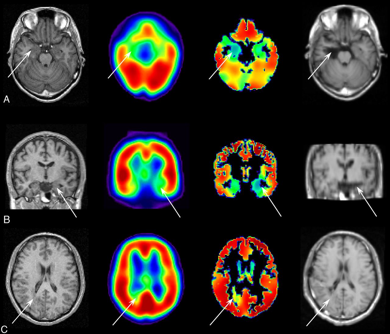
  • Fig 3.
    • Download figure
    • Open in new tab
    • Download powerpoint
    Fig 3.

    MR imaging acquired before surgery (far left column), uncorrected SPECT (the second column from the left), PVE-corrected SPECT (the third column from the left), and MR imaging acquired after surgery (far right column). Arrows indicate the location of resected lesions. A, A case of glioma without any remarkable findings in the uncorrected SPECT, showing a count decrease detected in the PVE-corrected SPECT in the right mesial temporal region. Right mediobasal temporal corticectomy was performed and the postoperative outcome after a 2-year follow-up period was Engel class Ia. B, A case of mesial temporal sclerosis without any remarkable findings in the uncorrected SPECT, showing a count decrease in the PVE-corrected SPECT in the left mesial temporal region. Left selective amygdalohippocampectomy was performed and the postoperative outcome after 2-year follow-up was Engel class Ia. C, A case of heterotopias showing a slight increase in the uncorrected SPECT and a count decrease in the PVE-corrected SPECT in the right temporo-occipital subcortical region. Lesionectomy in the temporo-occipital subcortical area was performed, and the postoperative outcome after 2-year follow-up was Engel class Ia.

Tables

  • Figures
    • View popup
    Table 1:

    Demographics of the patients

    DemographicNo.
    Total75
    Focus laterality
        Right35
        Left40
    Focus location
        Temporal57
        Frontal13
        Parietal1
        Occipital2
        Temporo-occipital1
        Parieto-occipital1
    Histologic diagnosis
        MTS34
        MCD
            Cortical dysplasia8
            Tuberous sclerosis2
            Heterotopia1
        Tumor
            Glioma8
            Ganglioglioma3
            Amygdalar hamartoma3
            Angioma3
            DNT3
        Nonspecific
            No remarkable abnormality8
            Atrophy2
    Prognosis (Engel classification)a
        Ia44
        Ib9
        Ic-d0
        IIa0
        IIb22
        IIc-d0
        III0
        IV0
    Medicationb
        CBZ52
        PHT34
        PB18
        VPA11
        CLB14
        DZP1
        CZP4
    Application of invasive EEGc
        No52
        Yes23
    • Note:—DNT indicates dysembryoplastic neuroepithelial tumor; CBZ, carbamazepine; PHT, phenytoin; PB, phenobarbital; VPA, sodium valproate; CLB, clobazam; DZP, diazepam; CZP, clonazepam.

    • ↵a Class I: Free of disabling seizures. a, Completely seizure-free since surgery. b, Nondisabling simple partial seizures only since surgery. c, Some disabling seizures after surgery, but free of disabling seizures for at least 2 years. d, Generalized convulsions with antiepileptic drug discontinuation only. Class II: Rare disabling seizures. (“almost seizure-free”) a, Initially free of disabling seizures but has rare seizures now. b, Rare disabling seizures since surgery. c, More than rare disabling seizures since surgery, but rare seizures for the last 2 years. d, Nocturnal seizures only. Class III: Worthwhile improvement. a, Worthwhile seizure reduction. b, Prolonged seizure-free intervals amounting to greater than half the follow-up period, but not <2 years. Class IV: No worthwhile improvement. a, Significant seizure reduction. b, No appreciable change. c, Seizures worse.

    • ↵b Use of anticonvulsants at the time of I-123 iomazenil SPECT examination.

    • ↵c Group of patients who underwent 2-step surgery: intracranial EEG electrode implantation and foci resection. This group comprised 5 cases of MTS, 6 cases of MCD, 7 tumors, and 5 healthy patients.

    • View popup
    Table 2:

    Evaluation criteria for visual assessment of SPECT

    Resected Brain RegionUnresected Brain Region
    SPECT positiveaTrue-positiveFalse-positive
    SPECT negativebFalse-negativeTrue-negative
    • ↵a SPECT count decrease detected in the brain region by visual assessment.

    • ↵a No count decrease in the brain region.

    • View popup
    Table 3:

    Results of the visual assessment of foci detectability

    No.Sensitivity (%)Specificity (%)Accuracy (%)
    Pre-PVCPost-PVCPPre-PVCPost-PVCPPre-PVCPost-PVCP
    Total754985<.0016985.0034384<.001
    Histologic diagnosis
        MTS347093.0049093NS6793.001
        MCD115090NS6080NS3080.06
        Tumor202883<.0015683.062883<.001
        Nonspecific104060NS4060NS3060NS
    Foci location
        Mesial temporal415690<.0017890NS5190<.001
        Neocortex344179<.0015979.033276<.001
    Invasive EEG
        Noa525690<.0017388.014888<.001
        Yesb233574.0046178NS3074.002
    • Note:—PVC indicates partial volume correction; NS, not significant.

    • ↵a Application of the 1-step resection surgery without invasive EEG.

    • ↵b Application of both invasive EEG and resection surgery.

    • View popup
    Table 4:

    Results of the quantitative assessment based on asymmetry of SPECT counts

    Histologic DiagnosisNo.Mean AI (%) (SD)Mean AI (%)/CV (%) (SD)
    Pre-PVCPost-PVCPPre-PVCPost-PVCP
    MTS3423 1622 16NS0.76 (0.52)1.1 (0.80)<.001
    MCD1118 (17)25 (17).010.55 (0.49)1.3 (0.82)<.001
    Tumor2014 (15)17 (16)<.0010.44 (0.43)0.89 (0.81)<.001
    Nonspecific1016 (22)14 (16)NS0.62 (0.98)0.72 (0.83)NS
    • Note:—NS indicates not significant.

PreviousNext
Back to top

In this issue

American Journal of Neuroradiology: 33 (11)
American Journal of Neuroradiology
Vol. 33, Issue 11
1 Dec 2012
  • Table of Contents
  • Index by author
Advertisement
Print
Download PDF
Email Article

Thank you for your interest in spreading the word on American Journal of Neuroradiology.

NOTE: We only request your email address so that the person you are recommending the page to knows that you wanted them to see it, and that it is not junk mail. We do not capture any email address.

Enter multiple addresses on separate lines or separate them with commas.
MR Imaging–Based Correction for Partial Volume Effect Improves Detectability of Intractable Epileptogenic Foci on Iodine 123 Iomazenil Brain SPECT Images: An Extended Study with a Larger Sample Size
(Your Name) has sent you a message from American Journal of Neuroradiology
(Your Name) thought you would like to see the American Journal of Neuroradiology web site.
CAPTCHA
This question is for testing whether or not you are a human visitor and to prevent automated spam submissions.
Cite this article
H. Kato, K. Matsuda, K. Baba, E. Shimosegawa, K. Isohashi, M. Imaizumi, J. Hatazawa
MR Imaging–Based Correction for Partial Volume Effect Improves Detectability of Intractable Epileptogenic Foci on Iodine 123 Iomazenil Brain SPECT Images: An Extended Study with a Larger Sample Size
American Journal of Neuroradiology Dec 2012, 33 (11) 2088-2094; DOI: 10.3174/ajnr.A3121

Citation Manager Formats

  • BibTeX
  • Bookends
  • EasyBib
  • EndNote (tagged)
  • EndNote 8 (xml)
  • Medlars
  • Mendeley
  • Papers
  • RefWorks Tagged
  • Ref Manager
  • RIS
  • Zotero
0 Responses
Respond to this article
Share
Bookmark this article
MR Imaging–Based Correction for Partial Volume Effect Improves Detectability of Intractable Epileptogenic Foci on Iodine 123 Iomazenil Brain SPECT Images: An Extended Study with a Larger Sample Size
H. Kato, K. Matsuda, K. Baba, E. Shimosegawa, K. Isohashi, M. Imaizumi, J. Hatazawa
American Journal of Neuroradiology Dec 2012, 33 (11) 2088-2094; DOI: 10.3174/ajnr.A3121
del.icio.us logo Twitter logo Facebook logo Mendeley logo
  • Tweet Widget
  • Facebook Like
  • Google Plus One
Purchase

Jump to section

  • Article
    • Abstract
    • ABBREVIATIONS:
    • Materials and Methods
    • Results
    • Discussion
    • Conclusions
    • Acknowledgments
    • Footnotes
    • References
  • Figures & Data
  • Info & Metrics
  • Responses
  • References
  • PDF

Related Articles

  • No related articles found.
  • PubMed
  • Google Scholar

Cited By...

  • No citing articles found.
  • Crossref
  • Google Scholar

This article has not yet been cited by articles in journals that are participating in Crossref Cited-by Linking.

More in this TOC Section

  • Quiet PROPELLER MRI Techniques Match the Quality of Conventional PROPELLER Brain Imaging Techniques
  • Predictors of Reperfusion in Patients with Acute Ischemic Stroke
  • Enhanced Axonal Metabolism during Early Natalizumab Treatment in Relapsing-Remitting Multiple Sclerosis
Show more Brain

Similar Articles

Advertisement

Indexed Content

  • Current Issue
  • Accepted Manuscripts
  • Article Preview
  • Past Issues
  • Editorials
  • Editor's Choice
  • Fellows' Journal Club
  • Letters to the Editor
  • Video Articles

Cases

  • Case Collection
  • Archive - Case of the Week
  • Archive - Case of the Month
  • Archive - Classic Case

More from AJNR

  • Trainee Corner
  • Imaging Protocols
  • MRI Safety Corner
  • Book Reviews

Multimedia

  • AJNR Podcasts
  • AJNR Scantastics

Resources

  • Turnaround Time
  • Submit a Manuscript
  • Submit a Video Article
  • Submit an eLetter to the Editor/Response
  • Manuscript Submission Guidelines
  • Statistical Tips
  • Fast Publishing of Accepted Manuscripts
  • Graphical Abstract Preparation
  • Imaging Protocol Submission
  • Evidence-Based Medicine Level Guide
  • Publishing Checklists
  • Author Policies
  • Become a Reviewer/Academy of Reviewers
  • News and Updates

About Us

  • About AJNR
  • Editorial Board
  • Editorial Board Alumni
  • Alerts
  • Permissions
  • Not an AJNR Subscriber? Join Now
  • Advertise with Us
  • Librarian Resources
  • Feedback
  • Terms and Conditions
  • AJNR Editorial Board Alumni

American Society of Neuroradiology

  • Not an ASNR Member? Join Now

© 2025 by the American Society of Neuroradiology All rights, including for text and data mining, AI training, and similar technologies, are reserved.
Print ISSN: 0195-6108 Online ISSN: 1936-959X

Powered by HighWire