Advertisement

Main menu

  • Home
  • Content
    • Current Issue
    • Accepted Manuscripts
    • Article Preview
    • Past Issue Archive
    • Video Articles
    • AJNR Case Collection
    • Case of the Week Archive
    • Case of the Month Archive
    • Classic Case Archive
  • Special Collections
    • AJNR Awards
    • Low-Field MRI
    • Alzheimer Disease
    • ASNR Foundation Special Collection
    • Photon-Counting CT
    • View All
  • Multimedia
    • AJNR Podcasts
    • AJNR SCANtastic
    • Trainee Corner
    • MRI Safety Corner
    • Imaging Protocols
  • For Authors
    • Submit a Manuscript
    • Submit a Video Article
    • Submit an eLetter to the Editor/Response
    • Manuscript Submission Guidelines
    • Statistical Tips
    • Fast Publishing of Accepted Manuscripts
    • Graphical Abstract Preparation
    • Imaging Protocol Submission
    • Author Policies
  • About Us
    • About AJNR
    • Editorial Board
    • Editorial Board Alumni
  • More
    • Become a Reviewer/Academy of Reviewers
    • Subscribers
    • Permissions
    • Alerts
    • Feedback
    • Advertisers
    • ASNR Home

User menu

  • Alerts
  • Log in

Search

  • Advanced search
American Journal of Neuroradiology

American Journal of Neuroradiology

ASHNR American Society of Functional Neuroradiology ASHNR American Society of Pediatric Neuroradiology ASSR
  • Alerts
  • Log in

Advanced Search

  • Home
  • Content
    • Current Issue
    • Accepted Manuscripts
    • Article Preview
    • Past Issue Archive
    • Video Articles
    • AJNR Case Collection
    • Case of the Week Archive
    • Case of the Month Archive
    • Classic Case Archive
  • Special Collections
    • AJNR Awards
    • Low-Field MRI
    • Alzheimer Disease
    • ASNR Foundation Special Collection
    • Photon-Counting CT
    • View All
  • Multimedia
    • AJNR Podcasts
    • AJNR SCANtastic
    • Trainee Corner
    • MRI Safety Corner
    • Imaging Protocols
  • For Authors
    • Submit a Manuscript
    • Submit a Video Article
    • Submit an eLetter to the Editor/Response
    • Manuscript Submission Guidelines
    • Statistical Tips
    • Fast Publishing of Accepted Manuscripts
    • Graphical Abstract Preparation
    • Imaging Protocol Submission
    • Author Policies
  • About Us
    • About AJNR
    • Editorial Board
    • Editorial Board Alumni
  • More
    • Become a Reviewer/Academy of Reviewers
    • Subscribers
    • Permissions
    • Alerts
    • Feedback
    • Advertisers
    • ASNR Home
  • Follow AJNR on Twitter
  • Visit AJNR on Facebook
  • Follow AJNR on Instagram
  • Join AJNR on LinkedIn
  • RSS Feeds

AJNR Awards, New Junior Editors, and more. Read the latest AJNR updates

Research ArticleBrain
Open Access

Reduced Diffusion in a Subset of Acute MS Lesions: A Serial Multiparametric MRI Study

P. Eisele, K. Szabo, M. Griebe, C. Roßmanith, A. Förster, M. Hennerici and A. Gass
American Journal of Neuroradiology August 2012, 33 (7) 1369-1373; DOI: https://doi.org/10.3174/ajnr.A2975
P. Eisele
aFrom the Department of Neurology, UniversitätsMedizin Mannheim, University of Heidelberg, Mannheim, Germany.
  • Find this author on Google Scholar
  • Find this author on PubMed
  • Search for this author on this site
K. Szabo
aFrom the Department of Neurology, UniversitätsMedizin Mannheim, University of Heidelberg, Mannheim, Germany.
  • Find this author on Google Scholar
  • Find this author on PubMed
  • Search for this author on this site
M. Griebe
aFrom the Department of Neurology, UniversitätsMedizin Mannheim, University of Heidelberg, Mannheim, Germany.
  • Find this author on Google Scholar
  • Find this author on PubMed
  • Search for this author on this site
C. Roßmanith
aFrom the Department of Neurology, UniversitätsMedizin Mannheim, University of Heidelberg, Mannheim, Germany.
  • Find this author on Google Scholar
  • Find this author on PubMed
  • Search for this author on this site
A. Förster
aFrom the Department of Neurology, UniversitätsMedizin Mannheim, University of Heidelberg, Mannheim, Germany.
  • Find this author on Google Scholar
  • Find this author on PubMed
  • Search for this author on this site
M. Hennerici
aFrom the Department of Neurology, UniversitätsMedizin Mannheim, University of Heidelberg, Mannheim, Germany.
  • Find this author on Google Scholar
  • Find this author on PubMed
  • Search for this author on this site
A. Gass
aFrom the Department of Neurology, UniversitätsMedizin Mannheim, University of Heidelberg, Mannheim, Germany.
  • Find this author on Google Scholar
  • Find this author on PubMed
  • Search for this author on this site
  • Article
  • Figures & Data
  • Info & Metrics
  • Responses
  • References
  • PDF
Loading

Article Figures & Data

Figures

  • Tables
  • Fig 1.
    • Download figure
    • Open in new tab
    • Download powerpoint
    Fig 1.

    Time course of intralesional ADC values during the first 90 days in all 10 patients. Note that measurements outside the 90-day range are not shown for better visualization of early time points.

  • Fig 2.
    • Download figure
    • Open in new tab
    • Download powerpoint
    Fig 2.

    Exemplary ADC, contrast-enhanced T1-weighted, DWI, and FLAIR images of the first 4 MRI time points of patient 4 during the first 14 days after new symptom onset. The initially low ADC is associated with only slight T2 hyperintensity. The increasing ADC signal intensity on follow-ups is paralleled by the development of prominent T2 hyperintensity. Contrast enhancement is minimal in the zone of reduced ADC initially and becomes very prominent at time points 3 and 4.
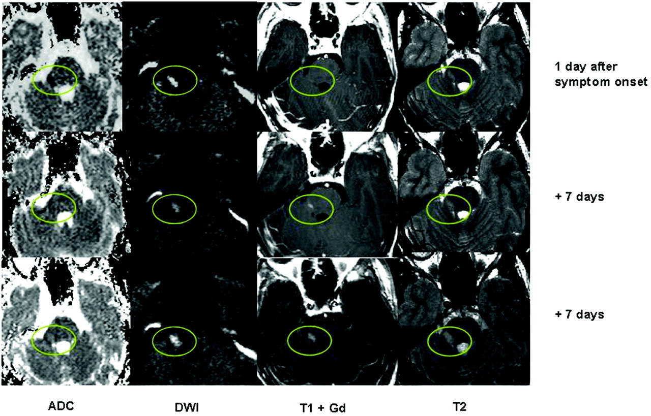
  • Fig 3.
    • Download figure
    • Open in new tab
    • Download powerpoint
    Fig 3.

    Exemplary ADC, DWI, contrast-enhanced T1-weighted, and T2-weighted images of the first 3 examinations of patient 9 in the first 15 days after new onset trigeminal sensory loss. The fascicular trigeminal fibers are affected by an acute lesion (circled in green). The lesion shows hyperintensity on DWI and a reduced ADC, while minimal T2 hyperintensity is noted without contrast enhancement on T1-weighted MRI initially. With time, the ADC pseudonormalizes and increases, while T2 hyperintensity and contrast enhancement become more prominent.

Tables

  • Figures
    • View popup
    Table 1:

    Summary of clinical data

    No.Age (yr)/SexDisease Duration/DiagnosisNew Clinical SymptomsSymptom Onset to MRI (days)Max. ADC ↓Treatment Acute/ProphylaxisOCB
    119/F2 yr/RRMSRight internuclear ophthalmoplegia1−41%IV cortisone/interferon+
    221/F1 yr/RRMSDysarthria0−66%IV cortisone/–+
    320/F1 yr/RRMSVertigo, left-sided numbness1.5−41%IV cortisone/–+
    445/F1.5 yr/RRMSRight-sided weakness2−20%IV cortisone/–+
    525/F1 yr/RRMSDysarthria, right-sided weakness4−17%IV cortisone/–−
    625/FInitial presentation/CISDysarthria, left-sided weakness and numbness2−51%IV cortisone/–+
    730/F1.5 yr/RRMSLeft-sided arm weakness2−16%IV cortisone/–+
    833/F0.5 yr/RRMSHemianopia3−22%IV cortisone/–+
    931/M1 yr/RRMSN.V. numbness1−24%IV cortisone/–+
    1044/M2 yr/RRMSRight-sided weakness3−41%IV cortisone/interferon+
    • Note:—OCB indicates oligoclonal bands; +, positive OCB; −, negative OCB.

    • View popup
    Table 2:

    Intralesional signal change on ADC and T2-weighted imagesa

    PatientDay
    Patient 1Day 1Day 8Day 17Day 55
        ADC−41102150
        T217182543
    Patient 2Day 0Day 7Day 14Day 21Day 49Day 83Day 207
        ADC−66−44−34−18723736
        T219203246644842
    Patient 3Day 1Day 8Day 35Day 42
        ADC−41−74741
        T215808635
    Patient 4Day 2Day 6Day 10Day 14Day 85Day 175
        ADC−20−15−115660125
        T23250548710543
    Patient 5Day 4Day 12Day 20Day 29Day 65
        ADC−175101830
        T23556726552
    Patient 6Day 2Day 6Day 14Day 28
        ADC−51−3563130
        T237629874
    Patient 7Day 2Day 10Day 21Day 30Day 66Day 106
        ADC−1654550102105
        T2234586736665
    Patient 8Day 3Day 6Day 9Day 21Day 64
        ADC−22−4255562
        T22545576366
    Patient 9Day 1Day 8Day 15Day 21Day 34
        ADC−2412325359
        T21520183145
    Patient 10Day 3Day 15Day 30Day 37Day 51Day 66
        ADC−41−2512275862
        T283246637854
    • ↵a Values are given relative to the NAWM in the contralateral hemisphere.

PreviousNext
Back to top

In this issue

American Journal of Neuroradiology: 33 (7)
American Journal of Neuroradiology
Vol. 33, Issue 7
1 Aug 2012
  • Table of Contents
  • Index by author
Advertisement
Print
Download PDF
Email Article

Thank you for your interest in spreading the word on American Journal of Neuroradiology.

NOTE: We only request your email address so that the person you are recommending the page to knows that you wanted them to see it, and that it is not junk mail. We do not capture any email address.

Enter multiple addresses on separate lines or separate them with commas.
Reduced Diffusion in a Subset of Acute MS Lesions: A Serial Multiparametric MRI Study
(Your Name) has sent you a message from American Journal of Neuroradiology
(Your Name) thought you would like to see the American Journal of Neuroradiology web site.
CAPTCHA
This question is for testing whether or not you are a human visitor and to prevent automated spam submissions.
Cite this article
P. Eisele, K. Szabo, M. Griebe, C. Roßmanith, A. Förster, M. Hennerici, A. Gass
Reduced Diffusion in a Subset of Acute MS Lesions: A Serial Multiparametric MRI Study
American Journal of Neuroradiology Aug 2012, 33 (7) 1369-1373; DOI: 10.3174/ajnr.A2975

Citation Manager Formats

  • BibTeX
  • Bookends
  • EasyBib
  • EndNote (tagged)
  • EndNote 8 (xml)
  • Medlars
  • Mendeley
  • Papers
  • RefWorks Tagged
  • Ref Manager
  • RIS
  • Zotero
0 Responses
Respond to this article
Share
Bookmark this article
Reduced Diffusion in a Subset of Acute MS Lesions: A Serial Multiparametric MRI Study
P. Eisele, K. Szabo, M. Griebe, C. Roßmanith, A. Förster, M. Hennerici, A. Gass
American Journal of Neuroradiology Aug 2012, 33 (7) 1369-1373; DOI: 10.3174/ajnr.A2975
del.icio.us logo Twitter logo Facebook logo Mendeley logo
  • Tweet Widget
  • Facebook Like
  • Google Plus One
Purchase

Jump to section

  • Article
    • Abstract
    • ABBREVIATIONS:
    • Materials and Methods
    • Results
    • Discussion
    • Conclusions
    • Footnotes
    • References
  • Figures & Data
  • Info & Metrics
  • Responses
  • References
  • PDF

Related Articles

  • No related articles found.
  • PubMed
  • Google Scholar

Cited By...

  • Monoclonal Antibodies: What the Diagnostic Neuroradiologist Needs to Know
  • Risk of stroke in multiple sclerosis and neuromyelitis optic spectrum disorder: a Nationwide cohort study in South Korea
  • Reduced diffusion in acute cervical cord multiple sclerosis lesions
  • MRI criteria differentiating asymptomatic PML from new MS lesions during natalizumab pharmacovigilance
  • Quantitative MRI for Analysis of Active Multiple Sclerosis Lesions without Gadolinium-Based Contrast Agent
  • Direct MRI detection of impending plaque development in multiple sclerosis
  • Crossref
  • Google Scholar

This article has not yet been cited by articles in journals that are participating in Crossref Cited-by Linking.

More in this TOC Section

  • Enhanced Axonal Metabolism during Early Natalizumab Treatment in Relapsing-Remitting Multiple Sclerosis
  • Progression of Microstructural Damage in Spinocerebellar Ataxia Type 2: A Longitudinal DTI Study
  • SWI or T2*: Which MRI Sequence to Use in the Detection of Cerebral Microbleeds? The Karolinska Imaging Dementia Study
Show more Brain

Similar Articles

Advertisement

Indexed Content

  • Current Issue
  • Accepted Manuscripts
  • Article Preview
  • Past Issues
  • Editorials
  • Editor's Choice
  • Fellows' Journal Club
  • Letters to the Editor
  • Video Articles

Cases

  • Case Collection
  • Archive - Case of the Week
  • Archive - Case of the Month
  • Archive - Classic Case

More from AJNR

  • Trainee Corner
  • Imaging Protocols
  • MRI Safety Corner
  • Book Reviews

Multimedia

  • AJNR Podcasts
  • AJNR Scantastics

Resources

  • Turnaround Time
  • Submit a Manuscript
  • Submit a Video Article
  • Submit an eLetter to the Editor/Response
  • Manuscript Submission Guidelines
  • Statistical Tips
  • Fast Publishing of Accepted Manuscripts
  • Graphical Abstract Preparation
  • Imaging Protocol Submission
  • Evidence-Based Medicine Level Guide
  • Publishing Checklists
  • Author Policies
  • Become a Reviewer/Academy of Reviewers
  • News and Updates

About Us

  • About AJNR
  • Editorial Board
  • Editorial Board Alumni
  • Alerts
  • Permissions
  • Not an AJNR Subscriber? Join Now
  • Advertise with Us
  • Librarian Resources
  • Feedback
  • Terms and Conditions
  • AJNR Editorial Board Alumni

American Society of Neuroradiology

  • Not an ASNR Member? Join Now

© 2025 by the American Society of Neuroradiology All rights, including for text and data mining, AI training, and similar technologies, are reserved.
Print ISSN: 0195-6108 Online ISSN: 1936-959X

Powered by HighWire