Advertisement

Main menu

  • Home
  • Content
    • Current Issue
    • Accepted Manuscripts
    • Article Preview
    • Past Issue Archive
    • Video Articles
    • AJNR Case Collection
    • Case of the Week Archive
    • Case of the Month Archive
    • Classic Case Archive
  • Special Collections
    • AJNR Awards
    • Low-Field MRI
    • Alzheimer Disease
    • ASNR Foundation Special Collection
    • Photon-Counting CT
    • View All
  • Multimedia
    • AJNR Podcasts
    • AJNR SCANtastic
    • Trainee Corner
    • MRI Safety Corner
    • Imaging Protocols
  • For Authors
    • Submit a Manuscript
    • Submit a Video Article
    • Submit an eLetter to the Editor/Response
    • Manuscript Submission Guidelines
    • Statistical Tips
    • Fast Publishing of Accepted Manuscripts
    • Graphical Abstract Preparation
    • Imaging Protocol Submission
    • Author Policies
  • About Us
    • About AJNR
    • Editorial Board
    • Editorial Board Alumni
  • More
    • Become a Reviewer/Academy of Reviewers
    • Subscribers
    • Permissions
    • Alerts
    • Feedback
    • Advertisers
    • ASNR Home

User menu

  • Alerts
  • Log in

Search

  • Advanced search
American Journal of Neuroradiology

American Journal of Neuroradiology

ASHNR American Society of Functional Neuroradiology ASHNR American Society of Pediatric Neuroradiology ASSR
  • Alerts
  • Log in

Advanced Search

  • Home
  • Content
    • Current Issue
    • Accepted Manuscripts
    • Article Preview
    • Past Issue Archive
    • Video Articles
    • AJNR Case Collection
    • Case of the Week Archive
    • Case of the Month Archive
    • Classic Case Archive
  • Special Collections
    • AJNR Awards
    • Low-Field MRI
    • Alzheimer Disease
    • ASNR Foundation Special Collection
    • Photon-Counting CT
    • View All
  • Multimedia
    • AJNR Podcasts
    • AJNR SCANtastic
    • Trainee Corner
    • MRI Safety Corner
    • Imaging Protocols
  • For Authors
    • Submit a Manuscript
    • Submit a Video Article
    • Submit an eLetter to the Editor/Response
    • Manuscript Submission Guidelines
    • Statistical Tips
    • Fast Publishing of Accepted Manuscripts
    • Graphical Abstract Preparation
    • Imaging Protocol Submission
    • Author Policies
  • About Us
    • About AJNR
    • Editorial Board
    • Editorial Board Alumni
  • More
    • Become a Reviewer/Academy of Reviewers
    • Subscribers
    • Permissions
    • Alerts
    • Feedback
    • Advertisers
    • ASNR Home
  • Follow AJNR on Twitter
  • Visit AJNR on Facebook
  • Follow AJNR on Instagram
  • Join AJNR on LinkedIn
  • RSS Feeds

AJNR Awards, New Junior Editors, and more. Read the latest AJNR updates

Getting new auth cookie, if you see this message a lot, tell someone!
Research ArticleBrain

Quantitative Assessment of External Carotid Artery Territory Supply with Modified Vessel-Encoded Arterial Spin-Labeling

Y. Dang, B. Wu, Y. Sun, D. Mo, X. Wang, J. Zhang and J. Fang
American Journal of Neuroradiology August 2012, 33 (7) 1380-1386; DOI: https://doi.org/10.3174/ajnr.A2978
Y. Dang
aFrom the Academy for Advanced Interdisciplinary Studies (Y.D., X.W., J.Z., J.F.)
  • Find this author on Google Scholar
  • Find this author on PubMed
  • Search for this author on this site
B. Wu
cDepartments of Radiology (B.W., X.W.)
  • Find this author on Google Scholar
  • Find this author on PubMed
  • Search for this author on this site
Y. Sun
bCollege of Engineering (Y.S., J.Z., J.F.), Peking University, Beijing, China
  • Find this author on Google Scholar
  • Find this author on PubMed
  • Search for this author on this site
D. Mo
dNeurosurgery (D.M.), Peking University First Hospital, Beijing, China.
  • Find this author on Google Scholar
  • Find this author on PubMed
  • Search for this author on this site
X. Wang
aFrom the Academy for Advanced Interdisciplinary Studies (Y.D., X.W., J.Z., J.F.)
cDepartments of Radiology (B.W., X.W.)
  • Find this author on Google Scholar
  • Find this author on PubMed
  • Search for this author on this site
J. Zhang
aFrom the Academy for Advanced Interdisciplinary Studies (Y.D., X.W., J.Z., J.F.)
bCollege of Engineering (Y.S., J.Z., J.F.), Peking University, Beijing, China
  • Find this author on Google Scholar
  • Find this author on PubMed
  • Search for this author on this site
J. Fang
aFrom the Academy for Advanced Interdisciplinary Studies (Y.D., X.W., J.Z., J.F.)
bCollege of Engineering (Y.S., J.Z., J.F.), Peking University, Beijing, China
  • Find this author on Google Scholar
  • Find this author on PubMed
  • Search for this author on this site
  • Article
  • Figures & Data
  • Info & Metrics
  • Responses
  • References
  • PDF
Loading

Article Figures & Data

Figures

  • Tables
  • Fig 1.
    • Download figure
    • Open in new tab
    • Download powerpoint
    Fig 1.

    A, Arrangement of the right ICA, left ICA, VAs, and ECA within the labeling plane seen with MRA. B, Location of the labeling plane (white) and image sections (gray).

  • Fig 2.
    • Download figure
    • Open in new tab
    • Download powerpoint
    Fig 2.

    Left: Perfusion territory maps. Images a, b, and c represent the results of 3 scans (right) with different spatial gradient-encoding methods. Image d indicates the integrated results of the perfusion image with encoded vessels. Blood flows contributed by the vessels of the right ICA, left ICA, VAs, and ECA are red, green, blue, and yellow (white arrows), respectively. Center: The histograms (shown in blue) of the calculated tagging efficiencies for each encoding step. Gaussian fits to the peaks in the histograms are red; the center of the Gaussian fits, as the estimated tagging efficiencies for each vessel, are used to reconstruct the modified encoding matrix. The orange arrows originating from the histograms indicate the corresponding labeling vessels whose tagging efficiencies are estimated by the abscissa values of the center of the Gaussian fits (eg, in the first row of the histograms, the center of the Gaussian fit in an abscissa of −1 represents the tagging efficiency of the right ICA, in which the center of the Gaussian fit in nearly an abscissa of 1 represents the tagging efficiency of the left ICA and ECA). Right: The modified tagging geometries. The red and blue lines represent the locations that are marked as contrast (one represents the vessel inverted; the other, the vessel relaxed). A, The right and left ICAs are separated. B, Carotid and vertebral arteries are prominently distinguished. C, The VAs and ECAs are contrasted.

  • Fig 3.
    • Download figure
    • Open in new tab
    • Download powerpoint
    Fig 3.

    Perfusion territory maps of a healthy subject by using modified VE-ASL. The first row is the perfusion territories of the right ICA (red), left ICA (green), and VAs (blue); the second row is the perfusion territories of intracarotid (gray) and extracarotid (yellow) arteries. Note that the bottom section shows flow in the frontal brain region, which appears to be artifacts caused by magnetic inhomogeneities of B0 or eye motion. In the fourth image from the bottom, there is also a hint of ghosting in the white matter of the right hemisphere, which may be caused by the decoding process.
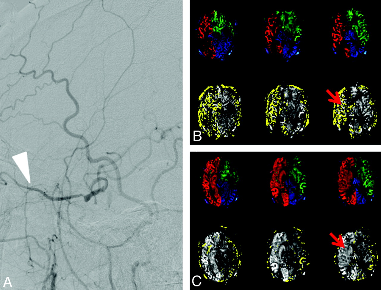
  • Fig 4.
    • Download figure
    • Open in new tab
    • Download powerpoint
    Fig 4.

    Sagittal DSA results (left) and vascular territories (right) in a patient undergoing carotid stent placement for right ICA stenosis. A, DSA results of the patient before surgery show that there is collateral flow from the ECA (white arrow). B, Preoperatively, the collateral supply is manifested as the ECA supply to the right ICA territory (red arrow). C, Postoperatively, there is normalization of the right ICA perfusion with a corresponding reduction of collateral supply (red arrow). Hyperperfusion of the right hemisphere after stent placement is also clearly shown.
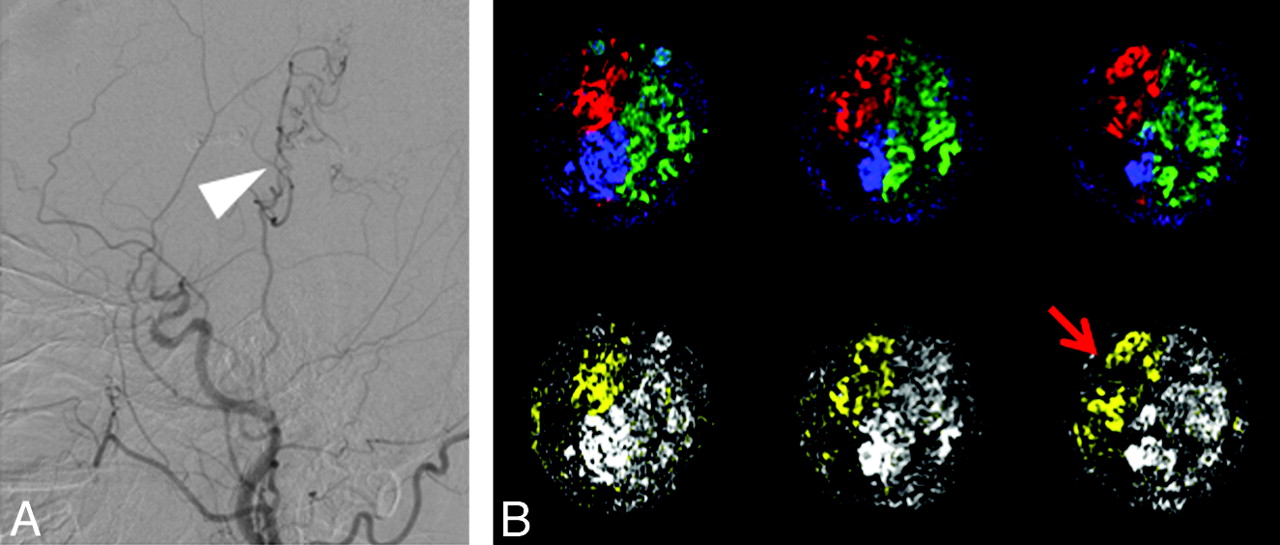
  • Fig 5.
    • Download figure
    • Open in new tab
    • Download powerpoint
    Fig 5.

    Sagittal DSA results (A) and vascular territories (B) in a patient with right MCA occlusion after ECIC bypass surgery. Postoperatively, the blood flow from the ECA is detected (red arrow), in agreement with the collaterals shown on DSA (white arrow).

Tables

  • Figures
  • Quantification of CBF of RICA, LICA, VAs, and ECA before and after surgery using modified VE-ASL

    SubjectSymptomScanning TimeCBFa
    RICALICAVAsECA
    1RICA severe stenosisBefore ICA stenting,14.328.026.425.2
        after ICA stenting42.231.329.12.0
    2bLICA occlusion, VA stenosisBefore VA stenting,15.80.016.518.9
        after VA stenting15.80.017.512.2
    3LICA occlusionBefore bypass,22.10.019.30.0
        after bypass20.50.019.112.3
    4RMCA occlusionBefore bypass,14.423.318.90
        after bypass25.821.517.519.1
    5RICA occlusionBefore bypass,21.917.623.60
        after bypass13.714.916.420.2
    6RICA occlusion, VA severe stenosisBefore bypass,030.831.50
        after bypass019.422.717.3
    7cRICA occlusionBefore bypass,
        after bypass17.317.221.511.8
    8cRMCA severe stenosisBefore bypass,
        after bypass015.214.220.5
    • Note:—RMCA indicates the right MCA; RICA, right ICA; LICA, left ICA.

    • ↵a CBF is mL/min/100 g.

    • ↵b Before VA stenting treatment, the patient was treated with bypass surgery.

    • ↵c Data of subjects 7 and 8 before bypass surgery are missing.

PreviousNext
Back to top

In this issue

American Journal of Neuroradiology: 33 (7)
American Journal of Neuroradiology
Vol. 33, Issue 7
1 Aug 2012
  • Table of Contents
  • Index by author
Advertisement
Print
Download PDF
Email Article

Thank you for your interest in spreading the word on American Journal of Neuroradiology.

NOTE: We only request your email address so that the person you are recommending the page to knows that you wanted them to see it, and that it is not junk mail. We do not capture any email address.

Enter multiple addresses on separate lines or separate them with commas.
Quantitative Assessment of External Carotid Artery Territory Supply with Modified Vessel-Encoded Arterial Spin-Labeling
(Your Name) has sent you a message from American Journal of Neuroradiology
(Your Name) thought you would like to see the American Journal of Neuroradiology web site.
CAPTCHA
This question is for testing whether or not you are a human visitor and to prevent automated spam submissions.
Cite this article
Y. Dang, B. Wu, Y. Sun, D. Mo, X. Wang, J. Zhang, J. Fang
Quantitative Assessment of External Carotid Artery Territory Supply with Modified Vessel-Encoded Arterial Spin-Labeling
American Journal of Neuroradiology Aug 2012, 33 (7) 1380-1386; DOI: 10.3174/ajnr.A2978

Citation Manager Formats

  • BibTeX
  • Bookends
  • EasyBib
  • EndNote (tagged)
  • EndNote 8 (xml)
  • Medlars
  • Mendeley
  • Papers
  • RefWorks Tagged
  • Ref Manager
  • RIS
  • Zotero
0 Responses
Respond to this article
Share
Bookmark this article
Quantitative Assessment of External Carotid Artery Territory Supply with Modified Vessel-Encoded Arterial Spin-Labeling
Y. Dang, B. Wu, Y. Sun, D. Mo, X. Wang, J. Zhang, J. Fang
American Journal of Neuroradiology Aug 2012, 33 (7) 1380-1386; DOI: 10.3174/ajnr.A2978
del.icio.us logo Twitter logo Facebook logo Mendeley logo
  • Tweet Widget
  • Facebook Like
  • Google Plus One
Purchase

Jump to section

  • Article
    • Abstract
    • ABBREVIATIONS:
    • Materials and Methods
    • Results
    • Discussion
    • Conclusions
    • Appendix
    • Acknowledgments
    • Footnotes
    • References
  • Figures & Data
  • Info & Metrics
  • Responses
  • References
  • PDF

Related Articles

  • No related articles found.
  • PubMed
  • Google Scholar

Cited By...

  • Accuracy of Vessel-Encoded Pseudocontinuous Arterial Spin-Labeling in Identification of Feeding Arteries in Patients with Intracranial Arteriovenous Malformations
  • Crossref
  • Google Scholar

This article has not yet been cited by articles in journals that are participating in Crossref Cited-by Linking.

More in this TOC Section

  • Fast Contrast-Enhanced 4D MRA and 4D Flow MRI Using Constrained Reconstruction (HYPRFlow): Potential Applications for Brain Arteriovenous Malformations
  • Multimodal CT Provides Improved Performance for Lacunar Infarct Detection
  • Optimal MRI Sequence for Identifying Occlusion Location in Acute Stroke: Which Value of Time-Resolved Contrast-Enhanced MRA?
Show more Brain

Similar Articles

Advertisement

Indexed Content

  • Current Issue
  • Accepted Manuscripts
  • Article Preview
  • Past Issues
  • Editorials
  • Editor's Choice
  • Fellows' Journal Club
  • Letters to the Editor
  • Video Articles

Cases

  • Case Collection
  • Archive - Case of the Week
  • Archive - Case of the Month
  • Archive - Classic Case

More from AJNR

  • Trainee Corner
  • Imaging Protocols
  • MRI Safety Corner
  • Book Reviews

Multimedia

  • AJNR Podcasts
  • AJNR Scantastics

Resources

  • Turnaround Time
  • Submit a Manuscript
  • Submit a Video Article
  • Submit an eLetter to the Editor/Response
  • Manuscript Submission Guidelines
  • Statistical Tips
  • Fast Publishing of Accepted Manuscripts
  • Graphical Abstract Preparation
  • Imaging Protocol Submission
  • Evidence-Based Medicine Level Guide
  • Publishing Checklists
  • Author Policies
  • Become a Reviewer/Academy of Reviewers
  • News and Updates

About Us

  • About AJNR
  • Editorial Board
  • Editorial Board Alumni
  • Alerts
  • Permissions
  • Not an AJNR Subscriber? Join Now
  • Advertise with Us
  • Librarian Resources
  • Feedback
  • Terms and Conditions
  • AJNR Editorial Board Alumni

American Society of Neuroradiology

  • Not an ASNR Member? Join Now

© 2025 by the American Society of Neuroradiology All rights, including for text and data mining, AI training, and similar technologies, are reserved.
Print ISSN: 0195-6108 Online ISSN: 1936-959X

Powered by HighWire