
Article Figures & Data
Figures
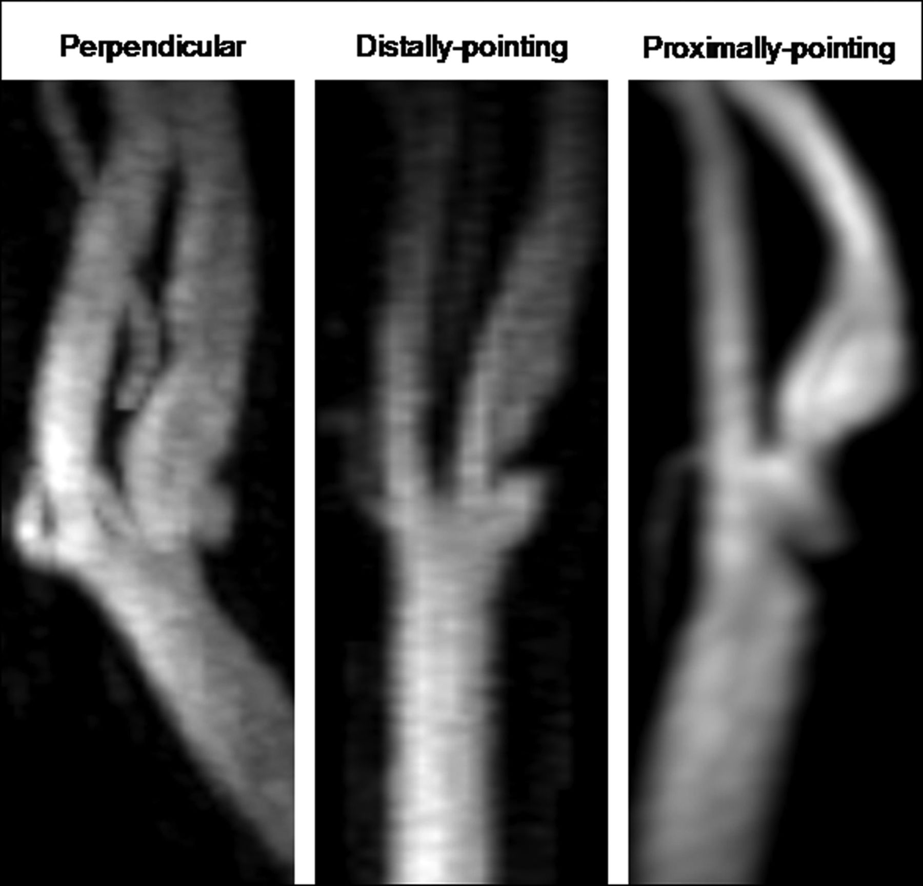
- Fig 1.
Examples of 3 ulcer orientations shown on CE-MRA MIP images. Perpendicular: If a line is to be drawn connecting the center of the ulcer opening and its tip, it will be perpendicular to the vessel axis. Distally pointing: Ulcer tip is distal to its opening. Proximally pointing: Ulcer tip is proximal to its opening.
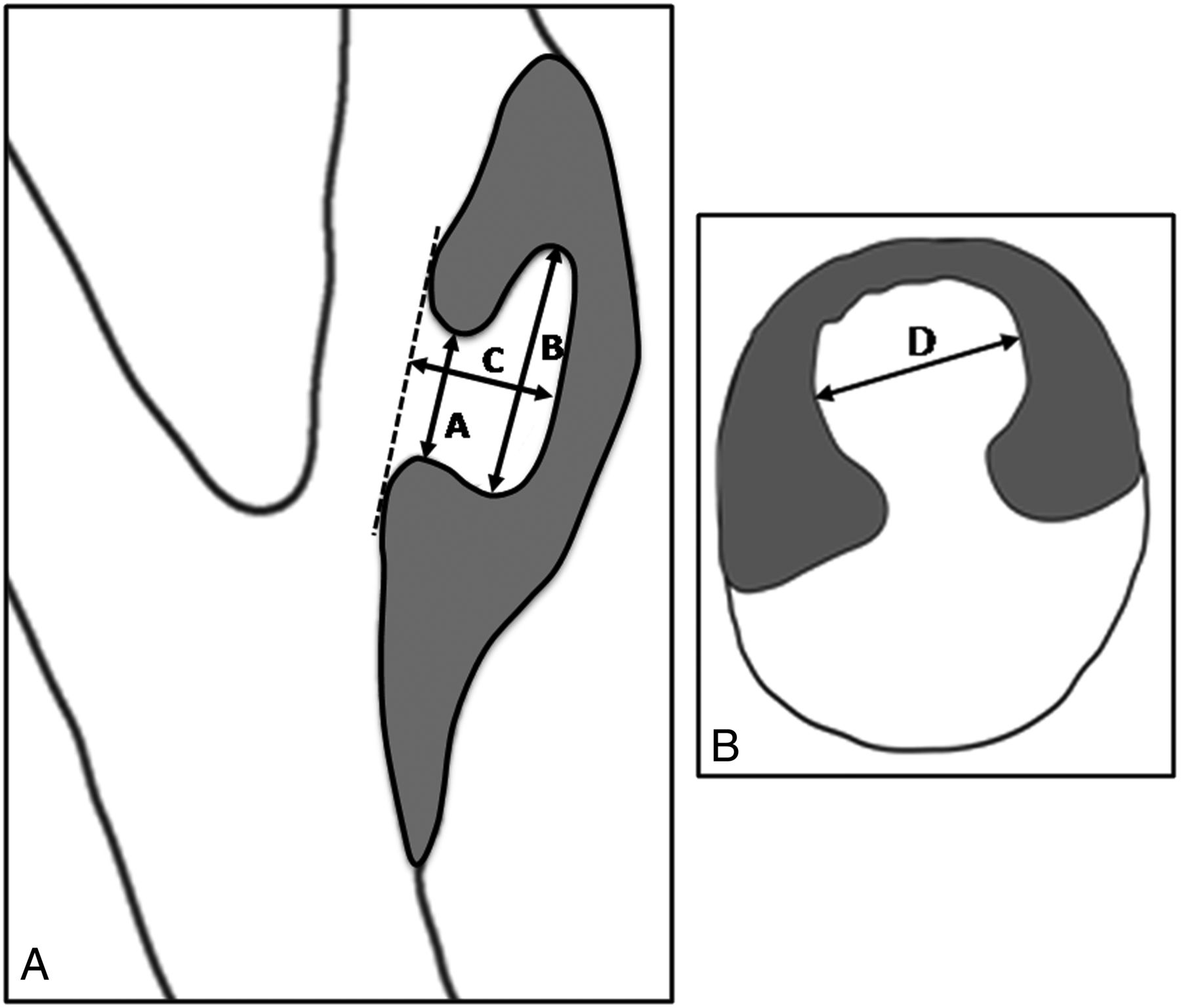
- Fig 2.
Technique for measuring ulcer dimensions. Ulcer neck (A), maximum craniocaudal dimension (B) and depth (C) were measured on CE-MRA MIP images based on the view that shows the largest dimensions (Fig 2A). Maximum transverse dimension (D) was measured on the reconstructed CE-MRA source image oriented perpendicular to the vessel axis that shows the largest transverse dimension (Fig 2B).
- Fig 3.
MIP images of a distally pointing ulcer (A) detected on both CE-MRA and TOF-MRA, and a proximally pointing ulcer (B) detected on CE-MRA but not on TOF-MRA.
- Fig 4.
Visualization of flow patterns computed in a 30%-stenosed carotid bifurcation with distally and proximally pointing ellipsoidal ulcers. The leftmost column shows the full CFD models and the initial distribution of particles seeded at end diastole. Subsequent columns show the distribution of particles remaining after each peak systole, with the amount of time since seeding (ie, residence time) also indicated. Colors are used to indicate particle velocity in cm/s, and size indicates the depth of the particles such that large particles are those closest to the symmetry plane and small particle are those closest to the walls.


Tables
- Table 1:
Ulcer detection by CE-MRA and TOF-MRA techniques for individual ulcers and vessels for 2 readers
Reader 1 Number (%) Reader 2 Number (%) Individual ulcer detection Both CE and TOF 45 (63) 33 (46.5) Only CE 24 (34) 29 (41) Only TOF 1 (1.5) 1 (1.5) Neither CE nor TOF 1 (1.5) 8 (11) Total (all MR sequences) 71 (100) 71 (100) Vessels detected as ulcerated Both CE and TOF 41 (73) 30 (54) Only CE 14 (25) 23 (41) Only TOF 1 (2) 1 (2) Neither CE nor TOF 0 (0) 2 (3) Total (all MR sequences) 56 (100) 56 (100) - Table 2:
Characteristics of 69 ulcers detected by CE-MRA by reader 1 and TOF-MRA ulcer-detection percent for each characteristic
Characteristics Detected by CE-MRAa Detected by TOF-MRAb (among Those Detected by CE-MRA) IQ score 1 28 (41) 18 (64) 2 41 (59) 27 (66) Ulcer orientationc Perpendicular 47 (68) 28 (60) Distally pointing 16 (23) 15 (94) Proximally pointing 6 (9) 2 (33) Ulcer location relative to point of maximum stenosisc Proximal 36 (52) 26 (72) Isolevel 14 (20) 5 (36) Distal 19 (28) 14 (74) Ulcer location relative to FD Proximal 18 (26) 13 (72) Isolevel 28 (41) 19 (68) Distal 23 (33) 13 (57) Ulcer circumferential location Opposite FD 44 (64) 29 (66) Along FD 0 (0) 0 (N/A) On sidewalls 25 (36) 16 (64) Ulcer neck-to-depth ratioc 1.61 (0.56) 1.71 (0.57) Ulcer volume index (mm3)c 67.63 (66.50) 81.83 (75.09) Degree of stenosis (%)c 27.16 (18.06) 23.4 (16.10) - Table 3:
Multivariable logistic regression model for ulcer detection by TOF-MRA among ulcers detected by CE-MRA
Characteristic Multivariable OR 95% CI P Orientation (distally pointing vs perpendicular) 5.57 1.08–28.65 0.04 Orientation (proximally-pointing vs perpendicular) 0.21 0.14–0.29 <0.001 Location (proximal vs isolevel)a 2.79 0.94–8.32 0.06 Location (distal vs isolevel)a 5.17 2.10–12.70 <0.001 Neck-to-depth ratio 1.96 1.11–3.45 0.02 Volume index 1.01 1.00–1.02 0.06 Degree of stenosis 0.97 0.94–1.01 0.23 Note:—Robust variance estimate was used to account for 2 readers' results for each ulcer.
↵a Ulcer location relative to point of maximum stenosis.
In this issue


Jump to section
Related Articles
Cited By...
- Unifying theory of carotid plaque disruption based on structural phenotypes and forces expressed at the lumen/wall interface
- Carotid Artery Wall Imaging: Perspective and Guidelines from the ASNR Vessel Wall Imaging Study Group and Expert Consensus Recommendations of the American Society of Neuroradiology
- Imaging Carotid Atherosclerosis Plaque Ulceration: Comparison of Advanced Imaging Modalities and Recent Developments
- Standards of practice and reporting standards for carotid artery angioplasty and stenting