Advertisement

Main menu

  • Home
  • Content
    • Current Issue
    • Accepted Manuscripts
    • Article Preview
    • Past Issue Archive
    • AJNR Case Collection
    • Case of the Week Archive
    • Classic Case Archive
    • Case of the Month Archive
  • Special Collections
    • Spinal CSF Leak Articles (Jan 2020-June 2024)
    • 2024 AJNR Journal Awards
    • Most Impactful AJNR Articles
  • Multimedia
    • AJNR Podcast
    • AJNR Scantastics
    • Video Articles
  • For Authors
    • Submit a Manuscript
    • Author Policies
    • Fast publishing of Accepted Manuscripts
    • Graphical Abstract Preparation
    • Manuscript Submission Guidelines
    • Imaging Protocol Submission
    • Submit a Case for the Case Collection
  • About Us
    • About AJNR
    • Editorial Board
  • More
    • Become a Reviewer/Academy of Reviewers
    • Subscribers
    • Permissions
    • Alerts
    • Feedback
    • Advertisers
    • ASNR Home
  • Other Publications
    • ajnr

User menu

  • Alerts
  • Log in

Search

  • Advanced search
American Journal of Neuroradiology

American Journal of Neuroradiology

ASHNR American Society of Functional Neuroradiology ASHNR American Society of Pediatric Neuroradiology ASSR
  • Alerts
  • Log in

Advanced Search

  • Home
  • Content
    • Current Issue
    • Accepted Manuscripts
    • Article Preview
    • Past Issue Archive
    • AJNR Case Collection
    • Case of the Week Archive
    • Classic Case Archive
    • Case of the Month Archive
  • Special Collections
    • Spinal CSF Leak Articles (Jan 2020-June 2024)
    • 2024 AJNR Journal Awards
    • Most Impactful AJNR Articles
  • Multimedia
    • AJNR Podcast
    • AJNR Scantastics
    • Video Articles
  • For Authors
    • Submit a Manuscript
    • Author Policies
    • Fast publishing of Accepted Manuscripts
    • Graphical Abstract Preparation
    • Manuscript Submission Guidelines
    • Imaging Protocol Submission
    • Submit a Case for the Case Collection
  • About Us
    • About AJNR
    • Editorial Board
  • More
    • Become a Reviewer/Academy of Reviewers
    • Subscribers
    • Permissions
    • Alerts
    • Feedback
    • Advertisers
    • ASNR Home
  • Follow AJNR on Twitter
  • Visit AJNR on Facebook
  • Follow AJNR on Instagram
  • Join AJNR on LinkedIn
  • RSS Feeds

Welcome to the new AJNR, Updated Hall of Fame, and more. Read the full announcements.


AJNR is seeking candidates for the position of Associate Section Editor, AJNR Case Collection. Read the full announcement.


Research ArticleBrain

4D Digital Subtraction Angiography: Implementation and Demonstration of Feasibility

B. Davis, K. Royalty, M. Kowarschik, C. Rohkohl, E. Oberstar, B. Aagaard-Kienitz, D. Niemann, O. Ozkan, C. Strother and C. Mistretta
American Journal of Neuroradiology October 2013, 34 (10) 1914-1921; DOI: https://doi.org/10.3174/ajnr.A3529
B. Davis
aFrom the University of Wisconsin School of Medicine and Public Health (B.D., E.O., B.A.-K., D.N, O.O., C.S., C.M.), Madison, Wisconsin
  • Find this author on Google Scholar
  • Find this author on PubMed
  • Search for this author on this site
K. Royalty
bSiemens AG (K.R.), Healthcare Sector, Hoffman Estates, Illinois
  • Find this author on Google Scholar
  • Find this author on PubMed
  • Search for this author on this site
M. Kowarschik
cSiemens (M.K., C.R.), Research Collaborations, AX, Forchheim, Germany.
  • Find this author on Google Scholar
  • Find this author on PubMed
  • Search for this author on this site
C. Rohkohl
cSiemens (M.K., C.R.), Research Collaborations, AX, Forchheim, Germany.
  • Find this author on Google Scholar
  • Find this author on PubMed
  • Search for this author on this site
E. Oberstar
aFrom the University of Wisconsin School of Medicine and Public Health (B.D., E.O., B.A.-K., D.N, O.O., C.S., C.M.), Madison, Wisconsin
  • Find this author on Google Scholar
  • Find this author on PubMed
  • Search for this author on this site
B. Aagaard-Kienitz
aFrom the University of Wisconsin School of Medicine and Public Health (B.D., E.O., B.A.-K., D.N, O.O., C.S., C.M.), Madison, Wisconsin
  • Find this author on Google Scholar
  • Find this author on PubMed
  • Search for this author on this site
D. Niemann
aFrom the University of Wisconsin School of Medicine and Public Health (B.D., E.O., B.A.-K., D.N, O.O., C.S., C.M.), Madison, Wisconsin
  • Find this author on Google Scholar
  • Find this author on PubMed
  • Search for this author on this site
O. Ozkan
aFrom the University of Wisconsin School of Medicine and Public Health (B.D., E.O., B.A.-K., D.N, O.O., C.S., C.M.), Madison, Wisconsin
  • Find this author on Google Scholar
  • Find this author on PubMed
  • Search for this author on this site
C. Strother
aFrom the University of Wisconsin School of Medicine and Public Health (B.D., E.O., B.A.-K., D.N, O.O., C.S., C.M.), Madison, Wisconsin
  • Find this author on Google Scholar
  • Find this author on PubMed
  • Search for this author on this site
C. Mistretta
aFrom the University of Wisconsin School of Medicine and Public Health (B.D., E.O., B.A.-K., D.N, O.O., C.S., C.M.), Madison, Wisconsin
  • Find this author on Google Scholar
  • Find this author on PubMed
  • Search for this author on this site
  • Article
  • Figures & Data
  • Info & Metrics
  • Responses
  • References
  • PDF
Loading

Article Figures & Data

Figures

  • Fig 1.
    • Download figure
    • Open in new tab
    • Download powerpoint
    Fig 1.

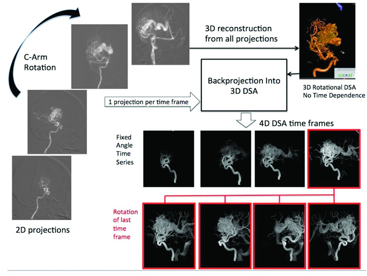
    Schematic of 4D-DSA reconstruction. After creation of the 4D-DSA timeframes, each one may be viewed in a dynamic form showing the inflow and outflow of contrast from the vasculature, just as with a standard 2D-DSA series. As shown on the 2 series of 4 images at the bottom of the illustration, any selected time (in this case, the image framed in red in the top row of 4 images) may be viewed at any desired angle. Four possible views of the selected image are shown in the bottom row of 4 images.

  • Fig 2.
    • Download figure
    • Open in new tab
    • Download powerpoint
    Fig 2.

    Images show a selected projection from a 3D-DSA rotational acquisition (top left), a 4D-DSA timeframe (center), and a standard 3D-DSA reconstruction (right). The SNRs of the 3 images at the region indicated by the yellow arrows are shown beneath each image. The single image at the bottom shows traces of the profile across the 2 arteries indicated by the red line. These images were obtained by use of an IA injection of contrast medium.

  • Fig 3.
    • Download figure
    • Open in new tab
    • Download powerpoint
    Fig 3.

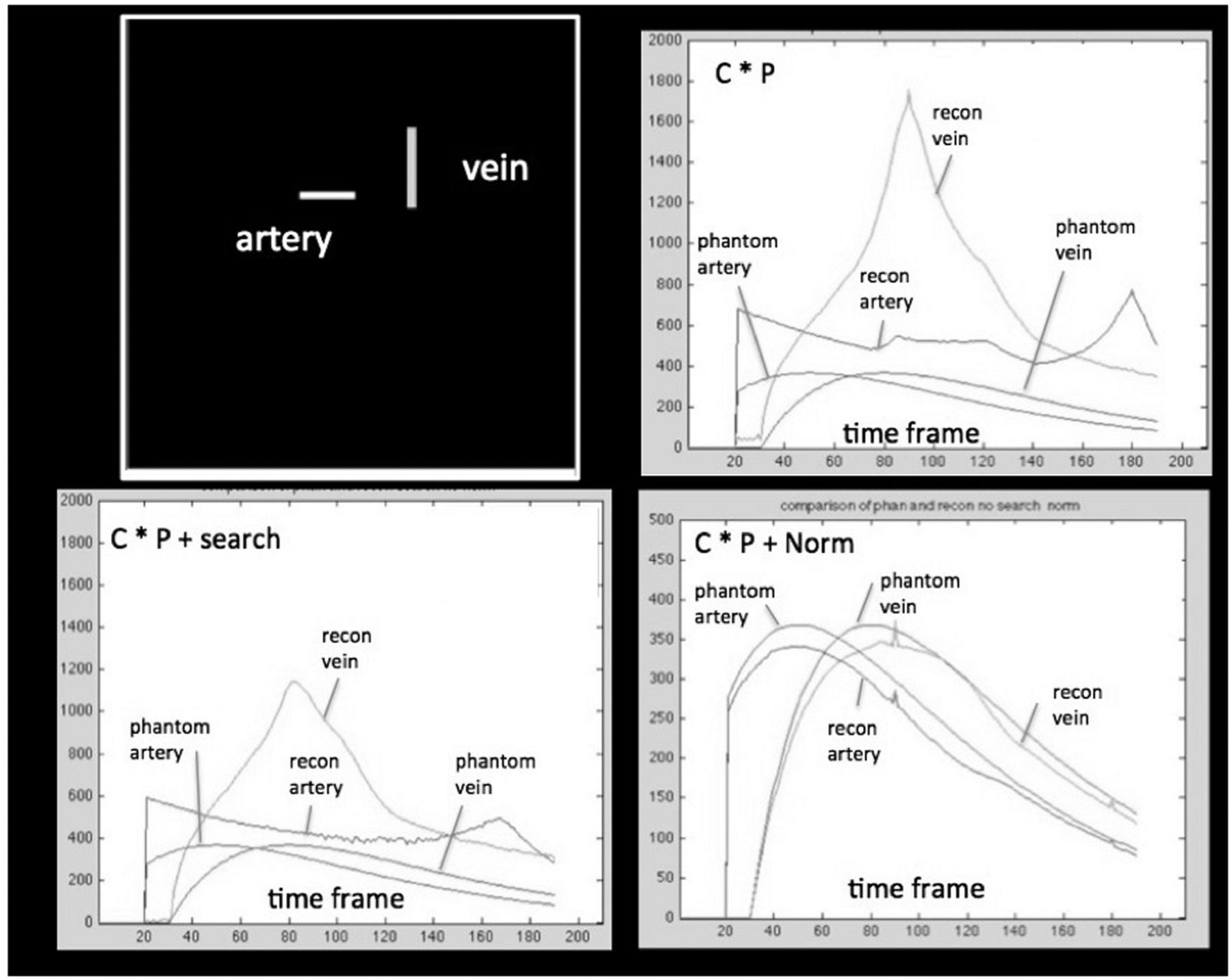
    Demonstration of the effects of reconstruction elements on accuracy of reconstructed simulated vessel signal curves. The digital phantom is shown in the top frame on the left. The vessels in the phantom (phantom artery and phantom vein) had specified time dependence. These are considered to represent ground truth. The input time curves for these vessels are shown in each panel for comparison. C indicates constraining image; P, projections. The reconstructed wave forms are shown: 1) after multiplication (represented by C × P) of the projections by a binary constraining image (top right); 2) multiplication of the projections by the binary constraining image plus an angular minimum search (represented by C × P + search, bottom left); and 3) multiplication of the projections by the binary constraining image followed by normalization by the estimate of the numbers of projected ray voxels obtained from the constraining image (C × P + Norm, bottom right). The y-axis shows arbitrary units of attenuation. The x-axis shows the projection number.

  • Fig 4.
    • Download figure
    • Open in new tab
    • Download powerpoint
    Fig 4.

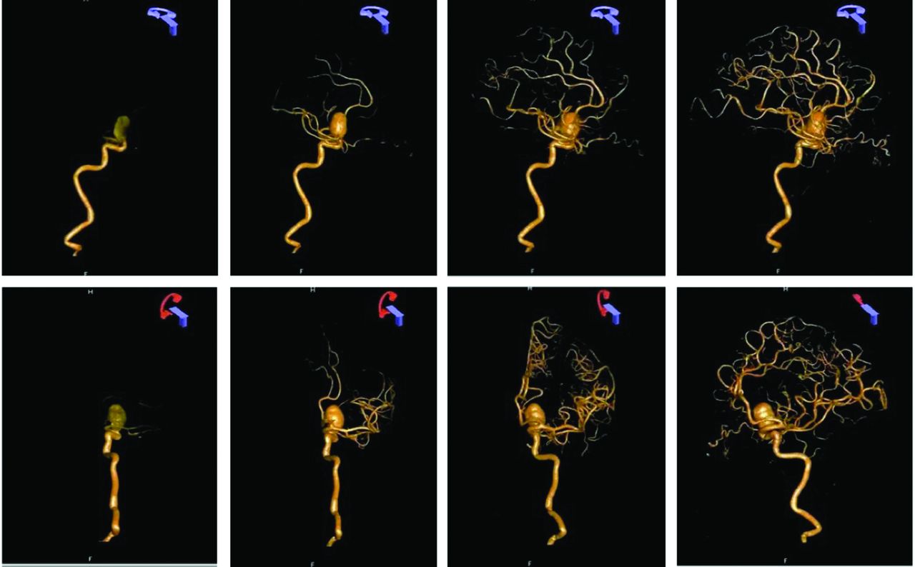
    Four-dimensional DSA reconstruction from a 3D-DSA reconstruction performed for evaluation of an unruptured paraclinoid aneurysm. An IA injection of contrast was used for this examination. The top row shows selected timeframes viewed at the rate of 6 frames per second at a fixed viewing angle. The bottom row shows the same timeframes viewed at 4 different angles, which would have not been obtainable in a biplane acquisition because of the mechanical inability to position the A-plane gantry (red icon).

  • Fig 5.
    • Download figure
    • Open in new tab
    • Download powerpoint
    Fig 5.

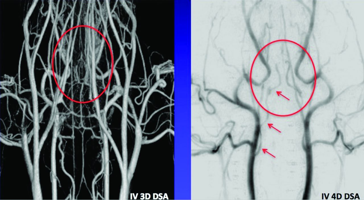
    Comparison of intravenous 3D-DSA and early arterial phase time from a 4D-DSA reconstruction. In the standard 3D-DSA on the left, the overlap of both arteries and veins obscures visualization of the internal carotid arteries. In this example, there is no viewing angle that will eliminate this overlap in the 3D-DSA. The image on the right is an early arterial timeframe from a 4D-DSA reconstruction viewed from the same angle as the 3D-DSA on the left. In this 4D image, the full course the right internal carotid artery is clearly visualized (red arrows). The red ellipse shows the position of the distal segments of the internal carotid arteries in both images.

  • Fig 6.
    • Download figure
    • Open in new tab
    • Download powerpoint
    Fig 6.

    Color-coded 4D-DSA and bolus arrival images. The sequence was displayed at a rate of 6 frames per second for visualization of arrival times between 1 and 4.5 seconds. On the top of the figure, binarized 4D-DSA timeframes multiply a time-of-arrival map providing a dynamic display in which each pixel in each timeframe is represented by a quantitative time of arrival value. In the bottom half of the figure, a sliding Gaussian display window is used to show the passage of the bolus through the AVM. The 4D-DSA TOA volume (static) and 4D-DSA TOA (dynamic) 3D timeframes can be viewed from any angle. The 4D-DSA TOA (dynamic) 3D timeframes allow viewing of the temporal dynamics of the 4D-DSA TOA from any angle at any point in time for which data were acquired. These images were obtained by use of an IA injection of contrast medium.
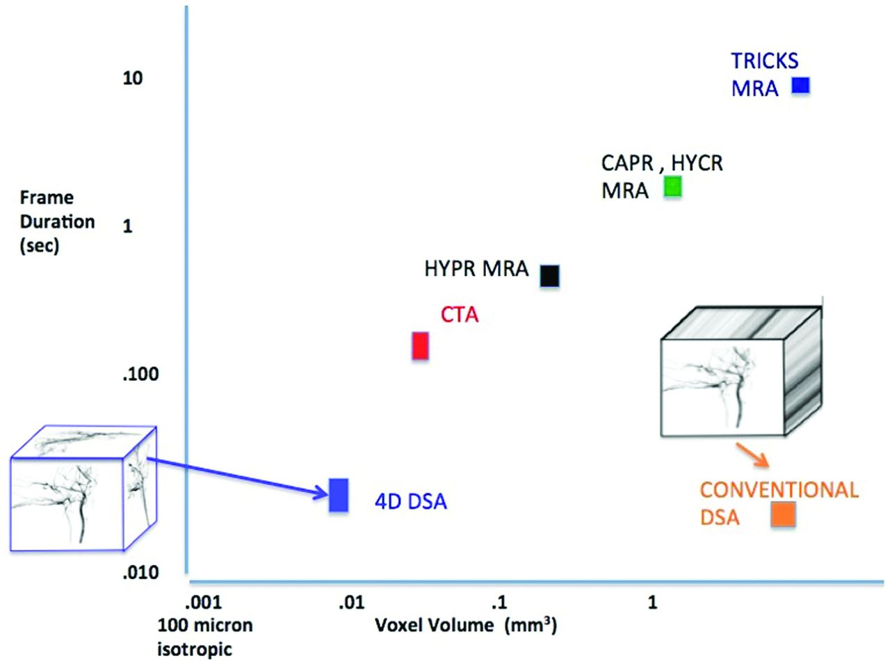
  • Fig 7.
    • Download figure
    • Open in new tab
    • Download powerpoint
    Fig 7.

    Relative spatial and temporal resolution for competing time-resolved angiography methods. The MRA values are based on the hybrid MRA method reported by Wu et al12 and provide 0.69-mm pixel dimensions, leading to a voxel volume of 0.33 mm3. Typical frame times are 0.3 seconds. The CTA estimate is based on the large area Aquilion system (Toshiba Medical Systems, Tokya, Japan)14 with a pixel dimension of 0.625 and frame rate of approximately 3/s. The 4D-DSA ultimate spatial resolution assuming a magnification factor of 1.5 and a pixel dimension 100 should easily support 200-μ pixel dimensions in a patient, leading to a voxel volume of 0.008 mm3.

PreviousNext
Back to top

In this issue

American Journal of Neuroradiology: 34 (10)
American Journal of Neuroradiology
Vol. 34, Issue 10
1 Oct 2013
  • Table of Contents
  • Index by author
  • Complete Issue (PDF)
Advertisement
Print
Download PDF
Email Article

Thank you for your interest in spreading the word on American Journal of Neuroradiology.

NOTE: We only request your email address so that the person you are recommending the page to knows that you wanted them to see it, and that it is not junk mail. We do not capture any email address.

Enter multiple addresses on separate lines or separate them with commas.
4D Digital Subtraction Angiography: Implementation and Demonstration of Feasibility
(Your Name) has sent you a message from American Journal of Neuroradiology
(Your Name) thought you would like to see the American Journal of Neuroradiology web site.
CAPTCHA
This question is for testing whether or not you are a human visitor and to prevent automated spam submissions.
Cite this article
B. Davis, K. Royalty, M. Kowarschik, C. Rohkohl, E. Oberstar, B. Aagaard-Kienitz, D. Niemann, O. Ozkan, C. Strother, C. Mistretta
4D Digital Subtraction Angiography: Implementation and Demonstration of Feasibility
American Journal of Neuroradiology Oct 2013, 34 (10) 1914-1921; DOI: 10.3174/ajnr.A3529

Citation Manager Formats

  • BibTeX
  • Bookends
  • EasyBib
  • EndNote (tagged)
  • EndNote 8 (xml)
  • Medlars
  • Mendeley
  • Papers
  • RefWorks Tagged
  • Ref Manager
  • RIS
  • Zotero
0 Responses
Respond to this article
Share
Bookmark this article
4D Digital Subtraction Angiography: Implementation and Demonstration of Feasibility
B. Davis, K. Royalty, M. Kowarschik, C. Rohkohl, E. Oberstar, B. Aagaard-Kienitz, D. Niemann, O. Ozkan, C. Strother, C. Mistretta
American Journal of Neuroradiology Oct 2013, 34 (10) 1914-1921; DOI: 10.3174/ajnr.A3529
del.icio.us logo Twitter logo Facebook logo Mendeley logo
  • Tweet Widget
  • Facebook Like
  • Google Plus One
Purchase

Jump to section

  • Article
    • Abstract
    • ABBREVIATIONS:
    • Materials and Methods
    • Results
    • Discussion
    • Conclusions
    • ACKNOWLEDGMENTS
    • Footnotes
    • REFERENCES
  • Figures & Data
  • Info & Metrics
  • Responses
  • References
  • PDF

Related Articles

  • No related articles found.
  • PubMed
  • Google Scholar

Cited By...

  • 4D-DSA for Assessment of the Angioarchitecture and Grading of Cranial Dural AVF
  • 4D DSA: technical addition or big revolution?
  • 4D-DSA: Development and Current Neurovascular Applications
  • Four-dimensional digital subtraction angiography for exploration of spinal cord vascular malformations: preliminary experience
  • Optimizing the Quality of 4D-DSA Temporal Information
  • Quantitative and Qualitative Comparison of 4D-DSA with 3D-DSA Using Computational Fluid Dynamics Simulations in Cerebral Aneurysms
  • Quantification of Blood Velocity with 4D Digital Subtraction Angiography Using the Shifted Least-Squares Method
  • Time-resolved 3D rotational angiography: display of detailed neurovascular anatomy in patients with intracranial vascular malformations
  • 4D DSA for Dynamic Visualization of Cerebral Vasculature: A Single-Center Experience in 26 Cases
  • Application of Time-Resolved 3D Digital Subtraction Angiography to Plan Cerebral Arteriovenous Malformation Radiosurgery
  • Comparison of the Diagnostic Utility of 4D-DSA with Conventional 2D- and 3D-DSA in the Diagnosis of Cerebrovascular Abnormalities
  • 4D DSA a new technique for arteriovenous malformation evaluation: a feasibility study
  • Technology developments in endovascular treatment of intracranial aneurysms
  • Intracranial Aneurysms: Wall Motion Analysis for Prediction of Rupture
  • A Comparison of 4D DSA with 2D and 3D DSA in the Analysis of Normal Vascular Structures in a Canine Model
  • Changes of Time-Attenuation Curve Blood Flow Parameters in Patients with and without Carotid Stenosis
  • Crossref (69)
  • Google Scholar

This article has been cited by the following articles in journals that are participating in Crossref Cited-by Linking.

  • Technology developments in endovascular treatment of intracranial aneurysms
    Robert M Starke, Aquilla Turk, Dale Ding, Richard W Crowley, Kenneth C Liu, Nohra Chalouhi, David M Hasan, Aaron S Dumont, Pascal Jabbour, Christopher R Durst, Raymond D Turner
    Journal of NeuroInterventional Surgery 2016 8 2
  • Flat-Panel Computed Tomography (DYNA-CT) in Neuroradiology. From High-Resolution Imaging of Implants to One-Stop-Shopping for Acute Stroke
    A. Doerfler, P. Gölitz, T. Engelhorn, S. Kloska, T. Struffert
    Clinical Neuroradiology 2015 25 S2
  • 4D DSA a new technique for arteriovenous malformation evaluation: a feasibility study
    Carolina Sandoval-Garcia, Kevin Royalty, Pengfei Yang, David Niemann, Azam Ahmed, Beverly Aagaard-Kienitz, Mustafa K Başkaya, Sebastian Schafer, Charles Strother
    Journal of NeuroInterventional Surgery 2016 8 3
  • Brain Vascular Imaging Techniques
    Bàrbara Laviña
    International Journal of Molecular Sciences 2016 18 1
  • Intracranial Aneurysms: Wall Motion Analysis for Prediction of Rupture
    A.E. Vanrossomme, O.F. Eker, J.-P. Thiran, G.P. Courbebaisse, K. Zouaoui Boudjeltia
    American Journal of Neuroradiology 2015 36 10
  • 4D DSA for Dynamic Visualization of Cerebral Vasculature: A Single-Center Experience in 26 Cases
    S. Lang, P. Gölitz, T. Struffert, J. Rösch, K. Rössler, M. Kowarschik, C. Strother, A. Doerfler
    American Journal of Neuroradiology 2017 38 6
  • 4D-DSA: Development and Current Neurovascular Applications
    K.L. Ruedinger, S. Schafer, M.A. Speidel, C.M. Strother
    American Journal of Neuroradiology 2021 42 2
  • Time-resolved 3D rotational angiography: display of detailed neurovascular anatomy in patients with intracranial vascular malformations
    Stephanie Lescher, Sonja Gehrisch, Sigrun Klein, Joachim Berkefeld
    Journal of NeuroInterventional Surgery 2017 9 9
  • Uterine Arteriovenous Malformation: A Pictorial Review of Diagnosis and Management
    Van Trung Hoang, Hoang Anh Thi Van, Cong Thao Trinh, Ngoc Trinh Thi Pham, Chinh Huynh, To Nguyen Ha, Phuong Hai Huynh, Hoang Quan Nguyen, Uyen Giao Vo, Thanh Thao Nguyen
    Journal of Endovascular Therapy 2021 28 5
  • Application of Time-Resolved 3D Digital Subtraction Angiography to Plan Cerebral Arteriovenous Malformation Radiosurgery
    K.-K. Chen, W.-Y. Guo, H.-C. Yang, C.-J. Lin, C.-H.F. Wu, S. Gehrisch, M. Kowarschik, Y.-T. Wu, W.-Y. Chung
    American Journal of Neuroradiology 2017 38 4

More in this TOC Section

  • Fast Contrast-Enhanced 4D MRA and 4D Flow MRI Using Constrained Reconstruction (HYPRFlow): Potential Applications for Brain Arteriovenous Malformations
  • Quiet PROPELLER MRI Techniques Match the Quality of Conventional PROPELLER Brain Imaging Techniques
  • Predictors of Reperfusion in Patients with Acute Ischemic Stroke
Show more Brain

Similar Articles

Advertisement

Indexed Content

  • Current Issue
  • Accepted Manuscripts
  • Article Preview
  • Past Issues
  • Editorials
  • Editors Choice
  • Fellow Journal Club
  • Letters to the Editor

Cases

  • Case Collection
  • Archive - Case of the Week
  • Archive - Case of the Month
  • Archive - Classic Case

Special Collections

  • Special Collections

Resources

  • News and Updates
  • Turn around Times
  • Submit a Manuscript
  • Author Policies
  • Manuscript Submission Guidelines
  • Evidence-Based Medicine Level Guide
  • Publishing Checklists
  • Graphical Abstract Preparation
  • Imaging Protocol Submission
  • Submit a Case
  • Become a Reviewer/Academy of Reviewers
  • Get Peer Review Credit from Publons

Multimedia

  • AJNR Podcast
  • AJNR SCANtastic
  • Video Articles

About Us

  • About AJNR
  • Editorial Board
  • Not an AJNR Subscriber? Join Now
  • Alerts
  • Feedback
  • Advertise with us
  • Librarian Resources
  • Permissions
  • Terms and Conditions

American Society of Neuroradiology

  • Not an ASNR Member? Join Now

© 2025 by the American Society of Neuroradiology All rights, including for text and data mining, AI training, and similar technologies, are reserved.
Print ISSN: 0195-6108 Online ISSN: 1936-959X

Powered by HighWire